Diseases General Health Skin Conditions

Archive for December, 2016

Green Tongue

Dec 14 2016 Published by under Diseases and Conditions

Why is my Tongue Green?

We all know that, in healthy individuals, the tongue has a common pink color, with a smooth texture. However, it is possible that certain conditions lead to the modification of both the color and the texture of the tongue. As you will have the opportunity to read below, the green tongue can appear as the result of an oral infection, after trauma or due to sores.

A healthy individual can be recognized after the state of the oral cavity. If a person’s tongue has gone green, this is suggestive of an underlying health problem. It is highly important to make the correct diagnosis, so as to recommend the best treatment.

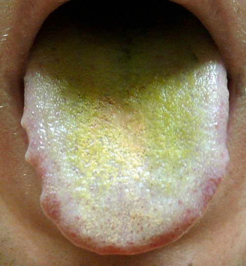
Pictures of a Green tongue

Here is how a green tongue looks like:
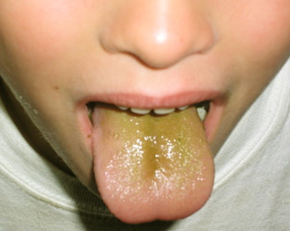
green tongue

green tongue pictures

Symptoms Signs of a Green Tongue

These are the most common symptoms associated with the green tongue:

In case of oral candidiasis:

  • White tongue in the initial stage
  • Color changes to green, after treatment or food
  • Underneath the initial colored layer, the tongue is red and may even bleed (upon scraping)
  • The dorsal surface of the tongue is affected (in the majority of the cases); however, the infection can be encountered in any part of the tongue
  • Bad breath
  • Burning sensation at the level of the mouth
  • Pain/discomfort (especially after eating food that is spicy or hot)

In case of the hairy tongue syndrome:

  • Hair-like structures are present on the surface of the tongue (dorsal surface)
  • Bacterial contamination can case these strands to become green or even black in color
  • Metallic taste in the mouth
  • Bad breath
  • Dry mouth.

What Causes a Green tongue?

These are the most common causes that can cause the tongue to become green:

Oral candidiasis (oral thrush)

  • Most common cause
  • Fungal infection (caused by Candida albicans)
  • Often encountered in individuals whose immune system is weak
  • Also seen in those wearing ill-fitted prostheses or dentures (prolonged wear)

Hairy tongue syndrome

  • Exact cause has yet to be identified
  • These are the risk factors associated with the appearance of this condition:
    • Smoking
    • Excessive mouthwash rinsing
    • Poor dental hygiene
    • Weak immune system

Other causes

  • Upper respiratory tract infection (sore throat) – extension of the infection to the tongue
  • Prolonged treatments with antibiotics
  • Toothpaste/mouthwash – the ingredients contained in these products can cause the tongue to become green (temporary)
  • Green colorants contained in candy and other sweets can lead to the appearance of a green tongue (temporary)
  • Drugs/marijuana consumption
  • Tongue piercing (green color of the tongue is suggestive of an infection, especially if there is associated discharge and pain).

Diagnosis

These are the most common methods used for the diagnosis of the green tongue:

Medical history of the patient

  • When did the color of the tongue change?
  • Are there other symptoms present?
  • Previous health problems (and treatments)

Physical examination

  • Microscopic examination of the tongue
    • Identification of abnormal long papillae strands (hairy tongue syndrome)

Laboratory testing

  • CBC (Complete blood count) – level of white blood cells
  • Tongue culture – identification of microorganisms (fungus or bacteria).

Green tongue Treatment

These are the treatments recommended for the green tongue, according to the main cause:

Oral candidiasis

  • Anti-fungal medication (topical administration)
  • Increased water intake
  • Balanced diet (fresh fruits and vegetables)

Hairy tongue syndrome

  • Treatment for the underlying cause
  • Lifestyle changes
    • Avoid smoking
    • Excellent dental hygiene (daily brushing)
  • Dislodgment of the surface epithelium of the tongue
    • Frequent brushing of the tongue surface
  • Avoid mouthwash or other products that may stimulate the growth of the tongue surface tissue (such as hydrogen peroxide or other oxygenating products)
  • Avoid products that cause the staining of the tongue (toothpaste, mouthwash, candy, chewing gum etc.).

Apart from the specific treatment for the underlying cause, one must also make a change in the diet. For example, you should make sure that the diet is rich in fiber, so that the colon is clean and free of toxins. Also, you need to maintain an excellent oral hygiene and quit smoking.

The water intake should be increased, in order to keep the tongue clean and prevent the food debris from depositing in the small papillae of the tongue. Gargling with warm water is also recommended, especially after a meal. Crunchy fruits and vegetables should be included within the diet, as they guarantee a natural cleaning process for the surface of the tongue. When brushing the teeth, make sure that you use the other side of the toothbrush in order to clean the tongue as well. A tongue scraper can be used at home, in order to eliminate the green coating and promote a faster healing process. Sugary foods and dairy should be eliminated from the diet, as these lead to increased mucus production at the level of the tongue.

Natural remedies

These are the most common natural remedies that are recommended for the symptoms associated with the green tongue:

  • Turmeric and honey
    • Mix the two natural remedies into a paste
    • Both have strong antibacterial properties
    • Can be used to keep the oral thrush under control
  • Mint/clove
    • Recommended for the bad breath (halitosis) associated with the green tongue
  • Vitamin C
    • Enhanced immune system
    • Prevention/elimination of oral thrush
  • Baking soda
    • Exfoliating properties
    • Maintain a good pH level in the mouth (acid neutralization)
    • Mix it with lemon juice and apply the resulting paste to the tongue
    • Rinse afterwards with water
  • Salt
    • Brush your tongue gently with salt (natural tongue scraper – remove debris and dead cells)
    • Antiseptic properties (elimination of bacteria)
  • Probiotics
    • A capsule should be mixed with water, the solution being used instead of mouthwash
  • Glycerin
    • Apply the vegetable glycerin to the tongue and brush it gently
    • Be sure to rinse with warm water afterwards
    • The procedure should be repeated until the tongue regains its natural color
  • Aloe vera
    • Anti-inflammatory and antibacterial properties
    • May remove the coating on the tongue, as well as eliminate the bad breath
    • Recommended choice – fresh aloe vera juice
  • Colloidal silver
    • Recommended in the situation of yeast overgrowth at the level of the mouth
    • Natural antifungal properties, also helps with the bad breath
    • Should be diluted with water before usage
  • Neem (Indian lilac)
    • Antifungal and antibacterial properties
    • Blood purifier (toxin removal)
    • Recommended – boiled in water, used for gargle
    • A thorough rinse with water is required to complete the process.

No responses yet

Enthesopathy

Dec 12 2016 Published by under Bone, Joints and Muscle

What is Enthesopathy?

Whenever a health problem occurs at the level of a tendon or a ligament’s insertion on the bone, this condition is known as enthesopathy. The name derives from the fact that the insertion site is presented in medical terms as entheses. When inflammation appears as part of the process, the condition will be known as enthesitis.

As you will have the opportunity to see below, enthesopathy can appear at different levels of the body, including in the spine, upper and inferior extremities. When it comes to occurs at the level of the vertebral column, it is generically presented as spondyloarthropathies (ankylosing spondylitis is one of the most common types of spondyloarthropathy, affecting primarily men).

enthesopathy

At the level of the upper extremities, enthesopathy can appear under the following forms: adhesive shoulder capsulitis (frozen shoulder), rotator cuff syndrome, periarthritis (shoulder involvement), fibrositis with involvement of the shoulder and scapula, hand/wrist synovitis, wrist periartritis. As for the inferior extremities, the most common forms include: tendinitis (gluteal, psoas or trochanteric) and spurs at the level of the iliac crest.

Enthesopathy Symptoms

These are the general symptoms of enthesopathy:

  • Inflammation (acute or chronic) → can modify the aspect of certain anatomical structures (if severe) → sausage-like aspect of the fingers or toes (for example)
  • Pain → nerve endings of entheses → enthesalgia → chronic musculoskeletal pain
  • Abnormal calcification/ossification → entheses level
  • Irritation at the level of the affected tendon or ligament attachment
  • Soreness can also be present among the symptoms
  • The symptoms can be intermittent (come and go)
  • Recurring stress → more inflammation → calcification and fibrosis
  • Because of the pain, inflammation and soreness, the patient might not be able to perform regular activities (for example, if the Achilles tendon is affected, he/she will have difficulties walking or running).

Causes of Enthesopathy

These are the most common causes that lead to the appearance of enthesopathy:

  • Inflammatory conditions
    • Psoriatic arthritis
    • Reactive arthritis
    • Rheumatoid arthritis
    • Osteoarthritis
  • Crohn’s disease
  • Reiter’s syndrome
  • Tennis elbow/golfer elbow
  • Postman’s heel
  • Iliotibial band syndrome
  • Physical injury or trauma
  • Excess load
    • Plantar fasciitis
  • Other causes
    • Diffuse idiopathic skeletal hyperostosis
    • Medication (side-effect)
    • Drug abuse
    • Infection
    • Metabolic change.

Enthesopathy Treatment

These are the general measures of treatment recommended for enthesopathy:

  • Anti-inflammatory medication
    • Topical or oral administration (depending on the severity of the pain and inflammation)
    • Reduce the inflammation and bring the necessary pain relief
    • Recommended choices – acetaminophen, ibuprofen, naproxen
  • Corticosteroid injections
    • Recommended in the situation that the above-mentioned anti-inflammatory medication has failed to provide the desired results
  • Anti-TNF agents
    • Recommended for patients who suffer for more serious cases, with severe inflammation
    • Indicated choices – Humira, Remicade and Enbrel
  • Radiotherapy
    • Beneficial effects for patients who suffered from affectation at the level of the heel
  • Other treatment solutions
    • Combination of sulfasalazine and methotrexate
    • Heel inserts (for patient who suffer from plantar fasciitis)
    • Physical therapy
    • Occupational therapy
    • Ultrasound
    • Electrical stimulation
    • Ice application (wrapped in a towel, not directly on the skin).

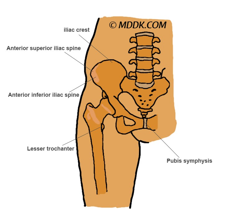
Enthesopathy of hip

enthesopathy of the hip

Picture of Enthesopathy of the hip

When a person suffers from the enthesopathy of the hip, this means that insertion of the tendons or ligaments at the level of the hip bone sockets have been affected. This is an arthritic disorder and it may impair the proper functioning of the hip, leading to reduced range of motion and pain. The patient might experience difficulties when trying to walk or move in general.

At the insertion of the hip muscles tendons or ligaments, one can either see calcification or ossification. The articular capsule is the least affected by the enthesopathy. X-ray investigations demonstrate characteristic changes at the level of the femoral head and also at the joint acetabulum. Severe cases of hip enthesopathy, such as the greater trochanteric bursal pain, can be successfully treated with platelet-rich plasma (provided other treatments have failed to provide the desired results).

Spinal enthesopathy

When talking about spinal enthesopathy, one talks about medical conditions, such as ankylosing spondylitis. Because of the chronic inflammation, in time, the ligamentous insertions to the spine are damaged. The more the ligaments are eroded, the more the bone will try to compensate through calcification or ossification. The treatment for spinal enthesopathy includes the administration of non-steroidal anti-rheumatic medication.

It is possible that a non-inflammatory form of enthesopathy appears at the level of the spine, this being known as ankylosing vertebral hyperostosis. This hyperostotic disease can cause the insertions of the spinal ligaments to become ossified, leading to the appearance of intervertebral bridges. Once these bridges have been formed, the patient will start to experience the first symptoms: pain and rigidity at the level of the neck and back, with reduced range of motion. In more severe cases, it is possible that the patient suffers from the compression of the spinal cord at cervical level or from the stenosis of the lumbar canal. The overall mobility of the spine can be reduced, with the combination of changes leading to the appearance of secondary osteoarthritis.

Spinal enthesopathy can also be present in patients who suffer from psoriatic arthritis, leading to back pain and a wide range of other symptoms. The back pain is chronic, having a gradual onset and improving with physical movement. Characteristic for this kind of problem is the fact that the pain does not improve with physical rest. The pain can keep the patient awake at night, the level of stiffness being at maximum in the morning. In general, this form of spinal enthesopathy is diagnosed in men who are over 40 years of age.

The treatment options include physical therapy, the administration of non-steroidal anti-inflammatory medication and intramuscular corticosteroid injections. Sulphasalazine might be used to provide relief from the pain but it is not effective in all of the patients. Anti-TNF therapy remains the standard treatment for those who suffer from more severe forms.

Achilles tendon enthesopathy

When the insertion of the Achilles tendon is affected, the condition is known as Achilles tendon enthesopathy. This condition occurs because of constant traction of the respective muscle tendon on the calcaneus bone. Being sedentary or obese are considered as risk factors for such health problems, as they lead to the shortening of the calf muscles (or to their chronic contracture). The patient presents pain at the level of the posterior heel, especially during walking. The pain is exacerbated by the palpation of the tendon at its insertion point. The same goes for the manual dorsiflexion of the ankle.

There are three treatment recommended for Achilles tendon enthesopathy, meaning physical therapy (stretching), splinting and heel lifts. Physical therapy is essential, as it can help one stretch the shortened calf muscles and eliminate the contractures that are present at this level. Night splints can also be used, having the purpose of passively stretching the shortened calf muscles during sleep. These are considered as one of the best ways to prevent additional contractures. The usage of heel lifts is recommended to be only temporary, as they can relieve the stress experienced by the heel during weight bearing. Heel lifts can alleviate the pain experienced by the patient, improving the gait and avoiding the compensatory movements of the hip (these can have a harmful effect, leading to low back pain).

No responses yet

Steatorrhea

Dec 10 2016 Published by under Diseases and Conditions

Steatorrhea can be defined as the present of excessive quantities of fat in the stool.

Clinical Presentation

The floating of the stool is also possible, due to the fact that excessive quantities of gas are present as well. The excess fat will cause the stool to have an oily aspect, such changes being accompanied by a characteristic foul odor. When one suffers from steatorrhea, fecal incontinence might be present. Some patients described oily anal leakage to be among the other symptoms they have experienced. When the main problem is caused by the lack of bile acids, the stool will turn a gray or pale color.

In patients who suffer from steatorrhea, abdominal pain and cramps are frequent. Patients complain of constant bloating, suffering either from constipation or diarrhea. In some cases, it is possible that blood is eliminated at the same time with the stool. Flatulence, nausea and vomiting complete the steatorrhea medical picture.

Apart from these symptoms, it is possible that the patients suffer from decreased density at the level of the bones. If there is a low light level, they may have problems with their vision (impaired vision). The more fat is eliminated through the stool, the more the patient will experience problems, especially when it comes to the blood clotting time (this will be reduced).

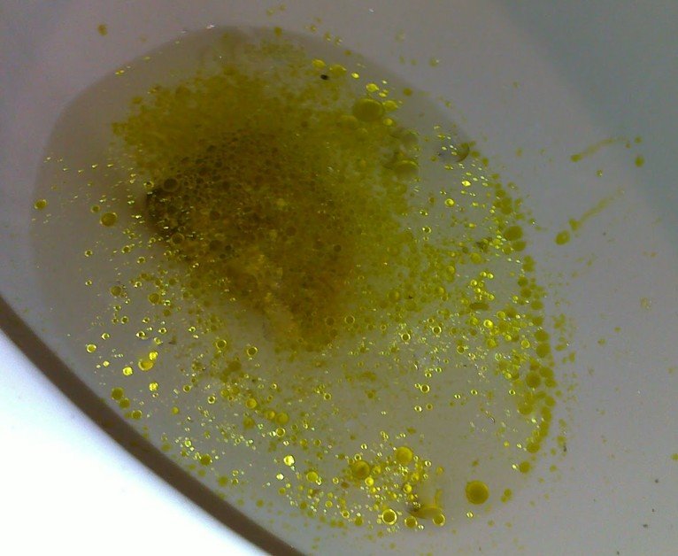
Steatorrhea Pictures

steatorrhea

Picture of Steatorrhea – floating of fat in the stool

Steatorrhea Causes

These are the main causes that can lead to the appearance of steatorrhea:

  • Inadequate production of bile (bile acids lacking)
    • Liver damage
    • Medication to reduce the level of lipids in the blood (hypolipidemic)
    • Surgical removal of gallbladder (cholecystectomy)
  • Pancreatic enzyme defect
  • Defect at the level of the mucosal cells
  • Medication that has as purpose the blockage of fat absorption
  • Fat rich diet (indigestible or excess oils in the diet might cause similar problems)
  • Acromegaly treatment
    • Somatostatin analogues – octreotide, lanreotide (adverse effect)
  • Other causes
    • Eating nuts in large quantities (such as cashews)
    • Jojoba oil (indigestible oil)
    • Escolar/oilfish consumption
    • Consumption of artificial fats.

Apart from the above-mentioned causes, it is worth mentioning that steatorrhea can be found in association with a number of medical conditions. For example, patients who suffer from different types of malabsorption also present steatorrhea. Among these conditions there are the celiac disease, the inflammatory bowel disease and the Bassen-Kornzweig syndrome (normal absorption of fat is prevented).

Patients who suffer from pancreatic conditions, such as exocrine pancreatic insufficiency or the inflammation of the pancreas (pancreatitis), can present steatorrhea. When the bile duct is obstructed by gallstones of different sizes, steatorrhea is going to occur as one of the consequences of this blockage. Excess fat in the stool is encountered in patients diagnosed with pancreatic cancer, if the biliary outflow is obstructed by the neoplastic growth. Patients who suffer from primary sclerosing cholangitis, a condition that affects the functioning of the bile ducts, present such problems as well.

Steatorrhea is encountered in patients who have been diagnosed with bacterial infections but also in those who suffer from the short bowel syndrome or cystic fibrosis. Excess quantities of fat in the stool are encountered in patients diagnosed with Zollinger-Ellison syndrome, parasitic infections (especially with giardia) and thyroid disorders (such as hyperthyroidism). Steatorrhea is often encountered in patients who abuse weight loss pills and in those who have been diagnosed with tropical sprue, a form of malabsorption which is characteristic only for certain tropical regions.

Diagnosis of Steatorrhea

These are the most common methods for diagnosis the underlying condition:

  • Medical history of the patient
    • When did steatorrhea appear for the first time
    • Apart from the oily appearance, are there blood, excess mucus or pus present in the stool
    • Stool color change (when)
    • Other symptoms presented
    • Diet (current and if there were any recent changes)
    • Current and past treatments (including surgical interventions)
    • Similar problems in the past
    • Family medical history (inherited conditions)
  • Physical examination
    • Palpation of abdomen
  • Lab testing
    • Hepatic enzymes
    • Leukocytes
    • Complete blood count
  • Imaging studies
    • CT scan/MRI
    • Ultrasonography
  • Other investigations
    • Function tests of intestinal absorption
      • D-xylose absorption
      • Schilling tests (I, II)
      • Bile acid breath test
    • Biopsy/barium study – identification of bowel abnormalities
    • Duodenal aspiration (giardia suspicion)
    • Serum gastrin (Zollinger-Ellison syndrome).

Steatorrhea Treatment

When it comes to steatorrhea, the treatment is concentrated on the underlying cause and not on the symptom per se. If the underlying cause is successfully treated, the symptomatology is going to improve as well. In some patients, digestive enzyme supplements can be administered, so as to improve the experienced symptoms.

In the situation that the patient suffers from a form of cancer, it is important to institute treatment as soon as possible (best survival rate). Chemotherapy and radiation therapy are among the treatment solutions that are recommended for such patients. If possible, the tumor can be surgically removed. If the steatorrhea is caused by liver cirrhosis, the only treatment available is the liver transplantation.

A dietary change is more than required for the patients who suffer from steatorrhea and other associated symptoms. Patients are advised to limit their fat intake, receiving replacements of pancreatic enzymes at the same time. These replacement pancreatic enzymes are also recommended in patients who suffer from chronic pancreatitis. Fat-soluble vitamins can also be administered, so as to help patients with chronic pancreatitis. Medium-chain triglycerides are recommended in patients who suffer from both steatorrhea and weight loss, while antibiotics are prescribed to those who suffer from bacterial infections at the level of the gastrointestinal tract. Last, but not least, if the steatorrhea is caused by bile acid malabsorption, bile acid sequesters such as cholestyramine can be administered.

How to Prevent Steatorrhea?

It is possible to prevent such health problems by maintaining an adequate diet, one that is rich in fresh fruits and vegetables, with a reduced fat intake. The alcohol intake should be reduced or even completely eliminated.

Are there any Complications of Steatorrhea?

Left untreated, the underlying conditions that have caused steatorrhea can lead to a number of complications. It is possible that these patients will suffer from anemia, intestinal obstruction and weight loss. If no treatment is undertaken in pediatric patients, these can suffer from failure to thrive and also from growth problems.

No responses yet

Burnt Tongue

Dec 09 2016 Published by under Oral Health

How to Deal with a Burnt Tongue?

No one wants to burn their tongue but the truth is that such things happen. Often times, we do not wait for our food or beverages to become cold. We drink our coffee scalding hot and we taste our food just as it is ready. Unfortunately, it is not that pleasant to have a burnt tongue. If you have gone through such an experience recently, be sure to try out the remedies suggested in this article. Keep in mind that, as soon as the burnt tongue has occurred, the standard first aid treatment will do.

Natural Remedies to treat Burnt Tongue

burnt tongue
These are the most common natural remedies that you can try for the burnt tongue:

  • Ice cube/ice pop
    • Sucking on an ice cube or an ice pop can be quite useful when it comes to a burnt tongue
    • Soothing effect on the recently burned tongue surface
    • Alternative – drinking a cold beverage (milk is perfect, as it has a soothing effect on its own)
  • Yoghurt
    • Excellent remedy for the burnt tongue
    • The yoghurt has a cooling effect, soothing the surface of the tongue
    • Do not swallow the yoghurt immediately, let it sit on the tongue, so as to soothe the burned tissues
    • Recommended – natural Greek yoghurt
  • Sugar
    • Recommended – sugar sprinkled on the tongue
    • Make sure that the sugar is placed on the burned surface of the tongue
    • Excellent remedy, helps you deal with the pain
  • Honey
    • Natural soothing properties
    • Can reduce the painful sensation caused by a burnt tongue
    • Keep the honey on the tongue for a short period of time, before swallowing it
    • Remedy not recommended in children under one year of age (toxic effect)
  • Vitamin E
    • Soothing effect on the burnt tongue
    • Faster healing process
    • Helps the tissues of the tongue to regenerate
    • Vitamin E oil applied directly to the tongue (from vitamin E capsule)
  • Mouth breathing
    • Soothing effect on the burnt tongue
    • Constant stream of air to the tongue (colder than the oral temperature)
  • No acidic/salty foods
    • Acidic foods to avoid – vinegar, tomatoes, citrus fruits, acidic fruit juice
    • Allowed – orange juice diluted with water (preferable cold, so that it has a more soothing effect)
    • Salty foods to avoid – potato chips (further irritation of the burnt area)
  • Aloe vera
    • Natural remedy – soothing effect on the burnt tongue
    • Can also promote a faster healing process
    • Recommended choices – aloe vera gel or natural aloe vera juice
    • Alternative – freezing the aloe vera juice in the form of an ice cube (ice cube sucking – cold application, delivers the necessary pain relief)
  • Baking soda
    • You can dilute the baking soda with water and swish it around the mouth
    • Soothing and calming effect, plus it has antibacterial properties
  • Tabasco sauce
    • Dilute the Tabasco sauce with water and rinse your mouth with the solution
    • Pain relieving effect, due to capsaicin, the active ingredient in the Tabasco sauce
  • Glycerin
    • Direct application to the burned surface of the tongue
    • Reduces the intensity of the burning sensation
  • Echinacea
    • Herbal drops applied directly to the tongue
  • Lavender oil
    • Applied directly to the tongue, with the help of a cotton dab
    • Pain relief and faster healing.

Methods for Pain Relief

Apart from that natural remedies that you can use, there are also specific methods to try out for pain relief:

  • Cough drops
    • Sucking on cough drops can reduce the discomfort or pain caused by the burnt tongue
    • Recommended cough drops – those that contain substances with soothing effect, such as menthol, phenol or benzocaine
    • Cough drops have an anesthetic effect on the tongue, thanks to the above-mentioned substances
    • Alternative – mouthwash (anesthetic effect – numbness and pain relief)
  • Chewing gum
    • Recommended – chewing gum with menthol (peppermint, spearmint)
    • Soothing and cooling effect on the tongue
  • Pain medication
    • Anti-inflammatory medication recommended – acetaminophen, ibuprofen or naproxen
    • Pain relief and reduction of inflammation
    • This medication should be taken with caution by those who suffer from chronic liver disease (processed through the liver)
  • No burn cream/ointment
    • Such products are designed exclusively to be used on the skin and not on the oral mucosa or the tongue
    • May contain ingredients that have a toxic effect upon ingestion
    • Only exception – cream/ointment designed intentionally for oral usage.

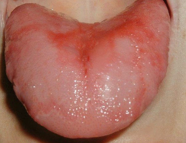
Pictures of a Burnt Tongue

burnt tongue pictures

burnt tongue pictures 2

When should you consider going to the doctor?

In the situation that the burns at the level of the tongue do not disappear within one week, you should consider seeing a specialist. The doctor might prescribe pain medication or other drugs, in order to speed up the healing process and improve the symptoms you are experiencing. In the situation that you present a burning sensation, without having an actual physical burn, you might be suffering from what is known as the burnt tongue syndrome. This medical problem can cause a lot of pain, especially since it is not restricted to the oral mucosa. A visit to the doctor becomes more than essential in this case, as you might also be suffering from an underlying condition, such as depression, hyperthyroidism or food allergies.

Prevention

Even though it may sound like the obvious thing to do, if you do not want to burn your tongue in the future, you need to be more careful. Avoid drinking beverages when they are too hurt, as they can hurt not only your tongue but also your esophagus, increasing the risk of esophageal cancer. The same goes for hot foods, as it can have a similarly damaging effect. If you cannot help it, then at least try to take small sips of your drink or, respectively, small bites of the food. In this way, you will reduce the risk of thermal burns at the level of the tongue and you will be able to enjoy your food/drink at the same time.

Future solutions

A team of researchers at the University of Texas have developed dissolvable oral strips that are recommended especially for the burnt tongue. According to the researchers, the strips will be available for application not only on the tongue, but also on the roof of the mouth or inside of the cheek. The oral strips contain benzocaine, a local anesthetic, which is released as they begin to dissolve.

No responses yet

Acrocyanosis

Dec 09 2016 Published by under Diseases and Conditions

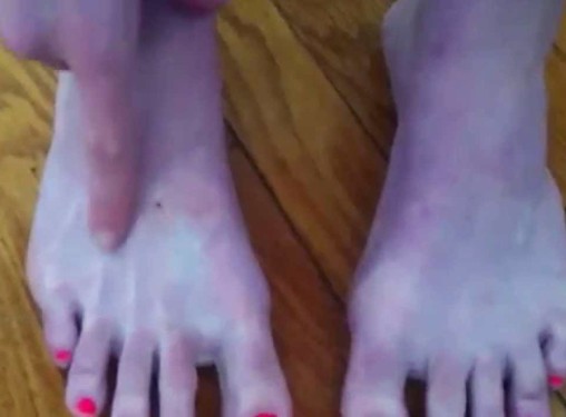
Acrocyanosis can be defined as the discoloration that occurs at the level of the extremities. In the majority of the situations, the hands become cyanotic, having a characteristic bluish tinge. However, it is possible that the acrocyanosis also affects the feet and even the face (distal parts). Even though this condition was identified a long time ago, scientists are still not certain about its exact cause. As you will have the opportunity to read below, one of the most commonly incriminated causes is represented by the neurohormonal disorder. In the situation that the patient is diagnosed with the benign form of acrocyanosis, no treatment is going to be necessary.

acrocyanosis

Acrocyanosis in Toddlers

This change should not be confused with the frostbites that appear after prolonged exposure to cold temperatures. The main difference between acrocyanosis and frostbite is pain. There are numerous other conditions that can mimic the symptomatology of acrocyanosis, such as Raynaud’s phenomenon, the blue finger syndrome or erythromelalgia.

It is possible that acrocyanosis appears in association with more severe medical problems, such as the connective tissue disease. In the situation that the acrocyanosis is caused by an underlying medical condition, such as cancer or infection, it is presented as secondary acrocyanosis. Acrocyanosis can be found in newborns, whether they are preterm or born on term. Even though there is no danger associated, it is possible to administer oxygen for prophylactic reasons.

Epidemiology

This condition is more often encountered in children and young people. There are a number of factors that increase the risk for acrocyanosis, such as living in a cold climate, working in the open outdoors or having a low BMI. Given the connection with the body mass index, it should come as no surprise that acrocyanosis is more often diagnosed in women than in men. No matter the climate, the incidence of acrocyanosis decreases with age. In women, the symptoms of acrocyanosis disappear, once they have reached menopause (suggestive of hormonal influence).

Acrocyanosis Symptoms

These are the most common symptoms that appear in association with acrocyanosis:

  • Peripheral cyanosis – hands, feet and face; rare situations, also the nipples
  • Cold and clammy extremities
  • Inflammation appears as a secondary effect of the cyanosis (especially when the temperature is not cold anymore)
  • Sweating – from moderate to severe (at the level of the extremities)
  • Normal peripheral pulse maintained
  • Symptoms are aggravated with the exposure to cold and improved when the temperature is warmer
  • Discoloration of the skin (bluish or cyanotic tinge).

Pathophysiology

The cyanosis that appears at the level of the extremities is believed to be caused by the vascular spasms in the superficial arteries of the skin. Because of these vasospasms, the small veins of the skin will dilate in a compensatory manner, which will cause the moderate-to-severe sweating at the level of the skin.

The secondary acrocyanosis is found in association with a wide range of medical conditions, such as hypoxemia (lung disease/chronic smoking), connective tissue diseases (systemic lupus erythematosus, rheumatoid arthritis, Wegener granulomatosis), cancer (ovarian cancer, Hodgkin’s lymphoma), peripheral vascular disease, malnutrition, atopic dermatitis, genetic disorders (Down’s syndrome), spinal cord injury and hematological disorders.

Diagnosis of Acrocyanosis

These are the most common methods used for the diagnosis of acrocyanosis:

  • Medical history of the patient
    • When did the symptoms appear for the first time
    • Similar problems in the past
    • Current and past treatments
    • History of illnesses
    • Family medical history (genetically-inherited disorders)
  • Physical examination
    • Examination of extremities
    • Identification of specific symptoms (cyanosis, sweating etc.)
  • Pulse oximetry
    • Normal result in the majority of cases (oxygen saturation)
  • Capillaroscopy
    • Recommended for the complementing of the clinical diagnosis
    • Often performed in patients who also suffer from disorders of the connective tissue
  • Other investigations
    • Urinalysis
    • Complete blood count
    • Liver and kidney function testing
    • Antibodies
    • Imaging studies (chest X-ray, CT scan, MRI)
    • Measurement of arteriosus and venous blood gases
    • Biopsy of the skin.

The differential diagnosis is highly important in this situation, being often made with Raynaud’s phenomenon. It should be highlighted that acrocyanosis is present all the time, as opposed to the manifestations of Raynaud’s phenomenon, which can come and go (intermittent). Acrocyanosis is not accompanied by ulcerations or other changes at the level of the skin, which can be useful for the differential diagnosis as well.

Apart from the differential diagnosis, medical specialists make avail of Kroq’s sign, which can be used to guide the diagnosis in the right direction. Even though this sign is characteristic for acrocyanosis, it is important to mention that it is not also specific. In order to bring out to surface the problems related to acrocyanosis, the doctor will apply pressure to the skin. The sign is considered positive when the blood returns to the pressed area from the periphery but not from beneath.

Acrocyanosis Treatment

As it was mentioned above, in the majority of the situations, no treatment is recommended (nor available). Those who suffer from acrocyanosis are advised to avoid prolonged exposure to cold or go out wearing protective garments (gloves, hats etc.). In more severe cases, a procedure that is known as sympathectomy can be performed, in order to decrease the activity of the sympathetic nervous system at local level. Alternative treatments, such as biofeedback or hypnosis, can help with the symptoms of acrocyanosis as well.

An alternative treatment consists of the administration of vasoactive drugs but this is rarely used. Some studies have tried to demonstrate the efficacy of these vasoactive drugs for acrocyanosis but further research is necessary to confirm this. Bioflavonoids can be administered in order to improve the symptoms of acrocyanosis. New studies are performed, so as to demonstrate the efficacy of the following treatment measures for patients with acrocyanosis: nicotinic acid derivatives, hexyl nicotinate (cream) and UVB (ultraviolet light).

Prognosis

Even though acrocyanosis cannot be cured, the prognosis remains positive. The prognosis is not as good, in the situation that there is an underlying condition causing the acrocyanosis, such as cancer (associated risk of death). The overall quality of life is not affected, as the extremities continue to function normally.

Pictures of Acrocyanosis

acrocyanosis pictures

acrocyanosis pictures 2

No responses yet

Lingering Cough

Dec 08 2016 Published by under Diseases and Conditions

A lingering cough can generate a lot of discomfort, especially if you are at work or in another public place. Often times, the cough is still present, due to the fact that the underlying condition has not been properly treated. So, if you want for your lingering cough to disappear, you need to seek out the adequate treatment for the medical condition that had let to it in the first place. In the situation that you are not certain about the diagnosis, do not hesitate to seek out the opinion of several medical professionals. In this way, you can be 100% sure that you will benefit from the right medical treatment.

Lingering Cough Symptoms

These are the symptoms that can accompany the lingering cough:

  • Runny nose
  • Watery eyes
  • Difficult swallowing (dysphagia)
  • Difficult breathing (dyspnea)
  • Pain with breathing or speaking
  • Throat irritation
  • Fever (from moderate to high-running; suggestive of infection)
  • Lack of appetite
  • Nausea and vomiting
  • State of general weakness
  • Pain at the level of the joints.

Causes of Lingering Cough

These are the main causes that stand behind a lingering cough:

  • Common cold – lingering cough because of irritated airways
  • Viral infection
    • Virus → inflammation + increased sensitivity at the level of the airways
    • Cough can be present for several weeks after the infection has cleared
  • Bacterial infection
    • Irritated airways → increased risk for bacterial infection
    • Type of bacterial infection → sinusitis, bronchitis, pneumonia etc.
    • Fever + pain accompany the lingering cough
  • Allergies
  • Asthma
  • Gastroesophageal reflux disease
    • Apart from the lingering cough, these are the symptoms that are present:
      • Heartburn
      • Indigestion
      • Burping and regurgitation
  • Obstructive sleep apnea
    • Symptoms present, apart from the lingering cough:
      • Lack of sleep (insomnia)
      • Snoring excessively or too loudly
      • Choking or gasping for breath during sleep
      • Because of the above-mentioned problems, the patient wakes up frequently during the night
      • Given the reduced hours of sleep during the night, the patient will feel sleepy throughout the entire day
  • Stress
    • Chronic stress → prolonged cold → lingering cough
  • Dehydration
    • Not drinking enough fluids during a cold/flu → chronic cough
    • Recommended fluids – water, juice, chicken soup
    • No alcohol, sugary or caffeine-based drinks → dehydration → aggravated cough
  • Excessive usage of nasal decongestants (sprays)
    • Excessive usage → worsening of the symptoms → rebound effect
    • Inflammation of nasal membrane → congestion + post-nasal drip + chronic cough
  • Air quality (too dry or too moist)
    • Dry air → winter → irritating effect → chronic cough
    • Moist air → excessive humidification → trigger of asthma attacks + dust mites/mold growth → allergies
    • Recommended humidity level in the home – 40-50% (both winter and summer)
  • Medication
    • Most commonly – medication to keep the blood pressure under control
    • Chronic and dry cough → side effect of medication
    • ACE inhibitors → used for high blood pressure → chronic cough, one of the most common side effects
    • Common prescribed ACE inhibitors include: Quinapril, Captopril, Benazepril, Lisinopril and Enalapril.

Treatment for Lingering Cough

These are the measures of treatment recommended for the lingering cough and the underlying conditions:

  • Anti-viral medication – recommended in case of viral infection
  • Non-steroidal anti-inflammatory medication
    • Recommended as symptomatic treatment for colds
    • Commonly prescribed NSAIDs include: acetaminophen, naproxen, ibuprofen
    • This medication should be administered with caution in patients who are suffering from chronic liver disease
  • Corticosteroids
    • Reduce the associated inflammation and bring the necessary relief from the cough
    • Cannot be administered for prolonged period of times, due to their negative side-effects over the general health
  • Antibiotics
    • Indicated for bacterial infections
    • The antibiotics should be taken for as long as they are prescribed, otherwise the bacteria will develop resistance to the treatment
    • Oral/intravenous administration (depending on the severity of the infection)
    • Probiotic supplements are administered at the same time with the antibiotic treatment, so as to maintain a healthy intestinal flora
  • Anti-cough medication
    • Administered as pills or as syrup (depending on the severity of the cough and also on the age of the patient; cough syrups are more commonly used in pediatric patients)
    • As the anti-cough medication contains codeine, the patient should refrain from driving a vehicle (codeine causes sleepiness)
  • Antihistamines
    • Recommended in case of allergies
    • Can reduce the inflammation and also improve the capacity of breathing
    • Can be administered as pills
  • Inhaler
    • Recommended in those who experience frequent asthma attacks
    • Improves the breathing capacity
  • Antacids
    • Recommended for patients who have been diagnosed with GERD
    • The improved of the GERD-related symptoms will lead to the disappearance of the chronic cough
  • Psychological counselling
    • Recommended for patients who are suffering from chronic stress and other associated problems (anxiety, depression)
    • The psychologist can recommend different methods to reduce stress:
      • Deep breathing
      • Relaxation techniques
      • Meditation
  • For obstructive sleep apnea, the following treatment solutions have been suggested:
    • Nasal CPAP therapy
    • BiPAP therapy
    • Oral appliance therapy
    • Surgical correction of upper airway
  • Natural remedies – turmeric, ginger, lemon, garlic, onion, hot milk with honey, cayenne pepper, carrot juice, grapes and almonds.

Self-care measures

These are the self-care measures that you can take in order to improve your symptoms:

  • Slowing down and reducing the amount of daily stress, so as to allow the body to recover
  • Sleeping at least 7-8 hours per night, so as to allow the body to regain its strength
  • Increasing the fluid intake – this is essential for a good recovery; as it was already mentioned, the fluids should include water, fruit juices and chicken soup
  • Ensure the necessary moisture in the room you are sleeping, using a humidifier (watch out for excessive moisture, as this can cause problems as well)
  • Keep the airways moisturized by using a saline nasal spray (once again, watch out, as the excessive usage of such products can cause a rebound effect)
  • In the situation that the chronic cough appears as a side-effect of medication, talk to your doctor about the possibility of changing it, with a drug that does not have such side-effects.

No responses yet

Pimple on Lip

Dec 05 2016 Published by under Oral Health

Do you see a pimple on your lip?

A tender spot on the lips can be easily noticed when a person is brushing his/her teeth. Women often notice such problems when applying lipstick or gloss. The first thing a person will do is analyze the spot, asking himself/herself if that is a pimple, a cold sore or something else. Unfortunately, the pimple on the lip can cause a lot of discomfort, preventing regular daily activities such as eating.

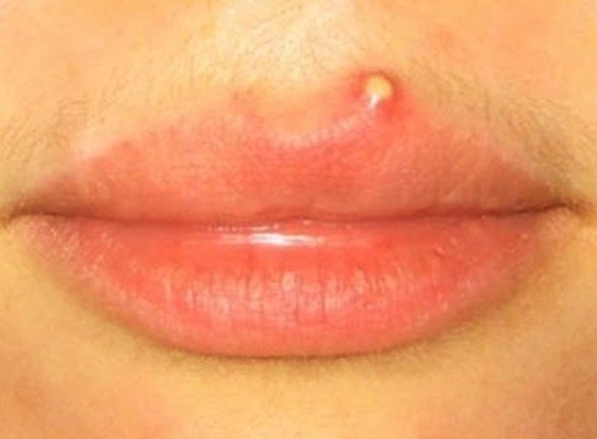
pimple on lip
First and foremost, you need to remember that the cold sores are actually caused by an infection with the herpetic virus (herpes simplex 1). Because of the fact that 90% of the adults suffer from such an infection at one point in their life, this can be easily mistaken for a pimple on the lip. However, knowing exactly what that tender spot on your lip represents is essential, so as to get the correct treatment. The treatment for the pimple on the lip is often symptomatic but there are remedies recommended for the underlying cause as well (hormonal imbalance, excessive sebum production at the level of the skin or viral infection).

The differential diagnosis is best made by a doctor or a pharmacist but, until you reach these specialists, you need to learn how to recognize such conditions on your own. Let’s find out more information on the subject.

Normal Pimple on Lip

pimple on lip pictures
Normal Lip Pimples that are slightly swollen and painful

Symptoms

  • Minor soreness
  • Tenderness can appear in the respective area
  • No shooting pain or burning sensation (such symptoms are more common for cold sores)
  • Firm texture in comparison to cold sores (content – skin oils, dead skin; not clear fluid)
  • Often appears as an individual lesion (as opposed to cold sores, that appear grouped)

Causes

  • Hormonal changes that occur during puberty (hyperactive glands)
  • Excess oil production at the level of the skin (oily skin type) – in combination with the dead skin cells, these can plug the pores and contribute to the appearance of pimples (+bacteria overgrowth)
  • Pregnancy (hormonal changes as well)
  • Medication (corticosteroids, lithium-based drugs)
  • Hyperglycemia
  • Sexual transmitted disease (herpes)
  • Low-quality or expired makeup products (lipstick, lip gloss, lip balm etc.)
    • Low-quality ingredients with harmful effects
    • Chemical substances plug the pores, promoting the buildup of harmful bacteria and thus the appearance of pimples
    • Keep in mind that certain makeup products can also become contaminated with bacteria
  • Allergic reaction – if you notice that the pimple appears every time you are using a certain product, you might be experiencing an allergic reaction to the said cosmetics
  • Diet – a diet that is rich in fats can lead to the appearance of pimples at the level of the lips
  • Poor personal hygiene
  • Chronic conditions
    • Bacterial infection
    • Dermatitis
    • Viral infection
  • Red pimple
    • Canker sore
    • Acne
  • Upper lip pimple
    • Upper lip hair waxing (irritation of hair follicles/ingrown hairs)
    • Threading
    • Lip piercing (infected)
  • Lower lip pimple
    • Threading
    • Waxing
    • Piercing
  • Near lip pimple
    • Stress
    • Sexual transmitted disease
    • Hormonal imbalance (puberty, pregnancy)
  • Lip crease pimple
    • Excess sebum in combination with bacterial overgrowth
  • Other causes
    • Smoking
    • Genetic inheritance (predominantly in relation to hormonal imbalance or oily skin type)
    • Stress
    • Pollution or climate (especially if dry and windy)
    • Toothpaste (high fluoride content).

Treatment

  • Acne treatment – recommended choice: Benzoyl peroxide, azelaic acid
  • Hydrocortisone cream – reduction of inflammation
  • Antibiotics – recommended choice: tetracycline
  • Medicated mouthwash
  • Vaporizing laser therapy (chronic pimple treatment)
  • Zinc – zinc supplements/topical treatments (cream, lotion)
    • Limit the side-effects
    • Quicken the pace of recovery
  • Avoid touching the pimple on the lip (risk of secondary infection and permanent scarring)
  • Diet change
    • Fruits (oranges, grapes, apples) – rich in alpha hydroxyl acid
  • Applying warm water compresses on the lips (reduce inflammation); in the acute period, when there is a lot of pain, it is for the best that a cold compress is applied (pain relief)
  • Avoid lip balms that contain chemical substances (plus, the constant application of lip balm can attract dust and other impurities, prolonging the presence of the pimple on the lips)
  • Wipe your mouth clean after eating or drinking (food residue on the lips increases the chance for pimples on the lips or aggravates them)
  • When brushing the teeth, clean the lips as well (a gentle brush will remove the dead skin cells and eliminate the debris accumulated throughout the day); the lip exfoliation should be performed twice per day
  • Increase the water intake (keep both the lips and mouth protected from bacteria)
  • Use high quality makeup products and throw out the ones that have expired (lip glosses and lipsticks should contain a reduced quantity of pigment, so as to reduce the chance of the pores becoming clogged; you also want to avoid those that contain coconut oil, for the same reasons as above)
  • Maintain excellent oral hygiene (salicylic acid toner for the cleaning of the lips
  • Apply herbal ointments to the lips (castor oil, neem, basil, eucalyptus).

How to squeeze the pimple on the lip

  • Before squeezing the pimple, make sure to apply a warm water compress for a couple of minutes; this will stimulate the circulation of blood in the area, helping you pop the pimple in an easier manner
  • Using sterile cotton swabs, apply gentle pressure to the pimple and start squeezing
  • The squeezing process will have to be continued until all the pus has been drained out; as you see blood coming out, this is the right time to stop (all the pus has been eliminated)
  • Refrain from washing the area immediately, as it will cause further irritation
  • Do not squeeze the pimple before it has formed the whitehead, otherwise you will only aggravate the infection and you will end up having a scar.

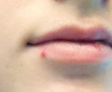
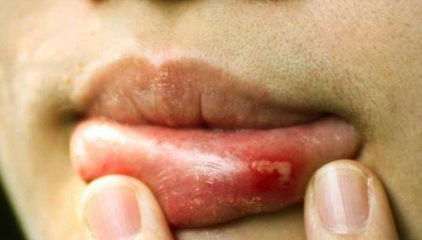
Cold sore on lip

cold sore on lip
Pimple of Lip due to of Cold sore

Symptoms

  • Pain or burning sensation at the level of the lip
  • Fluid-filled blisters (grouped)
  • Most common area – border of the lip
  • Upon healing the blisters can break, forming a crust or a scab (yellowish color)
  • Systemic symptoms can appear in more serious cases
    • Lymph nodes inflammation
    • Headaches
    • Muscle aches
    • Sore throat
    • Fever.

Causes

  • Infection with herpes simplex 1 virus – transmitted through direct contact (kissing, drinking sharing with an infected person)
  • Touching the personal objects of a person who suffers from this infection (eating utensils, towels)
  • Children present a high risk for cold sores
  • Virus remains dormant in the body throughout the entire life (may be re-activated in periods of reduced immunity)

Treatment

  • Over-the-counter treatments – recommended choice: Abreva, Docosanol (faster and better healing; pain relief)
  • Cold ice application (ice pack – not directly on the lips, better wrapped in a towel)
  • Prescription antiviral medication (oral or topical administration, such as cream) – recommended for patients who suffer from severe or frequent outbreaks; this medication can also prevent flare-ups in the future
  • Lip balm with lemon extract (quicken the healing process)
  • Diet change
    • Foods that are rich in lysine (chicken, fish, beans, turkey) – high amino acid content, useful for blister containment
  • Lysine cream/supplements
  • Breathing exercises/yoga – stress reduction (stress – risk factor for cold sores).

Canker Sore

canker sore on lip

Symptoms

  • Lesion appears inside the lip (not outside)
  • Soreness present in the area
  • Single patch, yellow or white in color (not grouped lesions or blisters present)
  • May also affect the inside surface of the cheeks or the roof of the mouth
  • Redness can surround the canker sore lesion

Causes

  • Suspected – stress, mechanical injury (biting the lip or the cheek), stress, vitamin/nutrient deficiency

Treatment

  • Oral numbing gel – pain relief.

Chapping

Symptoms

  • Intense dryness at the level of the lips
  • Peeling
  • Irritation
  • Pain
  • Bleeding
  • Generalized redness (no specific white spot, as it happens with pimples)

Causes

  • Dry winter air
  • Cold wind

Treatment

  • Lip balm
  • Avoiding picking the peeled skin or moisturizing the lips with saliva (aggravates the condition).

No responses yet

Duodenal Atresia

Dec 04 2016 Published by under Digestive System

What is Duodenal Atresia?

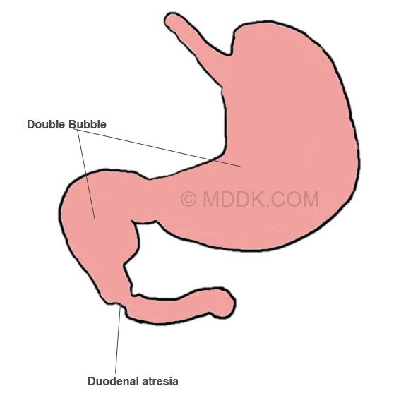
Duodenal atresia can be defined as a congenital problem, in which a part of the duodenum lumen is absent or completely closed. Also known as duodenojejunal atresia, this condition leads to the accumulation of amniotic fluid during the prenatal period. It is also responsible for complications such as intestinal obstruction, which can affect newborns. Upon performing imaging studies, such as X-rays, the stomach will appear distended and so will the duodenum. As you will have the opportunity to read below, the separation of the stomach from the duodenum is physiologically made by the pyloric valve, hence the double-bubble sign on the X-ray.

Duodenal atresia is believed to appear in 1 in every 5000-10000 live births, with 20-40% of all the born babies suffering from Down’s syndrome. No gender prevalence has been identified so far. Studies have shown that almost 8% of all the infants diagnosed with Down’s syndrome also suffer from duodenal atresia. Apart from Down’s syndrome, duodenal atresia is often associated with other birth defects (VACTERL, annular pancreas, jejuno-ileal atresia, ileal atresia or anal atresia). Other associated birth defects are present at the level of the heart or within the genitourinary tract. This condition is known to be one of the most common causes that leads to the appearance of fetal bowel obstruction. It can be however corrected in the neonatal period.

There are children who are born prematurely because of this condition. Upon being born, many of the children who suffer from duodenal atresia appear to be completely healthy. However, the symptoms because obvious when they are fed, with green vomiting raising the suspicion for the diagnosis. Duodenal atresia is often confirmed through imaging studies (X-rays in particular).

Symptoms

The symptoms of duodenal atresia are present during and after the pregnancy. It is important to understand that duodenal atresia can lead to increased quantities of amniotic fluid in the uterus, a condition which is known as polyhydramnios. This occurs as the fetus is unable to swallow the amniotic fluid, with a poor absorption at the level of the digestive tract.

Once the baby has been born, he/she will present abdominal distension and bilious vomiting as primary symptoms. The distension is mainly presented at the level of the upper abdomen, while the vomiting contains high quantities of bile acid. Due to the duodenum not having developed properly, the stomach contents cannot pass through the pyloric valve, leading to vomiting. The vomiting often occurs in large amounts and is not necessarily correlated to the feeding times (the infant can vomit hours after a meal). Apart from these symptoms, both the urination and the bowel movements can be absent (in the situation of urination, after the first few voidings; as for the bowel movements, the absence is noticed after the first few meconium stools).

If the condition is left untreated, serious dehydration can occur, with important electrolyte imbalance. Without treatment, duodenal atresia can lead to life-threatening symptoms.

Causes

Despite the extensive research made in the field, the exact cause that leads to the appearance of duodenal atresia has yet to be identified. At the moment, it is believed that the main cause behind this congenital problem is a defect in the development of the embryo. Basically, the duodenum does not change its structure, leading to the respective problems.

Other scientists consider that the main cause of duodenal atresia is the damage that occurs at the level of the duodenum in the fetal period, due to the loss of blood supply. This can cause the structure of the duodenum to become modified, as this part of the intestinal tract is narrower and obstructed.

Pictures of Duodenal Atresia

duodenal atresia
Picture of Duodenal atresia showing the Double bubble sign

Diagnosis

These are the most common methods used for the diagnosis of duodenal atresia:

  • Imaging studies
    • X-ray – double bubble sign – the air is trapped in the stomach and proximal part of the duodenum, with the pyloric valve separating them (due to the atresia, the distal part of the duodenum does not contain trapped air)
  • Physical examination
  • Patient history – information on the pregnancy
  • Blood testing – check for electrolyte imbalance.

During the pregnancy, duodenal atresia can be diagnosed with the help of the fetal ultrasound. Excessive amounts of amniotic fluid can be identified at the level of the womb; the ultrasound can also reveal the distension in the upper part of the abdomen. Unfortunately, this condition is cannot be diagnosed until the second half of the pregnancy. Moreover, its signs cannot be seen until the third semester of pregnancy. The increased quantities of amniotic fluid suggest a risk of pre-term labor, with the mother-to-be being closely monitored for such problems.

Differential diagnosis can be made with the following conditions: duodenal stenosis, duodenal web, intestinal malrotation, midgut volvulus (plain film appearance). In regard to the ultrasound appearance, the differential diagnosis can be made with: fetal choledochal cyst, fetal omental cyst and fetal enteric duplication cyst.

Treatment

These are the most common treatment approaches used for duodenal atresia:

  • Fluid removal from the stomach with the help of a nasogastric tube
  • Administration of intravenous fluids (to prevent dehydration and correct electrolyte abnormalities)
  • Surgical intervention
    • Open or laparoscopic approach
    • Surgical repair of the intestinal closure
    • Not an emergency intervention
    • Reduced morbidity and mortality rate with the initial repair
    • A second intervention might be necessary.

Aside from the surgical intervention, there aren’t other alternatives for correcting the defect at the level of the duodenum.

The risks of the surgical intervention include:

  • Small risk of bleeding
  • Damage to the nearby structures of the abdomen (very rare)
  • Leakage of bowel contents into the abdomen (risk of infection – antibiotics are administered as a prophylactic measure; might require a second surgical intervention)
  • Anesthesia-related complications.

After the surgery, it may require some time for the bowels to start functioning properly. During the recovery period, it is highly likely that the child will be fed with the help of parenteral nutrition. Immediately after the operation, the vital signs (breathing, heart rate and oxygen levels) are closely monitored. A ventilator might be connected, so as to assist the baby with the breathing process. Pain medication is administered through the intravenous infusion.

In the situation that this condition is identified during the pregnancy, genetic counseling is recommended – a specialist can discuss the most essential aspects related to genetic abnormalities and how they can influence the health of the future baby. Moreover, if there are increased quantities of amniotic fluid, a procedure known as amnioreduction can be performed. This procedure will reduce the risk of pre-term labor and it will also improve the breathing in the mother.

Prognosis and Complications

In the majority of the patients, the prognosis is positive, with no complications present. However, the risk of complications is higher when there are other congenital anomalies present. It is also possible that complications due to the duodenal atresia occur later in life, with a 6% mortality rate for these patients being identified. The late complications that can occur after the surgical intervention include: gastroesophageal reflux, problems regarding the bowel movements and inflammation of the proximal duodenum (condition known as megaduodenum). The complications of the surgery can also lead to significant feeding and absorption dysfunctions. After the surgery, the baby might present increased requirements for vitamins, such as those from the B12 complex.

Once a child has gone through surgical intervention for duodenal atresia, regular check-ups are essential with the passing of time. The check-ups are required, so as to ensure that the child grows and develops within standard. The loss of intestinal tissue can also lead to nutritional deficiencies, so the child has to be monitored for that as well. The progress of the child is influenced not only by the remaining length of the intestine but also by the association of other defects or abnormalities. A multidisciplinary team can assess the progress of the child as he/she grows older.

No responses yet

Sigmoid Volvulus

Dec 03 2016 Published by under Diseases and Conditions

Definition

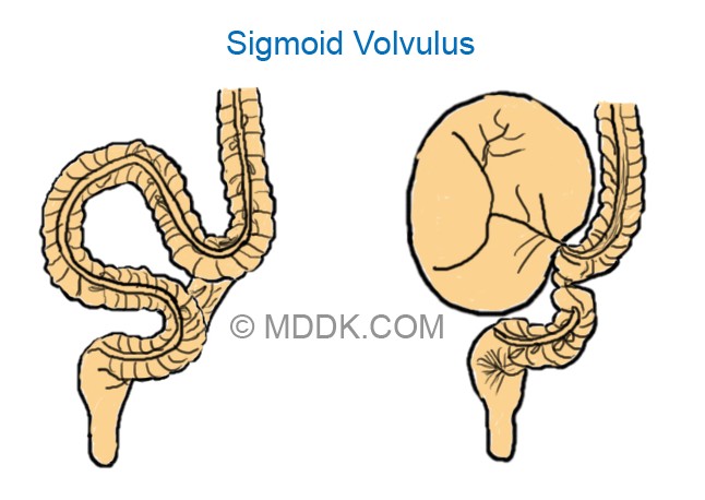
Sigmoid volvulus is a medical condition, in which the sigmoid colon practically twists on the sigmoid mesocolon. These changes frequently lead to the large bowel obstruction. It is estimated that, out of all large bowel obstructions, approximately 5% are due to the sigmoid volvulus. This condition is more often encountered in the elderly population, being one of the most common types of volvulus that can occur at the level of the gastrointestinal tract. If the conditions is left untreated, serious complications – such as venous infarction, perforation and fecal peritonitis – can occur.

According to the most recent research, the sigmoid volvulus is more often encountered in populations from South America, Africa, Asia and Eastern Europe. On the other hand, it is rarely encountered in developed countries, such as Japan, Australia, USA or the UK. While this condition is often encountered in adults, it is quite rare in children or infants. The same studies have shown that men are more predisposed to developing sigmoid volvulus than women.

Symptoms of Sigmoid Volvulus

These are the most common symptoms associated with the appearance of sigmoid volvulus:

  • Constipation
  • Bloating (obvious distension of the abdomen)
  • Nausea
  • Vomiting – appears at a later stage, when the distension of the abdomen is already severe
  • Abdominal pain – sudden, colicky and located in the lower part of the abdomen; in some patients, only some slight abdominal discomfort might be present (insidious form)
  • Failure to pass either flatus or stool
  • The patient might present a history of recurrent mild attacks – however, these have been relieved, as the patient has managed to pass a large quantity of flatus or stool
  • The examination of the abdomen can reveal the distention of the abdomen, as well as a palpable mass
  • In the situation of complications, such as the colonic perforation, the patient can present systemic symptoms (fever, shock)
  • The rectal ampulla is empty upon physical examination

The onset of the sigmoid volvulus can be acute or chronic. Complications such as the perforation of the colon and peritonitis can occur, especially if the diagnosis is delayed and the necessary treatment measures are not taken.

Causes

These are the most common causes that can lead to the appearance of the sigmoid volvulus:

  • Chronic constipation
  • Excessive or prolonged usage of laxatives
  • A diet that is too rich in fiber (geographic prevalence – Africa)
  • Chagas disease (geographical prevalence as well – Africa)

There are certain predisposing factors that can increase the risk for sigmoid volvulus, such as having a megacolon or suffering from an excessively mobile colon.

The sigmoid volvulus can be found in association with the following:

  • Chronic neurological conditions
    • Parkinson’s disease
    • Multiple sclerosis
    • Pseudobulbar palsy
  • Treatment for psychiatric conditions that are chronic, such as schizophrenia.

Diagnosis

The diagnosis of sigmoid volvulus can be made based on the following investigations:

  • X-ray
    • The abdominal X-ray can identify the site of the problem with specific signs, such as the coffee bean sign or the absence of rectal gas
    • Key radiologic feature – double-loop obstruction (present in 50% of all the patients)
  • Fluoroscopy/ barium enema examination
    • Water-soluble contrast enema – reveals the bird beak sign
    • Rarely performed nowadays
    • Not recommended in the situation that a gangrenous bowel is suspected, in the situation of pneumoperitoneum or peritonitis
  • CT
    • Identifies the closed-loop at the level of the sigmoid colon
    • Whirl sign present – the mesenteric vessels are twisted
    • Beak sign present – this appears only if rectal contrast has been administered
    • Recommended for the identification of the etiology and also to see the actual site of the obstruction (suggestive of other pathologies)
    • Least invasive imaging techniques that can be used for the assessment of complications, such as ischemia
  • MRI
    • Recommended for the assessment of large-bowel obstruction (not specifically for sigmoid volvulus)
  • Ultrasonography
    • Can be used for the diagnosis of large-bowel obstruction and, thus of sigmoid volvulus
    • Not the most faithful investigation whereas the correct diagnosis is concerned
  • Magnetic resonance angiography
    • Recommended in case of suspected complications, such as mesenteric ischemia
  • Sigmoidoscopy
    • This procedure is indicated for the suspicion of an ileosigmoid knot

The differential diagnosis can be made with the following conditions: large bowel obstruction of other etiologies, caecal volvulus and colonic pseudo-obstruction.

Sigmoid Volvulus Pictures

sigmoid volvulus
Sigmoid Volvulus Picture 1 : In the left side image, you can see the unattached loop of bowel getting twisted resulting in the obstruction of bowel lumen which you can see on the right image.
sigmoid volvulus pictures
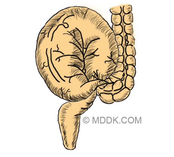
Picture 2 : Obstructed bowel lumen

Treatment for Sigmoid Volvulus

The main approach for the treatment of sigmoid volvulus is the insertion of a rectal tube. The decompression of the sigmoid volvulus can be guided through endoscopy or fluoroscopy. It is important to understand that the acute sigmoid volvulus is a medical emergency and that it should be treated as such. The risk of complications is quite high with the acute onset of the sigmoid volvulus. While non-operative decompression is successful in treating the majority of the patients, the surgical intervention is more recommended, as it can reduce the risk of recurrence.

For the decompression procedure, the patient is placed in a left lateral position. The decompression and untwisting of the sigmoid colon are performed with the help of a sigmoidoscope and a flatus tube. Basically, it is the flatus tube that will solve the problems, with a rush of liquid feces and flatus actually relieving the obstruction. Given the immediate results of this procedure, it should come as no surprise that the patient will experience immediate relief from the previous symptoms.

The flatus tube will remain in position for approximately one day, so as to maintain the decompression and reduce the risk for decompression. Another purpose of the flatus tube is to give the vascular supply to the bowel wall the necessary time to recover. After the intervention, it is essential to monitor the patient. In the situation that the patient suffers from constant pain in the abdomen or blood appears in the stool, a second surgery might be required (these symptoms suggest that ischemia has occurred at the level of the GI tract).

In the situation that the patient suffers from recurrent volvulus episodes, the surgical intervention is going to be recommended. The procedure refers to the resection of the redundant part of the sigmoid colon, being also indicated in the situation that tube decompression has failed or in those who have suffered from bowel ischemia.

Prognosis

The prognosis is influenced by the appearance of complications, such as bowel ischemia. The mortality rate for sigmoid volvulus varies between 20 and 25%, according to how much time has passed between the diagnosis and the actual treatment intervention. The earlier the diagnosis is made on the abdominal X-ray, the better the prognosis will be for this condition.

No responses yet

Sialolithiasis

Dec 02 2016 Published by under Oral Health

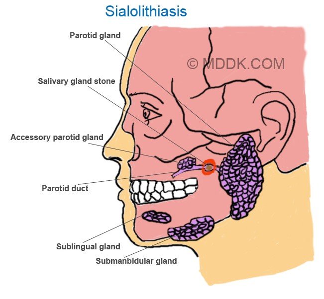
Sialolithiasis is a medical condition, in which calculi or stones form within the salivary glands. The most common appearance is at the level of the submandibular gland, this being also known as Wharton’s duct. It is also possible (however, it only occurs rarely) that the calculi appear at the level of the smaller salivary glands, as well as in the sublingual or parotid gland.

As you will have the opportunity to read below, two of the most important symptoms of this condition are inflammation and pain at the level of the respective salivary gland. The symptoms are usually aggravated by the stimulation of the salivary gland (seeing, thinking, smelling or tasting food), as well as by the mechanical actions in the area (chewing the food). Because of the association with food, sialolithiasis is often presented as the mealtime syndrome. There are different causes that can lead to the appearance of sialolithiasis, including chronic infections of the salivary glands, dehydration and autoimmune conditions (Sjögren’s syndrome). There are a lot of patients in which no exact cause can be identified.

The treatment, as you will see, requires the removal of the calculi through specific techniques. In the situation that the patient suffers from recurrent salivary stones, the surgeon might decide to remove the submandibular gland completely. This condition is quite common, affecting adults in general. Sialolithiasis is also more prevalent in the male population.

The salivary stones have different shapes and sizes; their texture and consistency vary from one patient to the other. They can also appear as a single calcified mass or in clusters (multiple salivary stones are identified in 25% of the patients diagnosed with this condition). In the majority of the situations, the salivary stones appear at the level of the submandibular glands, these being located at the back of the mouth, on both sides of the jaw. The calculi can also appear at the level of the parotid glands, these being located in front of the ears, on both sides of the face. In regard to the size, it is known that the calculi affecting the submandibular glands are larger than the ones appearing in the parotid glands.

Symptoms of Sialolithiasis

First and foremost, it should be mentioned that the symptoms of sialolithiasis depend on whether the obstruction caused by the salivary stones is complete or partial. The size of the calculi is also important, as it can cause more or less pressure on the respective salivary gland.

When the salivary glands have become infected due to the salivary stones, these are the symptoms that the patient is going to suffer from:

  • Pain – this comes and goes, being aggravated around mealtimes, due to the stimulation of the salivary glands. If the pain subsides in a short period of time, this might mean that the obstruction is partial.
  • Inflammation of the salivary glands – the swelling comes and goes as well, being aggravated around mealtimes and slowly subsiding afterwards (also suggestive of a partial obstruction).
  • The salivary glands become tender.
  • If the calculi are located at a close distance from the salivary ducts, these can be palpated (felt as a hard lump). In some situations, the patient can feel this lump right under the tongue (suggestive that the salivary stone is near the submandibular duct orifice).
  • If the obstruction is complete, no saliva is released from the duct.
  • In the situation that there is an infection, the floor of the mouth presents a distinct, red color; pus can also be discharged from the respective duct.
  • The lymph nodes in the cervical area can be swollen due to infection.
  • Dry mouth (due to the reduced flow of saliva)
  • Difficulty swallowing (dysphagia) or opening the mouth
  • Systemic symptoms (suggestive of a bacterial infection)
    • Fever
    • Nausea, vomiting
    • These symptoms are accompanied by local changes, such as foul taste in the mouth and redness in the affected area

It should be noted that, in case of chronic obstruction, the respective salivary gland can become atrophied. In such situations, the patient will not present any symptoms – the condition becomes obvious only in the situation of a secondary infection.

Causes

These are the most causes that lead to the appearance of sialolithiasis:

  • Abnormality in the metabolism of the calcium
  • Dehydration
  • Reduction in the regular flow rate of the saliva
  • Modified pH of the saliva (due to infection in the oropharyngeal area)
  • Precipitation of mineral salts (due to the altered solubility of crystalloids)
  • Accumulation of food debris, bacteria and foreign bodies at the level of the salivary gland ducts
  • The sphincter mechanism of the salivary duct presents abnormalities, these contributing to the formation of salivary stones
  • Salivary disease – chronic sialadenitis
  • Gout – main composition from uric acid crystals
  • Medication – taking certain medication, such as drugs to keep the blood pressure under control or allergy medication (antihistamines), can reduce the production of saliva at the level of the salivary glands (thus favoring the formation of calculi)
  • Reduced food intake – you might not be aware of this fact but not eating enough food contributes to the reduction of saliva production (same as above, such actions favor the formation of salivary stones)
  • Trauma and local inflammation due to different injuries
  • Chronic conditions – saliva stasis (with associated changes in the composition of the saliva)
  • Infection
    • With viruses – mumps
    • Bacterial – most commonly with: staphylococcus aureus, streptococcus viridans, streptococcus pneumonia, haemophilus influenza

Pictures of Sialolithiasis

sialolithiasis
Sialolithiasis Picture 1 – Diagram showing Salivary gland stond along with various other parts including Parotid gland, Accessory parotid gland, Parotid duct, Sublingual gland, Submanbidular gland.

Diagnosis

These are the main methods used for the diagnosis of sialolithiasis:

  • Patient medical history
    • Recurrence of salivary stones and/or infections
    • Onset and duration of symptoms
    • Recent operative history
    • Recent dental work
    • Thorough medication history
    • Immunization history – measles, mumps, rubella
    • History of autoimmune conditions
    • Surgical intervention history
    • History of radiation therapy
    • Recent health changes – weight loss, identification of a mass, swollen lymph nodes
  • Physical examination
    • Visual scanning – identification of inflammation and redness in the following areas: submandibular, preauricular and postauricular
    • Intraoral examination – analysis of the salivary ductal orifice (in case of salivary stones, this is inflamed and red in color)
    • Massage of the salivary gland – allows for the inspection of the saliva; in case of infection, a white secretion will appear through the salivary duct (purulence can be only identified through the examination of the ductal opening)
    • Manual palpation of the parotid gland – check the consistency of the gland (recommended – bimanual palpation)
    • Palpation extended to the floor of the mouth; the soft tissues of the tongue, cheek and neck should be examined as well
    • The examination should be made for all the salivary glands – identification of existent masses, analysis of symmetry, examination for any discharge.
  • Blood testing
    • While blood cell count – increased levels suggestive of infection
    • C-Reactive protein increased – sign of infection
    • Serum amylase increased – another sign of infection
  • X-ray – used for the confirmation of the diagnosis (the salivary stones are visible on the X-ray films)
  • Sialography – radiographic examination of the salivary glands (contrast substance injected into the salivary ducts); the result of the investigation is known as sialogram. It is important to mention that, if an active infection is suspected, this investigation is not recommended (the infection can become exacerbated). This investigation allows for the visualization of the ductal system, thus making it easier for the doctor to identify any strictures or dilatations. Filling defects can also be identified with the help of sialography.
  • Ultrasound – used for the confirmation of the diagnosis as well, recommended after the clinical examination of the patient. The advantage of the ultrasound is that it allows not only for the visualization of the salivary stones but also of the gland itself. If the obstruction is acute, the gland is going to appear enlarged and the salivary ducts dilated. The advantages of the ultrasound investigation relate to it being a non-invasive and non-painful measure. However, the ultrasound has a limited value when it comes to calculi that are located in the deep portion of the submandibular gland.
  • Computer tomography (CT) – useful investigation, allows for the salivary stones at the level of the glands and the ducts to be visualized. The CT can also be used for the evaluation of the respective salivary gland. Through the CT, one can determine whether the salivary gland is atrophied or not (sign of chronic obstruction). The smaller calculi might not be visible on the CT. However, this investigation is highly useful when it comes to the identification of calcifications, intraparenchymal stones included.
  • MRI – perfect investigation for the assessment of the salivary stones. Also recommended in order to make the difference between the acute and chronic obstruction of the salivary gland. If the obstruction is acute, the salivary gland appears enlarged and inflammation is present in the area. On the other hand, if the obstruction is chronic, the salivary gland presents a smaller size and signs of atrophy might already be present.

The radiographic investigation of the salivary stones is extremely useful in confirming the diagnosis. As it was already mentioned above, the salivary stones are visible on the radiographic films. However, it is important to keep in mind that not all the calculi are radiopaque. With the help of the X-rays, one can visualize submandibular stones (80-90%) and parotid duct stones (60%).

The differential diagnosis can be made with the following medical conditions:

  • Hemangioma
  • Phlebolith
  • Atherosclerotic calcification
  • Lymphadenitis
  • Dental abscess

It is important to understand that film defects might influence the results of investigations, such as the sialography. In general, these film defects are due to injecting a bubble of air along with the contrast substance. They can also appear due to a tumor or a blood clot being present in the respective area. Repeating the said investigation is always a good idea, so as to confirm the correct diagnosis.

Treatment

In general, the treatment for sialolithiasis refers to non-invasive measures. Depending on the size of the calculi, the doctor might recommend an increased intake of fluids, moist heat therapy and anti-inflammatory medication. Bitter or sour foods and beverages are recommended, as they can stimulate the flow of saliva and dislodge the salivary stones. For example, the doctor might encourage the patient to consume a piece of citrus fruit – the increased salivation will cause the salivary stones to become dislodged on their own.

A special massage can also be applied, in order to stimulate the dislodging of the salivary stones. Shock wave therapy has also been found to be highly effective in the treatment of sialolithiasis. This procedure is also known as extracorporeal shock wave lithotripsy, with the shock waves being actually used in order to break the salivary stones into smaller pieces. The procedure can be done with local or general anesthesia. More recently, symptomatic salivary stones have been removed through a procedure known as endoluminal balloon dilatation, followed by the extraction of the respective calculi.

In some situations, the doctor might recommend a minimally-invasive procedure, meaning sialendoscopy. For this procedure, the doctor will place a camera through the salivary duct and remove the calculi using specific techniques. In more serious cases, surgical intervention is recommended. The removal of the salivary stones is performed by the ENT or oral surgeon, the procedure being known as sialectomy. If the patient suffers from recurrent salivary stones, the surgeon will decide to remove the salivary duct completely. Antibiotics are administered, in order to prevent the infections that may be caused by the obstructing salivary stones. Among the recommended oral antibiotics, there are: Augmentin, Cefzil, Ceftin and Clindamycin. During the period of the treatment, it is recommended to avoid caffeine and alcohol – these have diuretic properties and they can add to the already existent dehydration.

Prognosis

In the majority of the patients, the prognosis is positive, as the salivary stones are removed without any complications. If the patient suffers from recurrent salivary stones or infections at the level of the salivary glands, the surgical removal might be essential. The surgical removal of the gland presents a series of risks, including related to nerve injury. The doctor will take you through the risks associated with each procedure, making sure that you are comfortable with it.

Prevention

Poor oral hygiene is considered a risk factor for the appearance of salivary stones, so maintaining an excellent oral hygiene is a good preventative measure. However, some of these calculi are the result of an infection and this cannot be prevented.

No responses yet

« Prev - Next »

© 2011-2019 MDDK.com - Medical Tips and Advice. All Rights Reserved. Privacy Policy
The health information provided on this web site is for educational purposes only and is not to be used as a substitute for medical advice, diagnosis or treatment.