Diseases General Health Skin Conditions

Archive for January, 2017

Sweet Taste in Mouth

Jan 23 2017 Published by under Oral Health

Have you been having sweet taste in mouth as if you took a sweet drink or licked some sugar? Do you experience a fruity taste that wouldn’t go away even after brushing? What you are experiencing is not new. A lot of people pass through similar experience without even understanding the cause of their problem.

sweet taste in mouth

There are several reasons why one experiences sweet taste in mouth. Whatever be the cause, the good news for you is that there are remedies for this condition. You can be treated and live your normal life again. In this post, we shall be sharing with you what sweet taste in mouth is, its causes, how to get rid of the condition, its relation with thyroid, etc. If you are currently passing through the experience or you have someone who is, then it is expedient that you pay attention to this post. Patiently read it to the end and make sure you share your concerns at the end, using the comment box.

Causes of Sweet taste in mouth

There are varied reasons why you experience sweet taste in mouth. We are going to look into few of the reasons why this phenomenon occurs.

Pseudomonas infection

This results from infection caused by a bacterium known as pseudomonas. This infection leads to sinusitis (inflammation of the sinuses) and occasionally alters the taste buds thereby causing sweet taste in the mouth. A more severe infection would bring about impairment in the functions of the taste receptors. Further attacks by the bacterium could lead to swimmer’s ear and itchy nose. Pseudomonas can also infect the lungs where it produces cough with or without sputum production, fever, chills, and difficulty breathing.

Indigestion

This symptom could come from acid reflux problem. When more acid is formed in the stomach, some acid are pushed back into the gut leading to sweet taste in mouth. Gastro Esophageal Reflux Disease (GERD) characterized by backup of particles of food into the mouth, with smell and taste of food. Other symptoms of GERD include chest pain and burning sensation in the stomach.

Diabetic neuropathy

Deposition of high blood sugar in diabetic disease as well as neuropathy that comes with condition can result in sweet taste in mouth. This can also be accompanied by dehydration and other symptoms. Sweet taste in mouth can persist even when the individual is fasting.

Epileptic attack

Some forms of epileptic attack, notably the grand mal, go through a warning stage (aura stage) which is characterized by unusual sound, flash of light, and taste in the mouth.

Diet

The type of diet you eat could be responsible for the sweet taste in your mouth. For instance if you ingest food containing artificial sweeteners like saccharin, there is the likelihood that you might experience sweet taste in the mouth hours after the main diet. Sometimes, you get the sweet taste after drinking water. So, before you start getting worried about what you are noticing, it is good to have a look at your diet again to be sure the taste is not coming from there. Foods with high sugar concentration can lurk around your mouth for a longer period. Sometimes, you can notice this taste after coughing especially if you have eaten the types of foods mentioned above.

Neurological disorder

The condition can also result from neurological disorder that may hinder the normal functioning of the nerve receptors that send out taste signals. The gustatory nerve (Glossopharyngeal nerve) is responsible for sending taste signals to the brain for interpretation. But when there is interference in the normal nerve signal resulting from nervous disorders such as seizure or stroke, this can affect the way you perceive taste. Serious degenerative nervous system disorders like Alzheimer’s disease, multiple sclerosis or Parkinson’s disease can also bring about a disturbance in electrical activities of the brain thereby impairing the normal functioning of the nerve responsible for sending gustatory signals. This can cause sweet taste in mouth.

Ketosis

When the body depends on fat for energy, the breakdown of the fats lead to the release of chemicals known as ketones. This condition is known as ketosis. Ketones also produce a byproduct referred to as acetone. It is this acetone that you breathe out, which is responsible for the fruity smell or taste that is experienced. Ketosis occurs in diabetes when there is a drop in the level of blood sugar and insulin. Conditions that give rise to drop in blood sugar level include going on weight loss diets with low carbohydrate and high protein contents, excessive exercise, anorexia (leading to starvation), etc. This is why there could be sweet taste in mouth after exercise or even when you are not exerting yourself physically.

Cancer

Some forms of cancer like lung carcinoma can lead to sodium depletion (hyponatraemia) due to the unbalance secretion of the antidiuretic hormone, a condition referred to as Syndrome of Inappropriate Antidiuretic Hormone Secretion (SIADH). This distortion results in a situation in which all foods are interpreted as sweet. When the sodium distortion is corrected, the condition is reversed.

How to get rid of Sweet taste in mouth

Do you want to know how to get rid of sweet taste in mouth? Then you need to pay proper attention to everything we are going to discuss here. Below are some of the things you could do to reduce the unpleasant symptoms you are experiencing.

Get rid of acid reflux

Since GERD has been fingered as one of the causes of sweet taste in mouth, one way to treat the problem therefore, is to treat the GERD. This can be done by employing a combination of medications such as antacids (Aluminium hydroxide, Magnesium trisilicate), H2 receptor antagonists (ranitidine, nizatidine, cimetidine), Proton Pump Inhibitors (lansoprazole, omeprazole, esomeprazole), etc. They play a major role in helping to reduce acid reflux thereby reducing the symptoms of GERD.

Treat diabetes

Taking care of diabetes will also eliminate the sweet taste in mouth if it is associated with the disease. The type of drug to use for diabetes management depends on the type of diabetes, whether insulin-Dependent or Non-Insulin Dependent. For insulin dependent diabetes, the use of insulin helps to put the high blood sugar under control.
Non-Insulin Dependent diabetes would need multiple drug therapy like Metformin (Glucophage), and Glibenclamide (Clamide) or Chlorpropamide (Diabinese), etc for the control of blood sugar.

The above treatments can be augmented with exercises and change in diet. Some forms of regulated diets are prescribed for diabetic patients as medications alone are hardly sufficient to take care of the condition.

Treat cancer

The offending cancer can be treated with chemotherapy, radiation therapy or surgery depending on the stage of the cancer. When this is appropriately handled, the sodium imbalance can be restored or other measures can be specifically taken to correct the hyponatraemia which is responsible for the sweet taste in mouth. Temporary measures aimed at controlling the sodium imbalance would only produce temporary relief until the underlying cause is pragmatically addressed.

Antibiotics

Pseudomonas infection should be treated with the right antibiotics. It usually responds positively to one or more of the following antibiotics – Ciprofloxacin or levofloxacin, ceftazidime, gentamicin, ureidopenicillins, carbapenems, cefepime and ticarcillin. It is good to run the necessary tests in order to determine the best antibiotics to use for the treatment of the infection.

Watch your diet

If the taste is coming from your diet, change the diet or try and rinse your mouth thoroughly after eating. This will get rid of those artificial sweeteners sticking to your tongue and gums. Avoid foods that contain high concentrates of sugar. In addition, avoiding concentrated sugars would also help put your blood sugar under control if you are a diabetic.

Alternative treatments

Sweet taste in mouth does not respond to orthodox treatment alone. It sometimes responds positively to alternative medicine especially when used to supplement the orthodox ones. However, you must be aware that alternative medicine may not be trusted when there are serious problems like diabetes type 2 and nerve damage. So, if you have ruled out the existence of the aforementioned disease conditions, you can now make use of any of the following alternative medicines:

  • Probiotic: Probiotic supplement are highly useful for improving digestion. They are bacteria that are highly beneficial for the preservation of healthy intestines that eventually assist in proper digestion. Probiotic can be found in foods like sauerkraut, yogurt, miso soup, kefir and kimchi.
  • Supplements: Supplements containing digestive enzymes are also useful for the management of problems resulting from indigestion. They assist the GIT in breaking down food and making digestion easier. You can easily order for any of them from online stores.

Sweet taste in mouth during Pregnancy

Occasionally, there could be sweet taste in mouth during pregnancy. Where this occurs, it is not an entirely strange phenomenon. A lot of women are known to experience metallic taste during pregnancy, which is oftentimes a sign of early pregnancy. This is one of the gustatory changes in early pregnancy.

However, some women have sweet taste in mouth during pregnancy as a result of gastrointestinal changes such as acid reflux. Like we noted earlier, when acid reflux occurs, some acid are pushed up into the gut leading to acid taste in mouth. This is the same way that the taste in mouth during pregnancy occurs.

This problem occurs in about seven percent of all pregnant women. The main prominent risk factors responsible for developing sweet taste in mouth during pregnancy include:

  • A history of type 2 diabetes in the family
  • History of previous pregnancy that resulted in gestational diabetes
  • Obesity or overweight
  • Being in pre-diabetic condition before pregnancy
  • Previous history of pregnancy resulting in a baby with birth weight of 10 pounds (4.5kg) or more
  • Being 25 years or older during the period of pregnancy

Though in majority of the cases the symptoms disappear on its own, it is advisable to see the physician to rule out pregnancy-induced diabetes mellitus which usually starts during pregnancy and is diagnosed during the same period.

Sweet taste in mouth in Early morning

sweet taste in morning during morning

Sweet taste in mouth in early morning can come from poorly controlled blood glucose in diabetics. Excess glucose gets deposited in between the gums/teeth thereby resulting in the sweet taste that you notice in the morning. This is one of the reasons while dental care is highly advocated for people suffering from diabetes.

The sweet taste in mouth in early morning often goes away after brushing. Sometimes, it gets less after drinking water, which helps to reduce the concentration of the sugar in the gums. In some cases, a visit to the dentist may be the first place to discover that you actually have diabetes even though the symptoms are not yet visible.

Relation with thyroid

Sugary taste in the mouth can be triggered by hyperthyroidism. In hyperthyroidism, there is an upsurge in metabolic rate which leads to carbohydrates being quickly used up by the body and an increase in the production of ketones and acetone. The acetone eventually results in sweet taste in mouth.
It is, therefore, not uncommon to see somebody with hyperthyroidism presenting with unusual sugary taste in the mouth. Other symptoms that may accompany this condition include hypersensitivity to heat, dehydration, nervousness, loss of concentration, weight loss, protruding eyes, rapid heartbeats, poor sleep, irritability, etc.

Conclusion

Sweet taste in mouth is a condition that results from different causes. When next you have such experience or you have a friend/relation who is going through it, remember to find out the exact cause before commencing any form of treatment. Where you are able to establish the cause of the problem, the treatments we have outlined in this post might be of help to you.

However, if for any reason your condition does not improve after trying the remedies, it is good to see the doctor for further investigations and proper medical management.

No responses yet

Pharyngitis

Jan 23 2017 Published by under Ear, Nose and Throat

What is Pharyngitis?

Pharyngitis is defined as the inflammation of the pharynx that often results in the condition known as a sore throat. The condition involves the pharynx which can be found immediately at the back of the mouth and the nasal cavity and just above the esophagus and the larynx.

The pharynx is an important part of the body for it is vital in respiration and deglutition. It serves as a channel from the mouth and nose down to the esophagus and the larynx and thus, a common channel for deglutition and respiration. The pharynx is also vital to the production of speech which enables communication.

Pharyngitis is a very common condition that is experienced internationally although this disease is seldom serious or life-threatening. It is prevalent among school age children from 5 to 18 years and rarely occurs in infants while adults may also get affected.

The onset of Pharyngitis can also be classified as either acute or chronic.

Acute pharyngitis

Acute pharyngitis is a type of pharyngitis that rapidly occurred although this usually lasts for a brief period of time. It is often the result of viral infection and commonly linked with acute nasal infection.

Chronic pharyngitis

Chronic pharyngitis on the other hand is more common in adults and is often associated with the disease of the upper respiratory tract.

Pharyngitis Symptoms

The symptoms of pharyngitis vary and greatly depend on the underlying condition that caused the inflammation. The symptoms also depend on the extent of the inflammation including the causative agent of the infection.

The sore throat is the primary and common manifestation of Pharyngitis. The terms sore throat and pharyngitis however are used interchangeably. Sore throat on the other hand is a manifestation of pharyngitis and is characterized by pain and irritation of the throat that is usually aggravated by swallowing.

The general symptoms of Pharyngitis on the other hand include the following:

  • Difficulty in swallowing
  • Onset of fever which is generally above 38 degrees Celsius in temperature
  • Difficulty in speaking
  • Swollen throat which is reddish and enlarged.
  • Itchiness of the pharynx
  • Visible white patches or pus on the covering of the tonsils or the throat
  • Hoarseness of voice
  • Difficulty in breathing
  • Dryness of throat
  • Coughing, which may cause the patient to expel mucus that may be clear, whitish, brownish, greenish or yellowish in color.

Other signs and symptoms of Pharyngitis may include the following depending on the condition that triggered the inflammation:

  • Fever and chills
  • Body aches
  • Weakness and fatigue
  • Onset of fever which may be high grade or low grade depending on the cause of the inflammation of the pharynx
  • Onset of runny nose
  • Frequent sneezing
  • Development of rashes
  • General malaise
  • Sudden loss of appetite
  • Problem with taste or an unusual taste in the mouth
  • Nausea and vomiting
  • Swollen lymph nodes
  • Earache and sore neck and jaw

Pharyngitis that is left untreated on the other hand may cause serious complications such as infection in the blood and rheumatic fever. The symptoms of pharyngitis associated with serious medical complications may be accompanied with life-threatening symptoms that require a medical emergency.

  • Onset of high grade fever that is greater than 101 degrees Fahrenheit
  • Sudden change in the levels of consciousness
  • Sudden loss of consciousness
  • Joint pain associated with an erratic movement of the body
  • Tachycardia or the rapid heart rate
  • Difficulty in breathing
  • Shortness of breath
  • Choking
  • Rapid swelling of the tongue or the throat
  • An extreme pain

Pharyngitis Causes

Various factors can trigger the onset of Pharyngitis although infectious organisms are the most common cause. Viral infection is regarded as the most common cause of Pharyngitis while bacterial infection is less in occurrence. Pharyngitis is often caused by viruses commonly causing colds and flu while most viral illnesses can result to the inflammation of the pharynx.

Numerous types of virus can cause Pharyngitis and such include the following:

Adenovirus

Adenovirus is the most common pathogen that causes inflammation of the pharynx among children and adults. It is regarded as the largest non-enveloped virus which can be transmitted through respiratory droplets or can also be transmitted via fecal means.

Orthomyxoviridae

Orthomyxoviridae is the type of virus that commonly causes influenza. This virus belongs to the family of RNA viruses that is composed of six genera. Influenza A and B are among the six genera of RNA viruses that cause influenza in humans and other vertebrates and other mammals.

Other viruses that can cause Pharyngitis are the following:

  • Herpes simplex virus
  • Coronavirus
  • Rhinovirus
  • Parainfluenza virus
  • Epsteinn-Barr virus
  • Cytomegalovirus

Bacterial infection is also another cause of Pharyngitis although this occurs less frequently. There are numerous types of bacteria that can infect the throat subsequent inflammation of the Pharynx.

Group A Streptococcus

Group A Streptococcus is the most common cause of throat infection and the condition is also referred to as strep throat. Pharyngitis with this etiology rarely occurs in children below the age of 3 years old and is common among adult patients.

Other bacteria that can cause Pharyngitis include the following:

  • Haemophilus influenza
  • Corynebacterium diptheriae
  • Neisseria gonnorrhoeaea
  • Moraxella catarrhalis
  • Bordetella pertussis
  • Bacillus anthracis
  • Mycoplasma pneumoniae
  • Chlamydophila pneumoniae

Pharyngitis can also arise from non-infectious disease and may be caused by several factors such as:

  • Irritation from thermal or chemical exposure
  • Cold climate or during cold season
  • Cold air
  • GERD or the gastroesophagegal reflux disease.

Pharyngitis Diagnosis

Pharyngitis is rather difficult to distinguish the cause whether it is from a viral infection or bacterial infection due to the similarities in signs and symptoms.

Laboratory studies to identify Pharyngitis include the following:

Throat culture

Throat culture is the standard diagnostic utilized in identifying Pharyngitis from Streptococcus infection. The test achieved through collection of culture from the secretions of the throat via the throat swabs. This test is generally done to rule out Pharyngitis caused by a bacterial infection.

Blood test

Blood test is also another diagnostic procedure for Pharyngitis that is caused by another factor aside from viral infection. The procedure is generally done by taking a small sample of blood drawn from the arms or from the hands.

Imaging test is not necessary in determining or diagnosing Pharyngitis except for cases where airway obstruction is occurring or there may be a suspicion of epiglottitis.

Pharyngitis Treatment

Pharyngitis is a common disease that generally affects children although adults may also get affected. It is however not a serious disease and is basically non-life threatening. The goal of treatment is directed towards the relief of symptoms and prevention of further medical complications.

Pharyngitis caused by bacterial infection and fungal infection are generally addressed through medications such as:

  • Antibiotics prescribed by doctors and which should be properly administered to avoid recurrence of infection
  • Analgesics and acetaminophen are both useful in relieving pain and reducing fever
  • Penicillin G benzathine and Penicillin V are both recommended therapies for Pharyngitis of bacterial infection
  • Cephalexin or Cefadroxil are the prescribed therapy for patients with allergy in Penicillin treatment.

Pharyngitis caused by viral infection usually does not respond to antibiotics and the treatment is basically symptomatic. Viral pharyngitis on the other hand usually resolves within 3 to 7 days from the time of infection. Home remedies can generally help viral pharyngitis.

Is Pharyngitis Contagious?

Pharyngitis is a condition characterized by the inflammation of the pharynx. It is mostly caused by viral infection and which can be transmitted from one person to another. The mode of transmission or spread of the disease is basically through respiratory droplets such as from sneezing and coughing. Fecal route is also another method of transmitting the disease. The spread of the disease is prevalent in close communities and to those living in places such as military barracks where close contact is common.

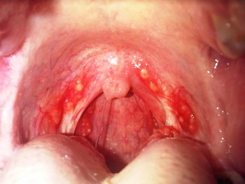
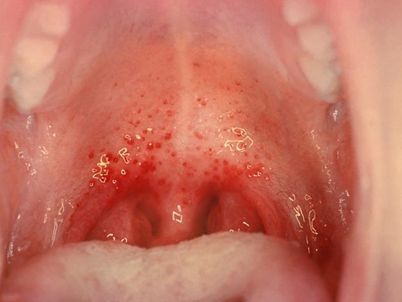
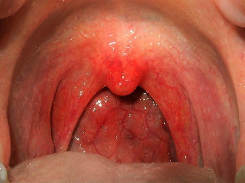
Pharyngitis Pictures

Pictures of Pharyngitis…

pharyngitis

pharyngitis pictures

pharyngitis pictures 2

pharyngitis pictures 3

No responses yet

Gastroschisis

Jan 22 2017 Published by under Diseases and Conditions

What is Gastroschisis?

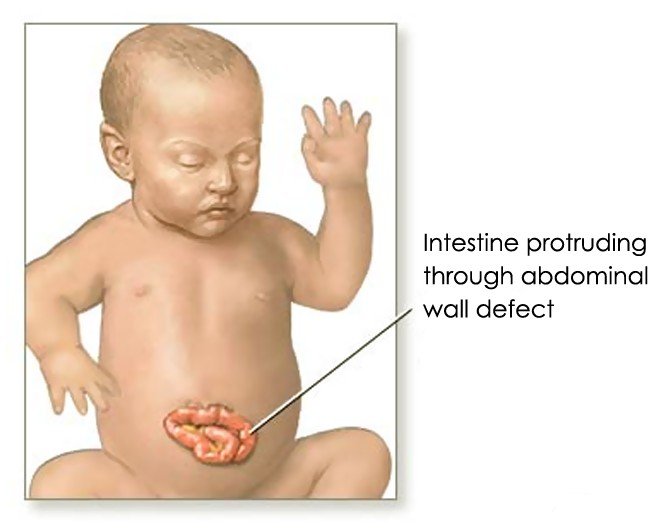
This is a congenital birth defect. When a baby has this birth defect their viscera, which are the internal organs in their abdominal cavity, protrude from their abdomen wall. The particular organ that is involved in this case is a protruding loop of small intestine on one side of their umbilical cord. It can also involve other organs like your liver. There are three other names that this medical condition is called which are:

  • Laparoschisis
  • Abdomionschisis
  • Paraomphalocele

The size of the defect is usually less than four centimeters. With the advances in surgical techniques today and better intensive care management the survival rate for infants born with gastroschisis is ninety percent. This birth defect is more frequent in males than females and most babies born with this defect have a low birth rate. Each year in the United States there is approximately one thousand nine hundred births with this congenital birth defect.

Gastroschisis Symptoms

There are only two symptoms associated with this birth defect, which are:

  • Having a lump in their abdomen.
  • Having the intestines sticking through the abdominal wall near their umbilical cord.

Causes of Gastroschisis

Gastroschisis is considered a type of hernia, which means rupture, This medical condition looks similar to having an omphalocele, which is when the baby’s intestines or other abdominal organs stick out of their belly button and is covered with a membrane, and not from around the umbilical cord. Gastroschisis is also not covered with a membrane. Gastroschisis is caused by incomplete fetal development. There also appears to be an inherited component but there other cases in which it just develops spontaneously without any reason.

The biggest risk factor that can cause gastroschisis is the age of the mother. It seems that it is seen in more mothers of a younger age. There is also evidence that suggest if the mother uses certain medications in the early stages of their pregnancy can have this happen to their baby. Other causes can include:

  • High risk pregnancies like those that are complicated by infection.
  • Smoking
  • Drug abuse
  • Anything that can contribute to low birth weight.

Clinical studies have also linked women taking aspirin during their pregnancy as to having a high risk of having a baby born with gastroschisis.

Diagnosis

Many times your obstetrician can diagnosis this birth defect when the blood work you had done shows abnormalities. It is more common for gastroschisis to be found when it shows up on the ultrasound. Once this birth defect is diagnosed you will most likely be referred to an obstetrician who treats high risk pregnancies. This is because both the mother and baby will need special care.

Treatment for Gastroschisis

When your obstetrician tells you that you are having or have had a baby with gastroschisis it can sound pretty frightening but in reality it is very treatable. If the pregnancy, delivering, and corrective surgery are handled right the prognosis can be excellent. In some pregnancies where this has been diagnosed the mother will undergo a caesarian section in order to reduce any complications that could happen during a vaginal deliver. If the parents know that their baby is going to be born with gastroschisis they should talk to a neonatal surgeon to make sure that their baby will get the right surgery and treatment after they are born.

If this birth defect is found before the baby is born, the mother will need to have special monitoring to ensure that her unborn baby stays healthy. At this time they will make plans for a careful delivery, most likely a caesarian section, and how they are going to handle the problem after the baby is born. No matter when gastroschisis is diagnosed, it is still considered a surgical emergency. Most babies will have to have more than one surgery. Of all the cases of gastroschisis only approximately ten percent could have it done on e with one surgery.

Once the baby is born the neonatal surgeon will tuck the intestine back on their body and close the incision. They will also use a pressure bandage to help promote healing. If the intestine is swollen because of an infection the neonatal surgeon will need to keep some of the small intestine outside their body in a sac that is called a silo. A silo is a mesh sack that stitched around the borders of the defect. Once they have it stitched they will pull the edges of the defect up. If the neonatal surgeon has to go this route he will gradually push the intestine back into their body over time. They may also have to wait if the abdominal cavity is too small to push all the intestines back in at the same time.

If the baby has difficulty breathing because of the abdominal pressure after surgery they will probably be put on a ventilator during the early stages of healing so they are able to breathe easier. Once the baby has healed they will usually experience no long term problems. Once the baby has had surgery they will need other treatments such as:

  • Being given nutrients intravenously.
  • Monitoring their temperature because if the intestine could not all be placed back into the proper place because with exposed intestine there can be a lot of body heat that escapes.

Gastroschisis Pictures

Pictures of Gastroschisis…

gastroschisis

gastroschisis pictures

No responses yet

Episcleritis

Jan 22 2017 Published by under Eye Health

What is Episcleritis?

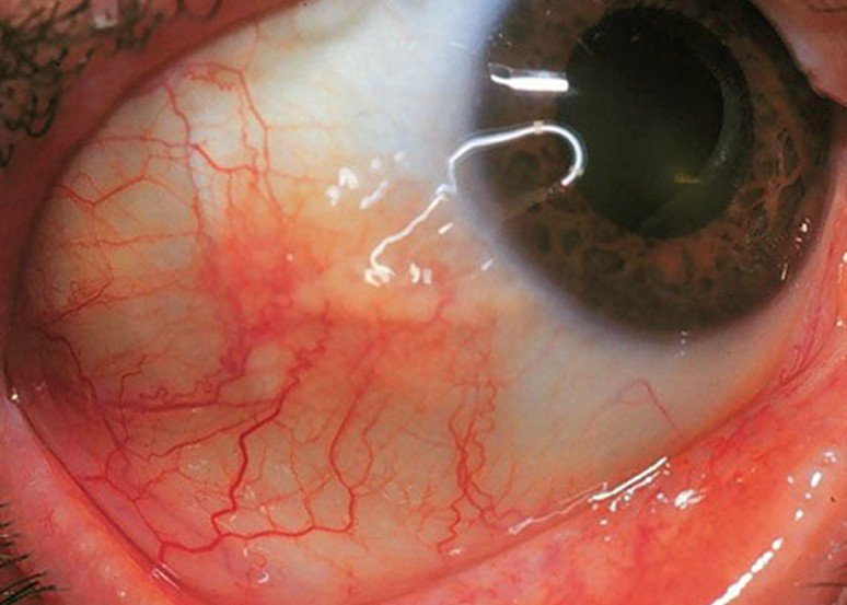
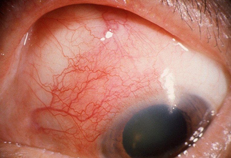
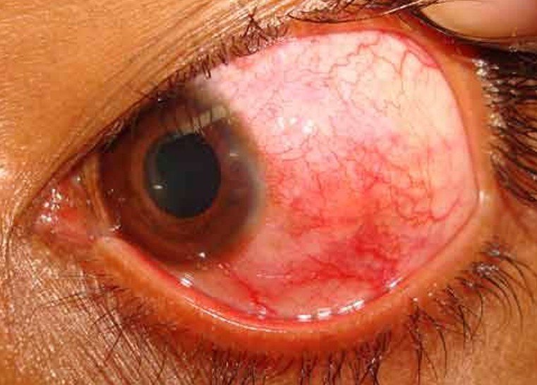
It is the abrupt onset of inflammation in the tissue that lies over the white part of your eye. It is a loose, elastic tissue that connects your part of the eye that is white and the mucous membrane that covers the front part of your eyelid along with lining the eyelid. It is normally a benign infection. When you see a person with this medical condition it is looks much worse that it really is. The reason is that what was the white part of your eye is now bright red and can appear quite alarming.

Episcleritis does not usually pose a threat to your vision if you make an appointment with your optometrist as soon as possible. If you do not seek medical attention and get it treated you could loose your vision. Approximately seventy percent of the cases of episcleritis occur in females. Although it can affect any age it is most frequently seen in middle-age and occurs between forty and fifty years of age but any one of any age or gender can get episcleritis. It is rare to see this medical condition in children younger than the age of five or in babies. If a child or baby does get episcleritis it will just be a inflammation that is mild and can last a week up to ten days. It is a common cause for someone to have red eye and can have episcleritis in one or both eyes.

Types of Episcleritis

There are different types of episcleritis:

Nodular episcleritis

This is the most painful type of episcleritis and have nodules in the loose connective tissue that you have between the white of your eye and the inside of your eyelids

Simple episcleritis

This is the most common type of episcleritis and can cause bouts of inflammation to recur with each recurrence lasting from a week to ten days. The duration can be longer if it is connected to another medical condition.

Episcleritis Symptoms

The symptoms will normally be mild. The main symptoms are usually pain in the eye and acute redness. Some of the symptoms that can be associated with episcleritis can include:

  • Tenderness in the eye.
  • Being sensitivity to light which is called photophobia.
  • Tearing of the eye but without discharge.
  • In the white part of the eye you may see a purple or pink color.
  • Itchiness

Episcleritis Causes

What actually cases a person to have episcleritis is still not known. This makes this medical condition an idiopathic medical condition. This just means that it is a medical condition that has no particular known cause. Although there is no exact cause known for episcleritis there are some things that might cause a case of episcleritis.

Inflammatory conditions are thought to be one of the causes of having this medical condition and some of these inflammatory conditions can include:

  • Having rheumatoid arthritis.
  • Having lupus
  • Having irritable bowel disease like Colitis.
  • Having Irritable bowel syndrome.

There are also a variety of other underlying medical conditions that could cause of a person having episcleritis which can include:

  • Herpes simplex infection
  • Herpes zoster
  • Tuberculosis
  • Having rosacea
  • Having gout
  • Being stressed out
  • Chlamydia
  • Menstruation
  • Addison’s disease
  • Autoimmune diseases and disorders
  • Allergies such as hay fever

In children under the age of five and babies there is no exact answer as to what can cause episcleritis but in children older than five it is thought to be associated with rheumatologic diseases. When a woman was it during the end of her menstruation or during her pregnancy it could be hormonal changes as the reason for cases of episcleritis during these times.

Episcleritis Pictures

Photos, Images and Pictures collection of Episcleritis…

episcleritis

episcleritis pictures

episcleritis pictures 2

Diagnosis

In addition to seeing your eye they optometrist will do an exam of your eye to positively diagnose episcleritis as there are other eye diseases that could cause these symptoms. They will also do a family history to see there is any medical condition or disease that could cause the episcleritis.

Episcleritis Treatment

Normally it is not necessary to have treatment done because it is just a mild inflammation that will generally go away with a week or two on its own. If a person has nodular episcleritis it can take a little longer to clear up as it is a more aggressive form of episcleritis. For discomfort you can get an over-the-counter saline solution. To make sure that the solution is going to help with you eye put in the refrigerator for an hour so the chill of the solution will help with the inflammation. If it is a severe case of episcleritis your optometrist may give you a prescription for mild steroid eye drops and/or anti-inflammatory medication. Either of these medications will help make the symptoms disappear quicker. Unfortunately with either of these two medications there are side effects that could be worse than actually having episcleritis. Other treatments can include:

  • Several times a day put in topical corticosteroid eye drops.
  • Topical lubricant eye drops like Artificial Tears.
  • Using cold compresses three to four times a day to give you dome comfort from the pain.

If you notice anything in or around your eyes or you are experiencing blurred vision, eye strain, or any other problems with you vision you should see your eye doctor as soon as you can.

No responses yet

Kyphosis

Jan 18 2017 Published by under Diseases and Conditions

Kyphosis Definition

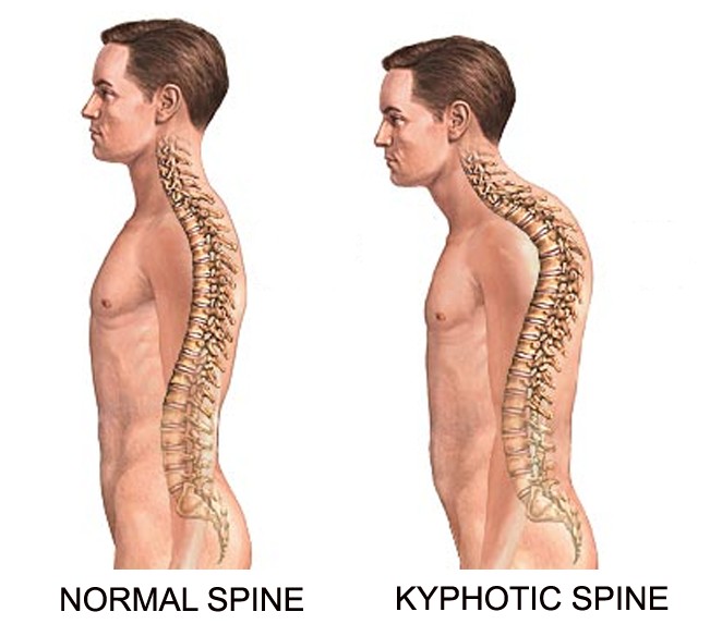
Kyphosis is the condition of the upper back or the thoracic vertebrae characterized by rounding or bowing of the back. The condition is also termed as round back or commonly called as a hunchback.

The human spine generally has a normal curve, particularly in the neck, upper back and the lower back. This curve however is naturally curved at an angle to help in absorbing shock during movements while also supporting the head. The curve in Kyphosis is defined at an arch which is rather excessive resulting to an over-curvature of the spine or the round back appearance of the spine.

Kyphosis can affect anyone at any age level except that it seldom occurs in newborns as Kyphosis is mostly caused by a poor posture. The normal curvature of the upper back is measured at 20 degree to 40 degree angle as measured by the Cobb angle. The abnormal Kyphosis on the other hand assumes an arch that is beyond the average degree of the arch which leads to an obvious slouching of the upper back. Most cases of Kyphosis are rather mild and lead to fewer problems. The severe case of Kyphosis can significantly affect the lungs, other organs and tissues of the body which can cause several medical problems.

Kyphosis Symptoms

Kyphosis is a condition involving the thoracic characterized by an exaggerated curvature of the upper back. This exaggerated curvature is the prominent manifestation of Kyphosis among patients and which may appear as hunchback or round back or a slouching posture. In mild cases of Kyphosis, signs and symptoms may not be noticed and may not cause discomfort to the patient although this case still requires regular monitoring for progression of thoracic curvature.

The common signs and symptoms of Kyphosis include the following:

  • Presentation of slouching posture or the hunchback or round back.
  • Back pain is also a common experience.
  • Stiffness of the back
  • Difficulty in assuming an upright position.
  • Weakness of the legs and the back.
  • Muscle fatigue

Severe cases of Kyphosis on the other hand have signs and symptoms associated with the affected organ and body tissues and such signs and symptoms include the following:

  • A more defined over the curvature of the back.
  • Possible onset of spinal cord compression due to the more exaggerated curvature of the back.
  • General weakness
  • Bowel and bladder incontinence.
  • Numbness or loss of sensation.
  • Chest pain
  • Shortness of breath
  • Difficulty in the digestion.
  • Severe back pain
  • Irregular heart beats

Kyphosis Causes

Kyphosis occurs when the normal and healthy structure of the spine deforms and develops a wedge like structure of the vertebrae instead of square stack columns. Kyphosis is more common in adults and rarely affects newborns while it may occur due to several reasons.

The potential causes of Kyphosis may include the following:

  • Disk degeneration that usually occurs with the normal aging process or the normal wear and tear process where the disk dry out and get smaller over time or as an individual advances in age.
  • Degenerative disease of the bone such as osteoporosis which can cause compression fractures.
  • Age advancement especially if associated with poor posture.
  • Neoplasm in the spine which can make the vertebrae more weak and potential for compression fractures.
  • Birth defects or a congenital deformity such as spina bifida where an error occurred during the development of the fetus in the womb.
  • Diseases and disorders of the endocrine system and of the connective tissues.
  • Other diseases and disorders that can affect the spine such as infection and muscular dystrophy.

Kyphosis can be identified either as postural or structural.

Postural kyphosis

Postural kyphosis is characterized by a smooth and very round hump. This type of kyphosis is caused by poor postures.

Structural kyphosis

Structural kyphosis is a type of kyphosis that occurred as a result of deformity in the spine. It is further classified as primary structural kyphosis or secondary structural kyphosis. Primary structural kyphosis is a kyphosis that directly resulted and not from other diseases and disorders while secondary structural kyphosis resulted as secondary to another existing condition.

The different types of Kyphosis under the structural kyphosis include the following:

Congenital kyphosis

Congenital kyphosis is among the main types of Kyphosis although the rare type of Kyphosis. It is characterized by a rapid progression that can result to severe deformity. This type of Kyphosis is further classified according to the degree of anomalies and the clinical feature is usually a peak in between T10 and L1. Congenital kyphosis develops during the fetal development where a malformation or fusion of the vertebrae occurred before the child is born.

Postural kyphosis

Postural kyphosis is the most common type and prevalent in girls and is usually first noticed during the age of adolescence and can occur in both the young and elderly individuals. This type of kyphosis is caused by a poor posture and a weakening ligament and muscles at the back. Postural kyphosis however develops slowly and does not worsen over time.

Scheuermann’s kyphosis

Scheuermann’s kyphosis is among the main types of kyphosis and is caused by a structural deformity of the vertebrae. This kyphosis is characterized by a severe curvature resulting in a cosmetic deformity and pain of varying degrees. It is regarded as a type of juvenile osteochondrosis of the spine and the apex of kyphosis is rather quite rigid. The pain is also felt at the apex of kyphosis and is aggravated by movements and long periods of inactivity. Scheuermann’s kyphosis is more prevalent among teenagers and is exhibited with an extreme deformity and affects the quality of life of the patient often causing an embarrassment. It is also potential for kyphoscoliosis which is a combination of kyphosis and scoliosis.

Gibbus deformity

Gibbus deformity is a structural kyphosis secondary to tuberculosis of the thoracic vertebral body. It is characterized by sharp angulations of the vertebral columns while the hump is easily observed when the patient is bending forward. Gibbus deformity is the result of hypoplasia of one or more neighboring vertebrae and is high potential for spinal cord compression.

Kyphosis Diagnosis

The initial step in diagnosing Kyphosis is through medical history taking where the physician will basically ask for any family member with similar medical problems, the time of the onset or when the signs and symptoms of the condition was first noticed.

The physical exam is the next step in assessing kyphosis in patients. Several physical tests may be done by the doctor to evaluate the deformity and such physical tests include the following:

Adam’s forward bending test

This is a test done to evaluate for any angulations and rounded curve of the thoracic which is indicative of kyphosis. In this test, the patient will be asked to bend forward up to the waist for easy examination and identification of abnormal kyphosis.

Range of motion

Range of motion is also part of the diagnostic test where a patient is asked to do a series of bend movements such as forward, sideways and backward including twisting. People with kyphosis usually have difficulties in achieving these specific movements.

Nervous system test

Nervous system test is also part of the diagnostic procedure where a patient is asked by the physician of significant pain, numbness, tingling and weakness especially in severe cases of kyphosis where other organs and body tissues are affected.

Lung function test

It is also a necessary test in determining the severity of kyphosis which may have affected the lungs and interfere with the ability of the patient to breathe.

Imaging tests

Imaging tests are also necessary depending on the presentation of signs and symptoms and such tests include the following:

X-ray is an imaging test useful in determining the degree of angulations or curvatures. Plain x-ray can also identify unusual shapes of the vertebrae which is wedge-like in kyphosis.

CT scans or computerized tomography scan may be required if cross sectional images of the structure are necessary.

MRI or magnetic resonance imaging is usually required if there is a suspicion of nerve problem, infection and tumor growth in the spine that is contributing to the incidence of kyphosis.

Kyphosis Pictures

Photos, Images and Pictures collection of Kyphosis…

kyphosis

kyphosis pictures

Kyphosis Treatment

The treatment for Kyphosis depends on the cause and the severity of the condition including the signs and symptoms presented.

Pharmacological therapy is best given to the patient suffering from pain and is also given to address the other signs and symptoms associated Kyphosis. Over-the-counter pain relievers such as acetaminophen, ibuprofen and naproxen are usually given. Prescribed pain medications are given if the pain is not responding well to over-the-counter medications and as such may be given with stronger dose.

Drugs to help strengthen the bone especially in the cases of kyphosis that resulted from osteoporosis can be given. Osteoporosis drugs are indicated to inhibit spinal fractures which can contribute or worsen Kyphosis. No medication can directly cure or treat Kyphosis but it can definitely help in relieving the symptoms to give patient comfort.

Physical therapy is useful in correcting posture and strengthening the muscles of the spine. It is also helpful in achieving flexibility and improve range of motions which have become difficulties for most patients of Kyphosis especially those with structural type of Kyphosis.

Orthosis or body brace is a treatment option for structural kyphosis such as Scheuermann’s disease while it is not necessary for postural kyphosis. The type of brace to be used depends on the pattern of the curves as there are different curves and angulations in Kyphosis.

Surgery

Surgery is usually recommended for severe case of Kyphosis although this form of treatment is usually the last option and if despite conservative treatments the patient is not responding well or no improvement has been achieved. The goal of doing surgical procedure is to reduce the deformity, reduce the pain and other signs and symptoms and to prevent progression of kyphosis and to prevent further damage and deformity. Several criteria are also being considered prior to opting for surgical procedures. The severity and degree of the curve, the progression, balance and neurological symptoms are all the criteria a patient has to meet prior to the procedure.

Surgical procedures for Kyphosis are the following:

Kyphoplasty is a minimal invasive procedure that requires only a small incision in the skin to allow a passage for the bone cement injection. The goal is to relieve back pain and restore the damaged vertebrae.

Osteotomy is a procedure of reshaping the bone by cutting bone wedges to correct the curvature and angular deformity and realigning bone ends. This procedure is usually combined with spinal instrumentation and fusion to stabilize the spine while in the process of healing.

Spinal instrumentation and fusion is a procedure where two or more affected bones are permanently connected by creating an environment where the connected vertebrae will fuse together over time. This is achieved with the use of metal wires, plates and screws to hold together or connect bits of bones placed between the affected vertebrae.

Surgery is generally followed by physical therapy to carefully and steadily regain the strength, flexibility and range of motion. A physical therapist is the usual person to create a program apt for the patient who has just undergone a surgery to correct Kyphosis.

No responses yet

Fifth disease

Jan 17 2017 Published by under Skin Conditions

What is Fifth disease?

Although adults can get this medical condition it is more commonly found in children. The disease is mostly seen in children between the ages of five and fourteen during spring and winter. Because of the rash that appears on their face it is also referred to as slapped-cheek disease because it looks as if the child has been slapped on the cheeks. It is an illness that is mild when a child or adult gets it but if you have a blood disorder or a weak immune system it can be a serious illness.

This illness got its name from a classification system that was developed in the 1890’s where was the fifth disease listed in a list of the most common rash-associated infections that a child got during that time. The other four on the list were measles, rubella also known as German measles, scarlet fever, and an illness that is referred to as fourth disease because it was an illness that was not known to the physicians today. Once you have been infected with the virus that causes fifth disease you will develop immunity to it and not get it again. In households where there are other people, especially children, there is about a fifty percent chance that other members of the house will get fifth’s disease.

Fifth disease Symptoms

After you are exposed to the virus you will start to show symptoms of fifth disease. Some of the early symptoms you see are similar to the flu and can include:

  • Sore throat
  • Runny nose along with nasal congestion.
  • Headache
  • Low-grade fever, which happens fifteen to thirty percent of the time.
  • Fatigue
  • Muscle aches
  • Swollen glands
  • Red eyes
  • Diarrhea

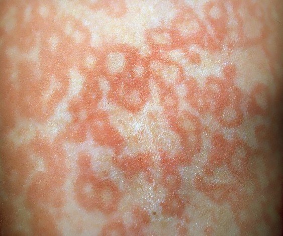
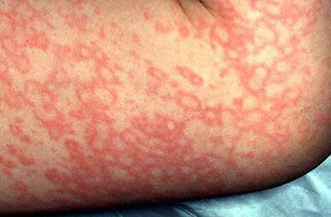
These symptoms will last for several days. There is even a chance that these first symptoms will be so mild that you do not even notice them or you may think you are coming down with the flu or cold. Seven to ten days later you will see the rash start to abruptly appear on your face and after four days it will slowly start to appear all over your body as a light pink rash. There is a possibility that you will feel itchy when you have the rash but within five days the rash will start to disappear along with the itch. It can take from seven to twenty-one days for the rash to completely disappear. For the next few weeks you may see the rash reappear when you get too warm, under stress, or out in the sun. Although the rash has come back does not mean that the illness is worse. Not everyone who has fifth disease will feel sick or get the rash.

The rash that appears on the face is reddish in color and appears to have a lacy look. Most children will not have any complications with this illness and will recover quickly. In some adult cases of fifth disease after having the disease it may be followed up by swelling or pain in your joints, especially in your wrists, ankles, hands, or knees.

Causes of Fifth disease

This disease is caused by a virus that is called human parvovirus B19 and can be spread by something as simple as a sneeze or cough. When people see the word parvovirus they immediately think of a disease related to animals but in this case this is not true. The virus can only be spread and caught by human. In 1975 the human parvovirus B19 was discovered as the cause of this illness. People are contagious when they have the flu-like symptoms and before you break out in the rash. By the time you see the rash you are no longer contagious and cannot spread the illness to anyone. The only people who may be able to spread the disease for a longer period of time may be the ones who have blood disorders and a weak immune systems. The rash that you get with fifth disease is the result of an immune system reaction that happens after the infection has passed and just before the rash.

Diagnosis

When you go to see your physician about the rash or flu-like symptoms they will do a physical exam and take down your medical history. If you only have flu-like symptoms it will be a little harder to diagnose but if you have the rash it will make it easier for the physician to diagnose fifth disease. Usually the physicians do not need to any tests run to make a diagnosis. If you or your child has been sick for awhile but do not have the rash that would tell the physician that it is fifth disease they may want to have blood tests done to see if their illness is caused by the parvovirus B19.

Fifth disease Treatment

At this time there is no vaccine for this illness. Most of the time when you have fifth disease it will disappear on its own as antibiotics will not help with the disease. The reason is that fifth disease is not caused by bacteria but a virus and antibiotics do not work on diseases caused by a virus. There are antiviral medications but at this time there none that are available to treat this disease.

Some home treatments that you can use to help with the symptoms can include:

  • Make sure that you get plenty of rest.
  • Use ibuprofen or acetaminophen to give you some relief for your headache, joint pain or fever.
  • Drink plenty of fluids

Fifth disease Pictures

Photos, Images and Pictures collection of Fifth disease…

fifth disease

fifth disease pictures

fifth disease pictures 2

fifth disease pictures 3

fifth disease pictures 4

fifth disease pictures 5

Fifth Disease and Pregnancy

Although it is not common if a woman who is pregnant gets fifth disease it can cause problems for the unborn baby. In some cases the unborn baby developed severe anemia, especially if the mother got fifths disease during the first half of her pregnancy. There have been instances where the anemia was so severe that the baby did not survive. Less than five percent of women who are pregnant will have serious complications if they become infected with fifths disease during their pregnancy. Half of the women who are pregnant are immune to fifth’s disease due to them having fifth disease when they were younger and are immune to the disease now.

No responses yet

Ascites

Jan 17 2017 Published by under Digestive System

Ascites Definition

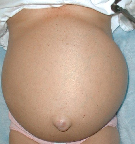
This is the build-up of additional fluid in your abdomen. It is also called your peritoneal cavity. This is not really a disease but more of a condition. When you have a mild case of ascites this extra fluid may not be easily noticeable. If you have a serious case your abdomen will protrude a lot. The fluid in your abdomen will either be pale, slightly straw colored, or clear. Your abdomen cavity can be found just below your chest cavity. Your diaphragm separates these two cavities.

Ascites Symptoms

If the case is mild you may have no symptoms. Ascites is considered a mild case if there is less than one hundred to four hundred ml in adults. As more fluid starts to accumulate you may have these symptoms.

  • An increase in your abdominal size and girth.
  • Abdominal discomfort
  • Shortness of breath due to the increased pressure on your diaphragm from all the fluid build-up.
  • Having pleural effusion which is fluid around your lung. This happens when the fluid gets pass your diaphragm and surround your lungs.
  • A cosmetically disfigured large belly.
  • Mental confusion
  • Easy bruising

If the accumulation of fluid measures more than five hundred ml you can see bloating and abdominal pain. If the ascites is severe you will see abdominal heaviness and abdominal distension. If it is caused by a bacterial inflammation you may have a fever, weight loss, and fatigue.

Most of the symptoms that you will see with ascites will be related directly to the reasons that are causing this disease.

Ascites Causes

When a person has ascites the cause is either advanced liver disease or advanced cirrhosis of the liver, which is marked by liver tissues that are scarred and results in impaired liver function and blocked blood flow to your liver. This disease is usually the result of alcoholism. Approximately eighty percent of all ascites cases are thought to be because of cirrhosis of the liver. Many people, especially men, will joke about having a “beer belly” but could be ascites.

You could also have this fluid accumulation due to congestive heart failure. With congestive heart failure you may also see an accumulation of fluid in your wrists, feet, hands, and ankles. You can also have ascites because of abdomen cancer, tuberculosis, or pancreatitis. Another cause is advanced kidney failure due to the generalized retention of fluid in your body.

Other causes of ascites can include:

  • Hepatitis
  • Portal vein thrombosis which means having clots in the veins of your liver
  • Liver cancer
  • Ovarian cancer

Although it is not completely known what the exact mechanism of the development of ascites most suggest that the main contributor is portal hypertension, which is an increased pressure in the blood flow from your liver. Two other contributors are water retention and salt.

Diagnosis

When you see your physician for the accumulation of extra fluid in your body the first thing that he will want to do is determine if ascites is present and what is causing you to have ascites. You will usually have to undergo many different blood tests including a complete blood count (CBC). This particular blood test will be able to gauge factors accurately such as your ability for your blood to clot and how your liver is function. Your physician may have a paracentesis done. This procedure is done by inserting a needle into your abdomen which is used to remove a tiny amount of asciitic fluid. This fluid will be evaluated so they can find out specifically what is causing your case of ascites.

The fluid will be analyzed to see what your serum ascites albumen gradient (SAAG) level is which can be either high or low. It will help to determine what is causing your case of ascites. If the level is low it can suggest tuberculosis or cancer as the underlying cause. If the level is high it can indicate heart failure or liver dysfunction.

Ascites Pictures

Pictures collection of Ascites…

ascites

ascites pictures

ascites pictures 2

Ascites Treatment

If you are diagnosed with a severe case of ascites your physician will use paracentesis to extract slowly the excess fluid that is in your abdominal cavity. If you have a mild case your physician will have you on a diet that is low in sodium. You should limit your salt intake (sodium) to less than two grams a day. They will also prescribe a diuretic, which is a pill that will help get the fluid out of your abdominal cavity. This medicine will usually work by increasing the amount of urine you put out.

Other treatments include:

  • For tuberculosis treatment you would be given antibiotics.
  • To treat some hepatitis types you would take anti-viral medications.
  • For a severe case of cirrhosis of the liver, having a liver transplant is the appropriate way to treat it.

When you know what is causing ascites and it is treated the pronosis is good. You will need to be monitored carefully to help prevent it from coming back. If the underlying cause cannot be found the treatment will focus on reducing the amount of discomfort you are in. Many times this condition will often signify an underlying cause that can shorten your life expectancy.

No responses yet

Numb Toes

Jan 16 2017 Published by under General Health

What are Numb toes?

This is when you loose the sensation in your toes and it is a condition that is abnormal. Numb toes can be experienced in just one toe or all of your toes. It is a discomforting condition and could make it hard for a person to get up and walk around. Numb toes can be just a temporary annoyance or it could be a symptom of a serious medical condition.

Numb toes Symptoms

Numb toes may happen with other symptoms such as if you have broken a toe or toes, you could also experience swelling and severe pain. Here are some of the other symptoms you may experience with numb toes.

  • A burning feeling
  • Anxiety
  • Frequent urination
  • When walking increased pain, toe numbness, or tingling.
  • Itchiness
  • Pain in your lower back
  • Muscle spasms
  • Rash
  • Having a prickling or pins-and-needles feelings.
  • Being sensitive to touch
  • Pain in the toes(s)

Symptoms that could indicate a condition that is life threatening

If you experience any of these symptoms you should get immediate help.

  • Loss of consciousness or confusion even for just a few seconds.
  • Having difficulty breathing.
  • Having difficulty walking.
  • Loss of bowel or bladder control.
  • Dizziness
  • Speech that is slurred
  • Paralysis
  • Following a back injury you experience numb toes.
  • Loss of strength or weakness.

Causes of Numb toes

Numb toes caused by circulatory problems

Having numb toes can be a symptom of many different conditions, disorders, or diseases that cause injury or restrict the blood flow to the nerves in your toes. Some of them include:

  • Having a tangled knot of veins and arteries called Arteriovenous malformation.
  • Having acute clotting and inflammation of veins and arteries called Buerger’s disease.
  • Having a blood clot in your leg that can break loose and cause a heart attack or pulmonary embolism in your lung called deep vein thrombosis. It could even cause a stroke.
  • Extremely cold temperatures or frostbite.
  • Having a narrowing of your arteries because of a buildup of cholesterol and fat on the walls of your arteries called Peripheral artery disease or Peripheral vascular disease.
  • Sitting in one position for a long period of time such as sitting cross-legged.

Numb toes caused by orthopedic problems

This could be caused by orthopedic conditions that range from moderate to serious and damage or injure the nerves. These may include:

  • Back injury
  • Toe or joint of the toe is fractured, dislocated, or broken.
  • Degenerative disk disease
  • Nerve pressure or entrapment that can happen because of shoes that are tight fitting.
  • Herniated disk
  • Tarsal tunnel syndrome
  • Osteoporosis
  • Bunions which is a deformity that affects your large toe caused by wearing shoes that squeeze all your toes tightly together. This cause mostly affects women because of wearing very high heels.
  • Having high arches or flat feet which could put stress on your toes when walking.

Numb toes caused by neurological problems

This could be caused by nerve damage or compression and may include:

  • Nerve damage due to blood sugar levels that are high called diabetic neuropathy.
  • Alcoholism
  • Any heavy metal poisoning like lead poisoning.
  • Having an under active thyroid called hypothyroidism.
  • Having the disease that affects your spinal cord and brain called multiple sclerosis.
  • Having a neuroma in your toe which is the unnatural growth of a nerve.
  • Having a disorder of the peripheral nerves called peripheral neuropathy.
  • Spinal cord tumor or injury
  • Stroke
  • Having a disorder which your body attacks its own healthy tissues and cells called systemic lupus erythematosus.
  • Having a neurological disorder that causes the inflammation of your spinal cord called transverse myelitis.
  • Having a deficiency of vitamin B12.
  • Having restless leg syndrome which is when you have an urge to move your body to stop odd sensations.

Numb toes Treatment

Before you can start any treatment you need to know what the underlying cause is to ensure you are getting the proper treatment.

  • If it is due to the footwear you are wearing, change to footwear that is more comfortable and fits better.
  • If it is due to flat feet or having a high arch you should talk to a podiatrist to find out which you have. The podiatrist may prescribe orthopedic supports to help with is the problem and can also suggest the right type of footwear you should wear.
  • If it is from a medical condition you need to talk to your physician and get on the right medication and treatment for the condition such as if it is due to diabetes, then your physician would address that medical condition with medication, healthy diet, etc.
  • Massages and physical therapy
  • If you are working in a job that requires a lot of sitting, get up and move around occasionally.

If the numb toes continues for several days or appears to get worse you need to talk to your physician to find out the cause.

No responses yet

Cellulitis

Jan 16 2017 Published by under Skin Conditions

What is Cellulitis?

This is a common bacterial infection in the deepest layers of your skin that can be serious. Cellulitis is not the same as cellulite, which is a dimpling in your skin that is caused by deep layers of fat. The skin on your lower legs is the area that is most commonly affected but you can get it on your face or anywhere on your body. In children you will usually find cellulitis around the anus or on the face.

Cellulitis Symptoms

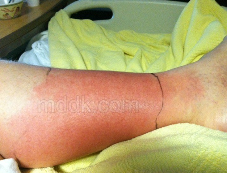
Many times when a person has cellulitis, the first symptoms you see are sometimes ignored because they do not think it is a serious infection and they just assume that the symptoms are what you have with an open wound. When you get cellulitis the first symptoms you will see in the infected area are:

  • Warm to touch
  • Red
  • Swollen
  • Tender

As the infection spreads you will experience:

  • Chills
  • Fever
  • Swollen glands
  • Pain in the affected area.
  • May have blisters because of the infection. The blisters are usually filled with pus that is an infectious fluid that is a yellowish-green in color and will leak out or drain out when you have cellulitis.
  • Swelling in the area and the spot begins to harden.
  • If the infection is not controlled it will start to spread into the blood and go throughout the body.
  • Nausea and vomiting
  • When touched you may experience some numbness.

Cellulitis Causes

A person will contact cellulitis when one or more type of bacteria enters through a break in your skin with the two most common bacteria being staphylococcus and streptococcus. There is another bacteria that is more serious and the cases of cellulitis being caused by this bacteria increasing. This serious bacteria is called methicillin-resistant staphylococcus aureus (MRSA). The bacteria can enter you body in a variety of ways such as through cuts, ulcers, dermatitis, puncture wounds, surgery, or athlete’s food. There are also some types of spider or insect bites that can transmit the bacteria. Two other places on your body that make good entrance points for bacteria is any area of swollen skin or areas of skin that is dry and flaky. If you have a cut or open area on your skin this does not automatically mean you are going to develop cellulitis. If you have any open areas just make sure that you are keeping the areas clean.

You can also get cellulitis if you have certain medical conditions such as a weak immune system, diabetes, or peripheral arterial disease. You can also get it is you injected illegal drugs under your skin, have edema in your arms or legs, or had liposuction to remove the excess fat from your body.

Diagnosis

If you have an injury that appears swollen, red, and does not appear to be healing up you should see your physician for a diagnosis. They will look at the area and may even take a sample so the specific bacteria that is causing the infection can be identified, They can even do a blood test to rule out any other medication conditions that could be causing the infection.

Cellulitis Treatment

The most common form of treatment is to give you a prescription for antibiotics to kill the bacteria that is causing your infection. The antibiotics can either be given by IV or by mouth. It depends on how advanced the infection is the type of bacteria that it is will determine how it is given to you. You may have to be hospitalized in some serious cases where the infection has spread into other parts of your body or it has spread throughout your skin. If the case is not serious you will usually take oral antibiotics in seven to fourteen days. To help relieve the swelling and pain you can take over-the-counter anti-inflammatory medication and pain relievers. So the swelling does not get worse you should keep the part of the body that has the infection elevated or apply cool compresses.

When your physician gives you an antibiotic you should call them back in three days to let them know if the infection is responding to the antibiotics and seem to be showing some signs of healing. In most cases cellulitis will disappear after a few days of starting the antibiotics. If the infection is not responding to the antibiotics or it has spread through your body you may need to be admitted to the hospital and be given a broad spectrum of antibiotics intravenously. Your physician may also admit you to the hospital if your blood pressure is real high or you are running a high fever.

Part of the treatment phase your physician will have you practice ways to prevent it from happening again and from spreading to other parts of your body. You will need to make sure that you are keeping the infected area clean using an antiseptic soap. When drying it make sure that you pat it dry, but do not scrub it dry. Talk to your physician to see if you could apply an antibacterial and/or anti-fungal cream to the area.

Cellulitis Pictures

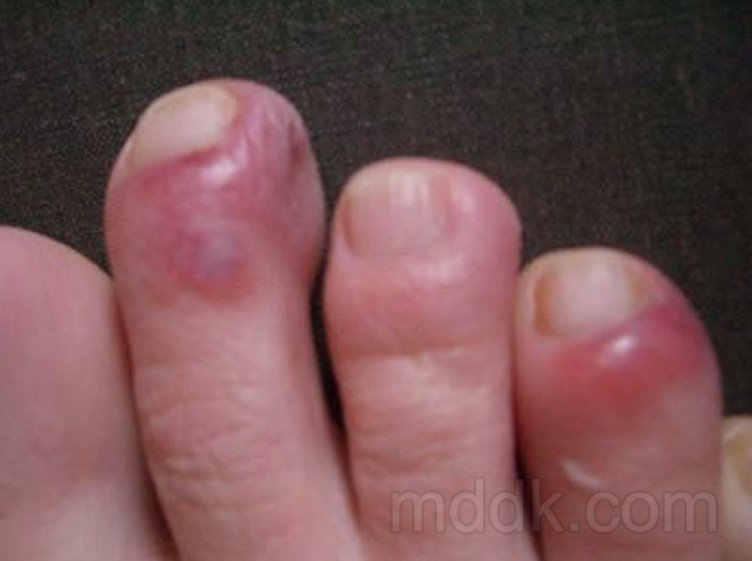
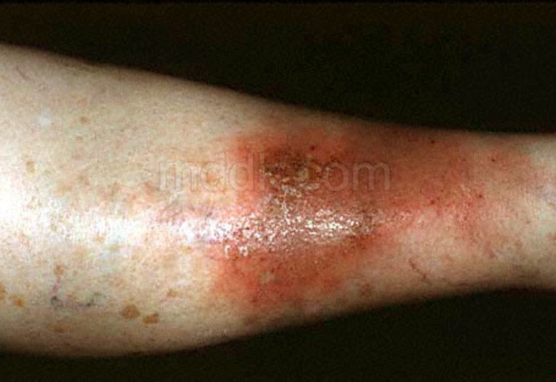
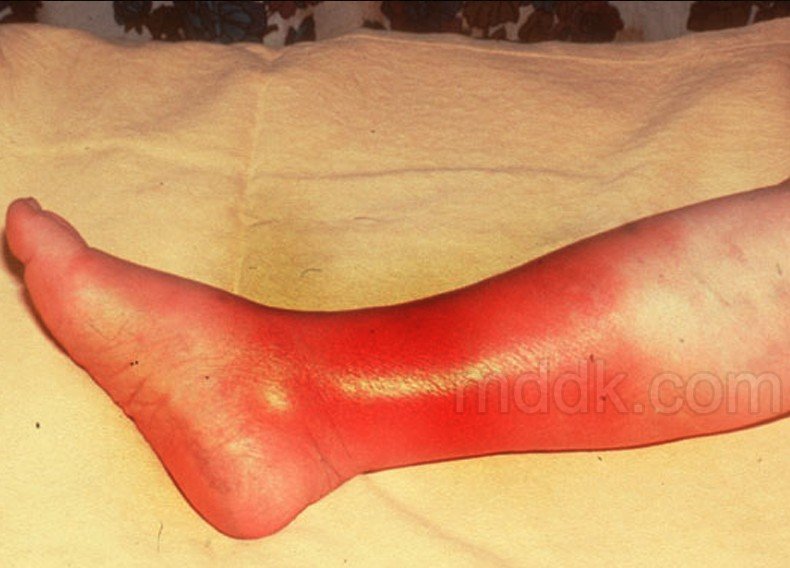
Pictures collection of the medical condition Cellulitis…

cellulitis

cellulitis pictures

cellulitis pictures 2

cellulitis pictures 3

Is cellulitis contagious?

This type of bacterial infection is usually not contagious. This is because the deeper layers of your skin are infected and not the outer layer of skin but if you have an open wound it is possible to pick up the bacteria from another person. This is true even if the bacteria is not making the other person is not showing any signs of infection.

Although it is not really contagious there are certain things that put you at risk for developing cellulitis, which can include:

  • Have an injury such as a burn, scrape, cut, or fracture.
  • Have a skin condition like eczema.
  • Participates in contact sports.
  • Have a weakened immune system or diabetes.
  • Us intravenous drugs

No responses yet

Necrotizing Fasciitis

Jan 16 2017 Published by under Diseases and Conditions

What is Necrotizing Fasciitis?

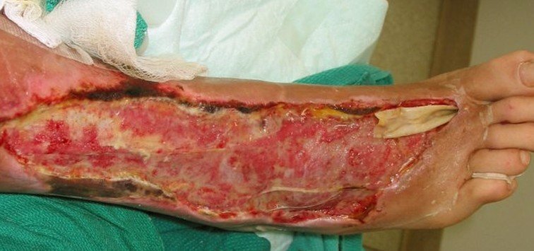
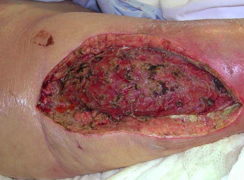
This medical condition is also referred to as the Flesh Eating Bacteria disease and it is very rare for a person to have this disease. It is a bacterial infection that is characterized by the breakdown of muscle tissue, fat, and skin. When this happens it looks as if the flesh has been eaten away, giving the disease its other name, flesh-eating disease. It is a very dreadful and severe disease and can target anyone anywhere. Necrotizing fasciitis life cycle is infection, growth, and quick reproduction. When you have these bacteria inside you the enzymes and toxins that are produced by the bacteria destroys the fascia (a layer of fibrous tissue) and tissues of your body causing the tissues to become gangrenous, which means death of soft tissue because of obstructed circulation. It is an infection that has a rapid course of progression along with a high mortality rate, which range from twenty-five to seventy-five percent. One in four people who get necrotizing fasciitis will die from this infecting.

Necrotizing Fasciitis Symptoms

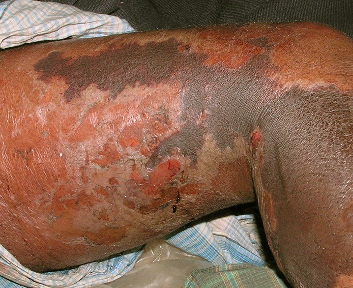
Necrotizing fasciitis can be visible on the surface around any cut or bruise or when the bacteria star destroying the fascia that is near the skin surface. It can also be invisible under your skin. If it is under your skin it is hard to tell all of the symptoms. The symptoms that a person might experience can include:

  • It will usually start out as a small red bump on your skin that is more painful that it should be for a wound that size.
  • Having pain that is unbearable in the area that is infected and where there is active bacteria.
  • Having nausea and vomiting.
  • Diarrhea
  • Lack of strength
  • Experiencing confusion
  • High fever
  • Due to the confusion and other problems you may have an extremely low blood pressure.
  • Due to the existence of the enzymes and toxins that are produced by the bacteria in your body you may be dehydrated.
  • The area will start to swell, and become red causing you to experience in that area a burning sensation.
  • Inflammation in the area when tissue death starts to occur

If it is not caught and treated in a matter of days the continuing infection will manifest as:

  • As blue, purple, or white skin discolorations.
  • Flakiness around the wound.
  • Blisters that are deep, black, and oozing.

At this point you might become unconscious because you have gone into toxic shock.

Necrotizing Fasciitis Causes

There are many different bacteria that can cause necrotizing fasciitis. The infections can be caused by one or more species of bacteria at same site. There are currently two species that are commonly found in a hospital, which are:

  • Methicillin-resistant Staphylococcus aureus (MRSA)
  • Streptococcus pyogenes

When either of these bacterial infections happen the symptoms that the toxins released by the infecting bacteria activates the immune system so potently that the bacteria will wreak mass destruction on the tissues around the toxins. The attacks keep happening as the toxin circulates through your blood. Many times necrotizing fasciitis is caused by the same bacteria that cause infections like impetigo and strep throat.

A person can get necrotizing fasciitis when it enters your body. Some of the ways that it can enter your body can include:

  • The bacteria can get into your body from having a trauma, surgery, cuts, touching anything that has the bacteria on it, burns, insect bite, and many other ways.
  • You can also get it through wounds that come in contact with raw saltwater fish, raw oysters, or ocean water. This includes any injuries that you get from handling any sea animals like crabs.
  • You can also get necrotizing fasciitis when you have gunshot injuries in your intestines, having tumors in your intestines, or having intestinal surgery.
  • Having a bruise or muscle strain, even if there is no break in your skin

Diagnosis

Necrotizing fasciitis is diagnosed based on the symptoms that you are experienced and doing a blood sample test. The physician may also take a sample of the infected tissues for a biopsy. Many times physicians will confuse the symptoms of this disease with symptoms of other diseases, especially if it is spreading under your skin and cannot be seen on the outside. If this happens it can lead to a delay in treatment and possibly cause you to die.

Necrotizing Fasciitis Treatment

It is very critical that if you have necrotizing fasciitis that you get early treatment because the sooner you get treatment the more likely you will recover and be able to avoid any serious complications such as having to have a limb amputated or even death.

Some of the ways that necrotizing fasciitis is treated can include:

  • Having surgery that will remove the fluids and infected tissues to stop the infection from spreading. When you have necrotizing fasciitis you will almost always need to have surgery. To control the spread of infection many times people will need to have several surgeries. In order to save your life the surgeon may have to remove organs or do amputations. Whether this is necessary or not will depend on where the infection has spread to and how severe the infection is. Because there is no vaccine available that can be used against necrotizing fasciitis, surgery is usually the option that is taken to remove the diseased tissue. Also, if the surgery to remove the infected tissues is done in the early stages when it was first diagnosed you would have a better change of surviving.
  • Taking antibiotics to help kill the bacteria that are causing the infection and also to strengthen and buildup your immune system. They usually use a broad spectrum of antibiotics because many times the disease is resistance to antibiotics so that is why it is necessary to try several antibiotics.
  • Doing procedures to treat any complications that come from having this infection such as treating the for breathing problems, organ failure, and toxic shock
  • To help prevent tissue death and promote healing they may use hyperbaric oxygen therapy. This is the procedure that will help to increase their bloods oxygen level which can also help to promote healing, fight infection, and help prevent tissue death. During this procedure you will be in a closed chamber having one hundred percent oxygen pumped in at high pressure.

Is Necrotizing Fasciitis contagious?

Yes, it is contagious. One way that you can get necrotizing fasciitis is it is passed from person to person. It can happen if you touch the wound of an infected person without wearing gloves or protecting yourself so you do not get the bacteria on you. Contacting necrotizing fasciitis from another person is rare and will usually only happen when the person who has been exposed to the bacteria has chickenpox, an impaired immune system, or an open wound somewhere on their body.

Prevention

If you are in close contact with a person who has necrotizing fasciitis, you have an immune system that is weakened, or suffering from a chronic disease seek immediate medical attention if you notice any symptoms of necrotizing fasciitis. If you are a nurse or a care taker of a person who has this infection make sure that you are wearing the proper protection such as gloves, sterile gowns, etc. You should also make sure that you are properly washing your hands after treating their wounds. You should also make sure that if you are around anyone with this infection and you have an open wound or sore that you keep it bandaged so the bacteria cannot get in the wound or cut. If your physician is aware that you have been around someone who has necrotizing fasciitis they may give you a prescription for antibiotics to help decrease your chances of getting the infection. In addition to keeping any open sores or cuts bandaged you need to make sure that you are keeping them clean.

Necrotizing Fasciitis Pictures

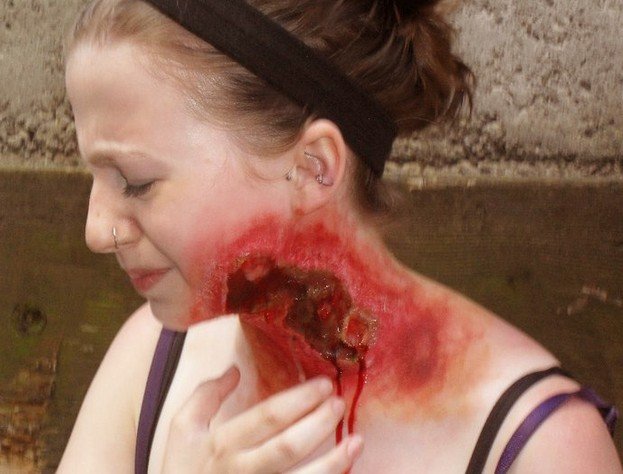
Photos, Images and Pictures collection of the Necrotizing Fasciitis disease…

necrotizing fasciitis

necrotizing fasciitis pictures

necrotizing fasciitis pictures 2

necrotizing fasciitis pictures 3

No responses yet

« Prev - Next »

© 2011-2019 MDDK.com - Medical Tips and Advice. All Rights Reserved. Privacy Policy
The health information provided on this web site is for educational purposes only and is not to be used as a substitute for medical advice, diagnosis or treatment.