Diseases General Health Skin Conditions

Archive for January, 2017

Nausea after Eating

Jan 31 2017 Published by under General Health

Nausea after eating is a common discomfort that plagues both the young and old. The feeling of irritation and being unsettled in the stomach shortly after eating is not something anyone feels cheerful about. This could be made worse sometimes when it comes with some sort of imbalance and secretion of excessive saliva. Nausea can be a real pain in the neck because it somewhat places a limit on the kind of food you can eat, upsets your mood, impinges interaction with people and overall affects your quality of life.

No matter how long you have had this problem, the good news for you is that it has a solution. Your problem is treatable and it can be handled in a professional manner and you will have the quality of your life back. So, you can even share this good news with any friend or relation you know of who might be suffering the condition too. You don’t enjoy the relief alone. Do you?

Nausea after eating, in most cases is nothing to worry about but at some other time, it could be a signal that all is not well in the body. In this post, a careful effort has been made to explain what nausea after eating is, the symptoms, what causes it, when to raise the alarm and the effective treatment. It is a very educative and well researched piece. We hope you find it quite useful.

What is Nausea after Eating?

nausea after eating

Nausea is an unpleasant feeling of wanting to vomit. This sensation usually comes before vomiting but in some cases it also occurs alone, that is, without vomiting. Nausea after eating can vary from just minor and temporary to severe and even protracted experience.

Nausea is a non-specific symptom which means there is a plethora of symptoms that accompanies the unpleasant irritation of nausea and they include:

  • The urge to vomit
  • Chest pain
  • Diarrhea
  • Feeling of dehydration
  • Weakness
  • Stomach pain
  • Dizziness
  • Fatigue
  • Headache

Causes of Nausea after Eating

There is a wide range of reasons why you may experience nausea after eating; it is almost impossible to discuss all the causes of nausea exhaustively. Some of the most common reasons have been clearly explained below.

Food poisoning

This has been established by the Centre of Disease Control and Prevention as one of the leading causes of nausea after eating in the United States. Food poisoning is the feeling of upset and irritation of the stomach as a result of eating stale food or food contaminated by microbial toxins. Eating such food causes nauseating feeling shortly afterward. Eating meat that are poorly cooked or not refrigerated can be harmful, as such meat may contain bacteria like C. botulinum or E. coli which are known causes of food poisoning. Other presenting symptoms related to food poisoning include vomiting, stomach cramps, fever and frequent stooling.

Gastroenteritis

This is also referred to as gastric flu. It is one of the most common causes of nausea; it is an infection that leads to the inflammation of the stomach and intestines. It is a very highly infectious illness, which spreads directly from infected food, water or when you get in contact with anyone who is infected and do not wash your hands afterward.. Besides nausea, other symptoms of gastroenteritis include diarrhea, stomach pain, fever etc.

Acid reflux

This is a condition in which food is regurgitated from the stomach to the esophagus. In normal condition, the stomach is structured in a way to prevent food that goes in from flowing back. This is made possible by a ring of muscle which acts as a valve. However, when it becomes defective, it allows content of the stomach to flow back causing nausea and heartburn.

Indigestion

We have all experienced this at one time or the other. Indigestion or dyspepsia as it is also called is associated with eating. Although not a disease, it could be caused by infections or certain medications. It is used to describe discomforting stomach upset or upper abdominal pain. In pregnant women this is a common occurrence that results because of hormonal change during pregnancy. Indigestion occurs in up to 80% of pregnant women at one stage or the other causing discomfort. The accompanying symptoms are heartburn, bloating, burping and nausea.

Food allergies

This is often responsible for a high rate of nausea after eating. The allergens which trigger such nauseating reactions could come from food preservatives and additives or even the food itself. Food allergies could have much more serious symptoms like vomiting, cramping and diarrhea. Foods which may cause allergy include peanut, egg, milk, shellfish, mustard, fish, walnut, some fruits and vegetables.

Meat allergy is also a well-known cause of nausea. A little bite of red meat Lone Star Tick is enough reason to trigger an irritating feeling of nausea. It is also possible to be allergic to other meat such as poultry.

Morning Sickness

This is a medical term used for ‘nausea and vomiting during pregnancy.’ Nausea during pregnancy is a familiar occurrence which is often associated with early signs of pregnancy and common symptom of the first trimester of pregnancy and sometimes beyond in some women. The symptoms are much more severe mostly in the morning.

Stress

Stress can trigger off a lot of chain reactions in the body, including nausea. In some cases, it could progress to vomiting itself. Stress is not a disease condition but the body’s reaction to stressors or any condition that exerts unusual pressure on it. Nausea caused by stress can also be accompanied by fatigue.

Peptic ulcer

Just like in acid reflux, there is excessive production of gastric acid in peptic ulcer. This sometimes flows back into the esophagus and triggers nausea after eating.

Blood Sugar

There is strong connection between sugar and nausea. Abnormal blood sugar can lead to a feeling of nausea. Infrequent eating can lead to low blood sugar or hypoglycemia causing nausea, headache and lightheadedness as though the body is starved of energy. In some occasion sugar malabsorption, the inability in some people to break down sugar in the small intestine can also result in low blood sugar. During hyperglycemia, when the blood sugar is too high, a person may also nauseated. This is usually common in diabetics.

Menstruation induced Nausea

Various reasons are responsible for nausea during period. For instance, a woman’s hormonal level changes during period and this sometimes prompts the secretion of gastric juice in higher volume in the stomach. The presence of hydrochloric acid in the gastric juice causes feeling of nausea.

Nausea could also result from dysmenorrhea occasioned by the production of prostaglandin by the lining of the uterus during menstruation, causing contraction, pain and consequently nausea.

There is also a more serious reason for nausea during period. It is called endometriosis. This is a condition in which the endometrium is found outside the uterus. When the lining, like other menstruation producing lining break down, because it has nowhere to go, it causes severe cramp and nausea.

How to get rid of Nausea after Eating

Sitting still in an airy place is a good way to stop nausea. Relax for about an hour after meal in a position where you can keep your head slightly propped up to ease the pressure on your stomach. This will definitely make you feel better. Moving around shortly after eating can aggravate the feeling of nausea

Find a way of ridding yourself of anxiety. Stretch, take a deep breath and take your mind off issues that cause anxiety.

Stay Away From Strong Odor

Certain smell can churn the stomach and irritate the bowel, and getting in contact with such odor could only be an invitation to trouble. It is better to stay away from strong irritating smells that trigger or worsen the condition. Typical nausea trigger include paint, perfume, caffeine etc.

When nausea is accompanied by vomiting and diarrhea, a feeling of dehydration and dry mouth sets in. It is important to rehydrate as quickly as possible to help check the risk of electrolyte imbalance and other complication from arising. Sipping water and juices slowly will help to get rid of the nauseous feeling. A number of juices to consider include flat soda, ice chip, ginger ale, apple juice. However, you should avoid acidic fruit juice like orange and grape juice, as well as caffeinated sport drinks.

As much as your stomach is being irritated from eating, avoiding food altogether is not a solution because that could worsen the situation. However, it is important to stay away from consuming heavy solid foods and fatty foods as well. Eat bland and light foods like salt crackers, bread, banana etc

Anti-Emetic drugs

Some drugs can be obtained over the counter to help suppress nausea and vomiting. Pink Bismuth, and emetrol and others which are available for pick up in local drug stores and supermarkets without prescription can give an almost instant relief.

Alternative Remedy

The good news about getting alternative ways to rid nausea is the fact that some of the natural remedies are readily available to us in our homes. They include:

Ginger

This highly medicinal rhizome has been used traditionally as an effective anti-nausea remedies for many centuries by different cultures. Several studies suggest that ginger works better than placebo in relieving nausea after eating. All you have to do is grab and slice ginger root into a pot of boiling water. Leave for a few minutes, strain and drink and nausea is rooted out.

However, should you need to combine with other herbs, you are advised to do so under the supervision of a healthcare provider as the possibility of interaction with other herbs cannot be ruled out.

Peppermint oil

From over 500 years ago when man first discovered the medicinal use of peppermint oil, this wonderful extract which nature has blessed man with, has been an important part of the household. Peppermint oil is a very effective treatment for nausea and other gastrointestinal disorders. It has wide range of applications which Includes rubbing the oil on the feet or taking a bath with few drops in a cool or warm water for quick relief. Adding two or three drops into a glass of drinking water after meal has also been proven to aid digestion which is often the cause of nausea. Peppermint oil is known to work effectively in relieving irritable bowel disease (IBD) which millions of people suffer from every year.

Drinking Lemon Juice

Lemon serves in a wide range of beneficial health use including as natural remedy for nausea. Sucking on a lemon or drinking lemon water can help alleviate nausea symptoms. It could also be used as aromatherapy; the sharp smell of lemon when inhaled can help.

Prevention

Nausea after eating as has been explained is caused by several factors. Identifying these factors can help prevent the unpleasant consequences that come with it. Any of the following or combination of factors can help prevent the malaise:

  • Identify the food to which you are allergic and avoid them
  • Eating right and healthy: including more fruits and vegetable in your menu as well as drinking plenty water can help prevent nausea after eating. Also avoid spicy and fatty foods
  • If it is pregnancy related, do not lie down immediately after meal. Avoid the intake of caffeinated drinks and alcohol
  • To prevent nausea that arises from acid reflux, you avoid eating late to bed
  • Being overweight might need you to shed a bit of the fat.

When to See a Doctor

You should seek medical attention if you start throwing up frequently and are not responding to home remedy.
Nausea after eating typically starts within 20-30 minutes after meal and lasts between 30 minutes to one hour. However, should it persist for up to a day and beyond, there is the need to visit the doctor urgently. When nausea is accompanied with a feeling of severe headache and confusion, you also need to see your physician

You should see the doctor if you have a feeling of weakness and experience issues with urination which are not normal. There is also the need to seek medical attention if there is coffee ground colored blood in your vomit.

Conclusion

Nausea after eating should not make you lose your shine. The tips we have provided in this post are enough to help you get rid of the nasty experience. However, if the suggestions fail to address your problem, it is advisable to see the doctor for further investigations and proper management.

No responses yet

Lichen Planopilaris

Jan 31 2017 Published by under Diseases and Conditions

What is Lichen Planopilaris?

This is an inflammatory condition that is rare and can result in having progressive patchy hair loss that occurs most of the time on your scalp that is permanent but can also affect other parts of your body that is hairy. Although it is a rare medical condition it is also one of the most common causes of scarring hair loss of your scalp.

Lichen Planopilaris Types

There are three forms of lichen planopilaris that are recognized which are:

Frontal fibrosing alopecia

This is scarring and hair loss in the frontal region of your scalp and usually affect post-menopausal women over the age of fifty.

Graham Little syndrome

This is a rare variant of lichen planopilaris and is an inflammatory form of scarring hair loss. It usually affects women in the age range of thirty to seventy with it particularly affecting the post-menopausal middle-age group.

Follicular lichen planus

This is the another name for the classic lichen planopilaris.

It is a medical condition that usually will affect women of all ages and it can also affect men. Many times with a person having lichen planopilaris it will be in connection with lichen planus that affects the nails, skin, and mucosa.

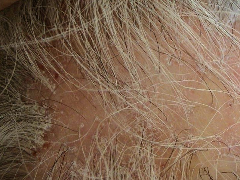
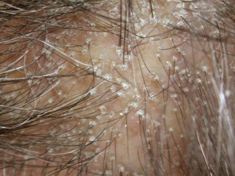
When a person has lichen planopilaris it will usually present itself as smooth areas hair loss from your scalp that is white. In the spots where you have the hair loss you cannot see any hair follicle openings. At the outside on the edges of the spots there could be redness and scales around each of your hair follicle. The hairs in these areas can be pulled out easily and it is also multifocal. The small patches where hair loss has occurred can come together to make bigger shapes that are irregular. The sites where hair loss is commonly found are usually on the front, lower back, and sides of your scalp and usually progress slowly.

Lichen Planopilaris Symptoms

There may be no symptoms at all but if there are they can include:

  • Itchiness
  • Pain
  • Discomfort
  • Tenderness
  • Burning

Lichen Planopilaris Causes

The reason that a person gets lichen planopilaris is not known but it is thought it could be the result of an allergic reaction to certain things like penicillin, gold, anti-malarial agents and more. The reason that some think these could be the cause of lichen planopilaris is that this inflammatory condition appears after some period of time which could be weeks or years after being exposed to these various things. Some even think that it has some hereditary or genetic characteristics which mean if you are related to someone who has had it there is a chance you will get it.

Diagnosis

If you are visiting your dermatologist or physician because you are noticing hair loss that is permanent and is causing any of the above symptoms they will carefully examine your skin, nails, and mouth for lichen planus evidence. An accurate diagnosis can be confirmed by having a biopsy from your scalp done which includes collecting hairs with scale and redness surrounding the sample at the bottom or top edge of the hair loss area. If the biopsy is taken from an already scarred area of hair loss it will not help make an accurate diagnosis. They may not even be able to make an accurate diagnosis is the biopsy is taken from an area where there is no evidence of inflammation and there is just scarring hair loss that is patchy.

Lichen Planopilaris Treatment

You should seek treatment immediately after getting an accurate diagnosis for lichen planopilaris because there is no treatment that will be able to recover the hairs that are already gone and scarring has taken the hairs place. The reason that you should treat this medical condition is to make sure the progression has been slowed and to help to relieve any symptoms that you might be experiencing. You may still notice continual hair loss but at a rate that is slower.

Some of the treatments that used include:

Potent corticosteroids

Topical which are effective anti-inflammatory preparations and applied directly to the skin, intralesional which is a steroid injection that is injected immediately below your skin, or oral which are synthetic derivatives of the natural steroids and taken by mouth.

Topical tacrolimus

It has immunosuppressant activity and is given by injection or orally. It works directly on skin cells and your immune system

Tetracycline

This is an oral antibiotic used to treat skin diseases

Acitretin

This is an oral retinoid which is a vitamin A derivative that is taken after eating.

Hydroxychloroquine

This is an anti-malarial medication and occasionally used in inflammatory skin conditions

Mycophenolate mofetil

It is the salt form of mycophenolic acid which is an immunosuppressive medication. It works by inhibiting antibody production.

Griseofulvin

This is a medication that is used to treat ringworm and stops fungal cells from dividing.

Ciclosporin

This is a strong immunosuppressant medication.

There have even been some cases where lichen planopilaris has cleared up on its own but most of the time it does need to be treated. For the moderate itching you can take over-the-counter antihistamines for relief.

Lichen Planopilaris Pictures

Collection of Pics, Images and Pictures of Lichen Planopilaris…

lichen planopilaris

lichen planopilaris 2

lichen planopilaris 3

lichen planopilaris 4

Natural Treatment

To treat lichen planopilaris you can use anti-inflammatory herbs that can inhibit your immune response that can trigger inflammation. You can also take over-the-counter analgesics to help reduce the pain. You can also use oats in your bath to help relieve the itchiness or make a paste of it and apply it to the itchy spots. Valerian is considered a calming herb so you can take it to help prevent and relieve the symptoms if there are any. There is no natural treatment that will cure or prevent permanent hair loss scarring from lichen planopilaris

No responses yet

Desmoplastic Melanoma

Jan 30 2017 Published by under Diseases and Conditions

What is Desmoplastic Melanoma?

This is a rare form of melanoma, which is a skin cancer that is usually malignant, which means cancerous. It is normally found in the neck region and head of older people who has skin that has been sun-damaged. It predominantly affects Caucasians and affects men more than women. It is also known as Spindled melanoma or Neurotropic melanoma. When a person has desmoplastic melanoma it usually does not have the ABCD melanoma warning signs which are

  • Asymmetry – one half is shaped differently than the other half
  • Border irregularity – the border is irregular
  • Color variation – the color is not uniform throughout the lesion
  • Large Diameter – it is usually larger than the diameter of the eraser on a pencil and can cause concern at this time.

Desmoplastic Melanoma Symptoms

When a person has desmoplastic melanoma the lesion looks like a slowly-enlarging area of thickened skin. It may look like an unusual mole, a keloid or hypertrophic scar but you have no history of having an injury at the site of this lesion, a neurofibroma, or dermatofibroma. As it grows it becomes more distinctive but it can take months, even years, before the lesion is recognized as desmoplastic melanoma.

The symptoms, or characteristics, of desmoplastic melanoma are:

  • Usually being more than six millimeters and one to two centimeters in diameter when it is diagnosed
  • Having an irregular or round shape
  • Having variable pigmentation that is most often a pink nodule or skin-colored
  • Being a firm scar-like nodule that has an irregular or smooth surface
  • Appearing as a flat nodule with a brown and bluish-red portion

Causes

A person gets desmoplastic melanoma because of the development malignant pigment cells called melanocytes within dermis, which is the layer of skin that is between the outer layer and the subcutaneous tissues. These cells can arise in skin that was normal-appearing previously or within another type of melanoma. It is not known what triggers these melanocytes to become malignant. It is thought to be a series of changes to a person’s DNA.

Diagnosis

Because it has a similar appearance to scars, cysts, fibromas, or sclerosing melanocytic nevi, it is very important that you have it diagnosed accurately so as soon as you notice an odd lump on your skin that is not going away and seems to be changing in size and appearance you should see your physician or dermatologist. They will ask your medical history and then do a physical exam of the area. The physician or dermatologist can make an accurate diagnosis by doing an excision skin biopsy where they take a sample of the lesion and look at it under a microscope to make sure that it is. If you have melanoma that is more than one millimeter thick they physician or dermatologist may advise you to have imaging studies, blood tests, a lymph node biopsy.

They may also use a dermoscopy which is when they examine your skin using a skin surface microscopy to evaluate pigmented skin lesions and can make it easier to diagnosis desmoplastic melanoma.

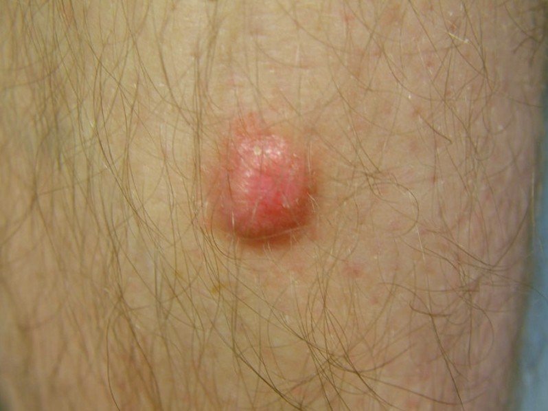
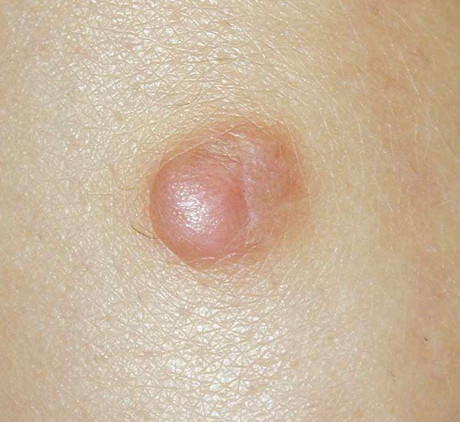
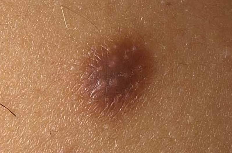
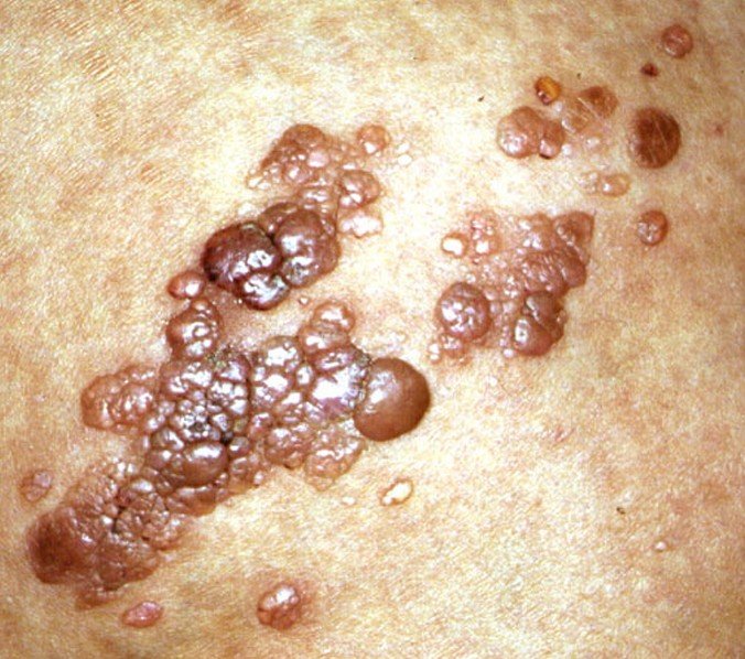
Desmoplastic Melanoma Pictures

Images, Photos and Pictures collection of Desmoplastic Melanoma…

  1. Picture 1 of Desmoplastic Melanoma
  2. Picture 2
  3. Picture 3
  4. Picture 4

Treatment

The initial treatment for a person who has desmoplastic melanoma is to cut it out and when this is done the lesion should be totally cut out with a one to two centimeter margin of tissue that is normal.

Survival rates

Surviving after being diagnosed with desmoplastic melanoma usually depends on several factors. The main factor is the measured thickness of the melanoma when it was removed surgically. There is an eighty-five percent survival rate of five years and an eighty percent survival rate of ten years.

No responses yet

Enlarged Papillae

Jan 29 2017 Published by under Diseases and Conditions

What are Enlarged Papillae?

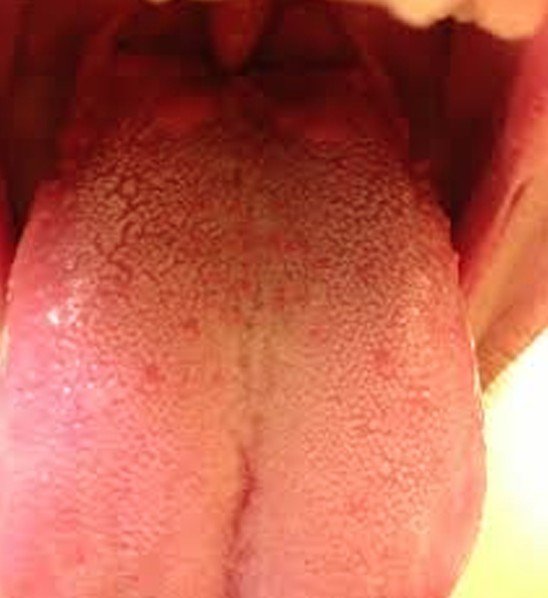
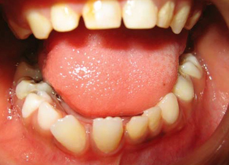
Papillae are small growths that appear on the surface of your tongue naturally. Although all not all papillae contain taste buds the ones found on your tongue do. In fact, the papillae on your tongue are usually called your taste buds. There are many different types of papillae and there are times that they do seem to be enlarged. The four types of papillae that are associated most often with your taste buds and have nerve endings that send messages to your brain about what type of food you are eating.

Enlarged Papillae Types

These four types of papillae are:

Fungiform papillae

On the surface of your tongue they are scattered around but most are located on the sides and apex of your tongue. They have nothing to do with fungus but are all that because of their shape which is the shape of a mushroom. These papillae detect sour and sweet tastes.

Foliate papillae

These papillae are found on the sides of your tongue and have an elongated fold. They are the ones that detect a sour taste

Filiform papillae

These papillae are thin, long, and V-shaped. They are also the type that is the most numerous and give your tongue the abrasive coating. It is the papillae that detect a sour taste.

Circumvallate papillae

These are papillae that detect a bitter taste and can be found in the back part of your tongue, arranged in a V-form that heads toward your throat. On average you will have from ten to fourteen of this type of papillae.

Symptoms

There are no symptoms

Causes of Enlarged Papillae

There are many different reasons that a person would have enlarged papillae which can include:

  • Happening naturally as you get older
  • Excessively smoking which can cause the tongue to become irritated
  • Having canker sores which can appear for no known cause but the canker sores can be more severe if you are under stress a lot.
  • GERD
  • Having ulcerative colitis
  • Having an infection from some type of trauma that can include eating something spicy or hot and scalding your tongue, eating foods that are very acidic, or biting your tongue.

Enlarged Papillae Pictures

Photos, Images and Pictures collection of Enlarged Papillae…

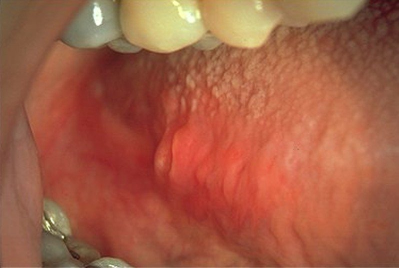
enlarged papillae

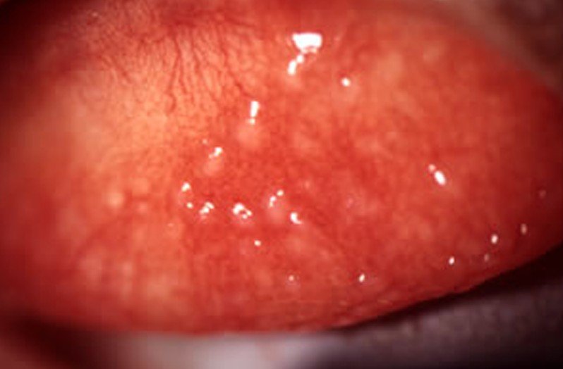
enlarged papillae 2

enlarged papillae 3

enlarged papillae 4

Treatment

Before your enlarge papillae can be treated you will need to know what has caused them so you can use the right treatment. Some of the treatments can include:
  • Stop smoking
  • Making sure that you are practicing good oral hygiene. When you are brushing and flossing your teeth make sure that you brush your tongue also.
  • Eating a diet that is healthy
  • Trying to avoid any foods that are too hot, spicy, or acidic
  • If you are having pain with your enlarged papillae gargle with salt water or with a mouth rinse to help relieve the pain.
  • Being careful when chewing or talking to help prevent biting your tongue
  • Having your dentist smooth down your teeth if you have sharp teeth so you will not scratch your tongue causing enlarged papillae
  • Increasing your intake of vitamins B 2, 1, 6, and 12 either by eating foods that contain these vitamins, taking vitamin B supplements or a combination of both. Doing this will help you manage any mouth ulcers or cold sores you might have.
  • In order to help balance the bacteria in your body you should eat yogurt every day and also increase the amount of dairy products you consume daily.
  • Taking an iron supplement daily or eating foods that are natural sources of iron
  • If you are wearing dentures make sure that fit your mouth correctly.

No responses yet

Albinism

Jan 28 2017 Published by under Diseases and Conditions

Albinism Definition

This is a medical condition that affects approximately one in seventeen thousand people. In animals it is often fatal but in humans it has almost no impact on their overall health or life expectancy but it can affect your vision to different degrees.

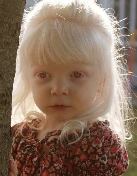
When a person has albinism they have hypo-pigmentation which is the loss or lack of natural skin color caused by a decreased level of melanin. This is the substance that gives your skin color, or pigmentation. It is either oculocutaneous which means it affects your eyes, hair, and skin or ocular, which means that it affects the eyes alone.

Ocular albinism accounts for ten to fifteen percent of all cases of albinism. If you have a child that has albinism you should be vigilant and if you notice any abnormal bruising or bleeding you should talk to their pediatrician.

The most factors that stem from having albinism is psychosocial because there is still some prejudice that exists toward anyone who is different from them. It can be particularly hard on someone who lives in a nation where most of the people are dark-skinned. In some cultures there are false beliefs that when a person lacks pigmentation it means that they are cursed or that having albinism causes sterility.

Albinism Symptoms

The symptoms of having ocular albinism can include:

  • Having eyes that are violet or light blue in color but not completely white as often believed
  • Vision that varies and can included far-sightedness or near-sightedness
  • Having nystagmus, which means that your eyes would move back and forth rapidly
  • Having lazy eye which means one of your eyes does not see with the same visual acuity as the other eye
  • Having crossed eyes
  • Having sensitivity to light
  • Legal blindness in some cases
  • Having strabismus which is when your eyes do not fixate and track together.

The symptoms of having oculocutaneous albinism can include:

  • Having hair and skin that are very light
  • Having hair that is blond and sometimes it can be red
  • Having vision conditions that are associated with ocular albinism

Albinism Causes

Albinism is an inherited medical condition and a person who has it will usually inherit a recessive gene that is responsible for this condition from each one of their parents. There are different gene defects that cause oculocutaneous albinism (OCA) and ocular albinism (OA).

Oculocutaneous albinism

Within this type of albinism there are four different types along with a subcategory. These types include:

  • OCA 1 – this is caused by a defect in the tyrosinase enzyme which helps to control the production of melanin. There are two subspecialties in this type.
  • OCA 1a – when a person has this subtype there is a complete absence of melanin and they have very pale skin, light eyes, and white hair.
  • OCA 1b – when a person has this subtype they are able to produce some melanin and have light-colored hair, eyes, and skin
  • OCA 2—this is a less severe form than OCA1 and is caused by a defect in the OCA2 gene that results in a decrease in the production of melanin. A person who has this type have light skin and their hair may be light brown, yellow, or blond. They do have slight coloring. This type is common in African Americans, Native Americans, and Sub-Saharan Africans.
  • OCA 3—this type is caused by a defect in your TYRP1 gene, which plays an important role in the melanin pathway, and will usually affect people who are dark skinned, particularly in black South Africans. This person would have brown or hazel eyes, reddish hair and reddish-brown skin.
  • OCA 4—this type is caused by a defect in the SLC45A2 protein, which provides the instructions for making the protein that is located in specialized cells that are called melanocytes. The melanocytes produce the pigment that is called melanin. This type results in a person having a minimal production of melanin. It is more commonly found in people of East Asian descent and people who have this type have symptoms that are similar to the ones found in OCA2.

Ocular albinism

This type is caused by a gene mutation on the X chromosome. It happens almost exclusively in men and only affects your eyes. People with this type will have normal skin, eye coloring, and hair but will have no coloring in their retina, which is found in the back of your eye.

Albinism Diagnosis

The best and most accurate way to diagnosis albinism is through genetic testing. This would be done to detect any defective genes that are related to albinism. There are other ways but they are less accurate. These ways would include having an electroretinogram test which is a test that measures the response of the light-sensitive cells in your eyes in order to reveal any problems with your eyes that can be associated with albinism or an evaluation of your symptoms by your physician.

Albinism Treatment

There is no known treatment for albinism at this time but there are treatments to help improve the quality of life for a person with albinism. The treatment for having ocular albinism or oculocutaneous albinism focuses on reducing, repairing and helping to prevent damage from the sun. It can include:

  • With light sensitivity when outside wear high quality sunglasses
  • For lazy eye training of the eye
  • Other vision problems can be addressed with wearing glasses
  • Having surgery on the muscles of your eyes to correct your vision problems.
  • Wearing the right SPF sunscreen and clothing to protect your skin from the UV rays of the sun

When a person has hypo pigmentation the most preventative care is to use sunscreen of at least twenty SPF or higher when you go outside, even if you are riding in a car. Some people with Type 2 may have the ability to tan but with the lack of melanin in Type 1 and 2 can lead to an increased risk of skin cancer later. This is why some people believe that when you have albinism it shortens one’s life span but in reality people with albinism often live longer. If you live in an area where sunscreen is not available or you do not use sunscreen or the right SPF, it will often result in having melanoma, which is skin cancer that is fatal.

Albinism Pictures

Photos, Images and Pictures collection of Albinism…

albinism

albinism 2

albinism 3

albinism 4

No responses yet

Dermatofibroma

Jan 27 2017 Published by under Skin Conditions

What is Dermatofibroma?

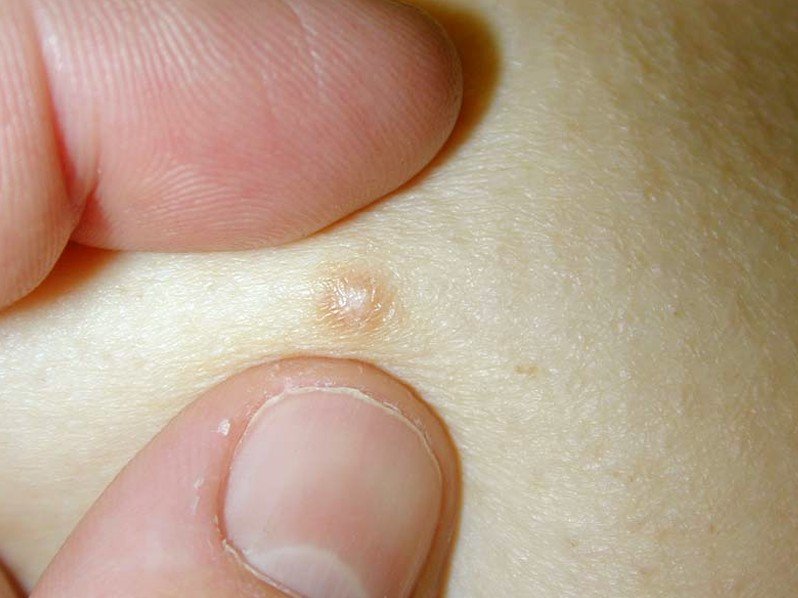
This is a growth on your skin, predominantly found on your lower legs or on your neck and face, but can develop anywhere on your body, that feels and looks like a hard lump. When you see them on your skin there is no cause for alarm because most are benign (non-cancerous) but there are a few cases where they have been malignant (cancerous). Some people will have a small cluster of growths but there are others that will only have one or two of them. There are some rare cases that there will ten or more growths. Dermatofibroma develop in an irregular pattern and are persistent in nature. Although they can happen to anyone it is mostly found in people who are middle-aged and rarely in children. Women are also more susceptible than men are. They are also called a “fibrous histiocytoma”.

Dermatofibroma Symptoms

Many times these dermatofibroma are asymptomatic, which means without symptoms, but there are others that can cause you tenderness, itching sensation, and pain. These are the ones that you will have to have removed. Other symptoms that may appear can include:

  • Ranging in color from brown, black, purple, and blue to yellow, red, orange, or pink. Many times the color of the growth is influenced by the underlying pigment of your skin.
  • May appear slowly and look like round bumps that are mostly red-brown in color.
  • When messed with they can bleed.
  • They are small in size ranging from five millimeter to ten millimeter.

Dermatofibroma Causes

The exact cause of dermatofibroma is not known but this overgrowth is a fibrous tissue that is present between the two layers of your skin, the dermis and epidermis resulting in a dermatofibroma. The dermis, where the overgrowth of cells takes place, is underneath the epidermis. They think that this medical condition is the result of insect bites, a minor injury to your skin, etc. The dermatofibroma is composed of collagen and spindle-shaped histiocytes, which is a type of immune cell that eats foreign substances in an effort to protect your body from infection.

Diagnosis of Dermatofibroma

Because of their size they can easily be diagnosed by the naked eye and when squeezing the bump it will form a dimple. If you are not sure what it is and you see your physician they may refer you to a dermatologist. They will review your medical history along with a physical exam of the growth. If they are not sure it is a dermatofibroma they will do a biopsy which is when they will look at a sample of the growth under the microscope. When the dermatologist gets the pathology report back and it is a dermatofibroma it can be a variant such as:

  • Aneurismal
  • Cellular
  • Epithelioid
  • Lipidized ankle-type
  • Atypical
  • Cholesterotic
  • Palisading

Treatment

Normally when a person has dermatofibroma it does not need any treatment unless it causes you discomfort. You may find it uncomfortable if it interferes with wearing clothing or shaving and may decide to have them removed for that reason. They will not heal themselves and they cannot be cured permanently. If you decide to have the dermatofibroma removed your dermatologist can surgically remove it. When you have this medical condition you should avoid exposure to the sun. You can also treat them at home by applying either cellulite cream or camphor spirit. The dermatologist may give you a steroid injection to reduce it in size. To help with the itching you can use an over-the-counter anti-itch medication.

Dermatofibroma Pictures

dermatofibroma

dermatofibroma 2

dermatofibroma 3

dermatofibroma 4

Dermatofibroma Removal

Removing the dermatofibroma is done under local anesthesia. One thing to know is that when you have them surgically removed it can lead to the formation of a permanent scar and it could look more unattractive that the presence of the dermatofibroma. The reason is that the scar will be larger in shape than the original dermatofibroma. Sometimes the dermatologist will use an external medication to treat this medical condition called cryosurgery.

Cryosurgery uses a liquid cooling agent that is applied to the bumps in order to destroy the defected cells by freezing them. The liquid cooling agent that is usually used is liquid nitrogen but they could use dry ice. Unfortunately with this treatment the bump is fixed superficially making it unnoticeable but over time it will be conspicuous again and the treatment will have to be repeated. When it re-grows it is not as noticeable as it was the first time. When using cryosurgery it will leave a white mark behind on your skin where the bump was removed. If you want it to be completely removed the dermatologist will also have to remove a part of your subcutaneous fat that surrounds the bump.

No responses yet

Lymphangioma

Jan 27 2017 Published by under Diseases and Conditions

What is Lymphangioma?

This is a benign, meaning non-cancerous, growth that can be found in you lymphatic system. This is the system that is an extensive interconnected system of nodes, vessels, and spaces allowing your body to circulate lymph which is a fluid that is made from other fluids and protein. Most of these abnormalities occur in children under the age of two. They can be there when the baby is born or it shows up later. Having lymphangioma does not pose a health threat unless it happens in the fetus. If they show up in the early part of fetal development the cystic lymphangioma can cause the accumulation of fluid and can result in the death of the fetus. When a child has lymphangioma it can form on any mucous membrane or the skin but it is mostly found on the neck and groin area.

Lymphangioma Symptoms

The symptoms vary according to the type of lymphangioma you have.

Cystic

These are fluid filled and large in size and may appear as bulges under your skin. This type is frequently found on your groin, armpits, and neck accompanied by mild pain when the swollen area is compressed.

Cavernous

These are similar to the cystic type but are usually found deeper under your skin of your mouth or neck and appear a thickening or bulge of your skin. They are also likely to spread. They vary in size from as little as a centimeter to several centimeters wide. There have been some cases in which they affect an entire extremity like your foot or hand. This type is usually painless but when pressure is exerted on the area you might feel pain that is mild.

Lymphangioma circumscriptum

These consist of various sized red or pink blister clusters on the surface of your skin.

They usually appear under your skin as a fluid filled reddish or yellowish bubble or series of bubbles.

Although these are not symptoms it is still something that a person needs to know which is that when you have lymphangioma it can cause complications like infections and bleeding. If it is large it could obstruct the mucus membranes and your respiratory system. This could cause distress breathing. They can also be an early indicator of chromosomal or genetic disorders.

Lymphangioma Causes

Lymphangioma is often formed during fetal development because of a blockage and subsequent tangling in your lymphatic system. Your lymphatic system is an important part of your immune system which is where the bacteria and any other foreign substances are destroyed. Having a lymphatic blockage can be caused a variety of factors like alcohol consumption by the mother while pregnant or an infection.

How is Lymphangioma Diagnosed?

To diagnosis lymphangioma in a fetus the gynecologist will have an ultrasound along with a testing done by taking some amniotic fluid from the uterus and testing it to see if the fetus has lymphangioma.

Lymphangioma Pictures

Collection of Photos, Images and Pictures of Lymphangioma…

lymphangioma

lymphangioma 2

lymphangioma 3

lymphangioma 4

lymphangioma 5

Lymphangioma Treatment

Because the lymphangioma does not have a chance of becoming malignant most them are treated for cosmetic reason. In cases that are not severe draining the fluid could give you short-term relief. Some patients have radiotherapy which is a form of medical therapy that involves using ionizing radiation. The most effective treatment to use that causes the least damaging to the surrounding healthy tissue is surgical removal.

No responses yet

Erythromelalgia

Jan 26 2017 Published by under Diseases and Conditions

What is Erythromelalgia?

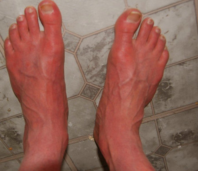
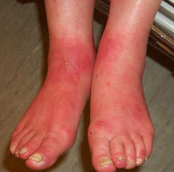
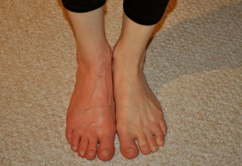
This is a rare neurovascular peripheral pain disorder that affects your skin. It will cause the skin on your feet and hands to become red and have painful burning sensation. Early onset erythromelalgia can develop before a person turns twenty-five and late onset erythromelalgia will usually develop between the ages of forty and sixty. Approximately sixty percent of the people worldwide are affected by late onset erythromelalgia. It is a disorder that can cause a person to become debilitated if it is severe enough. It can cause a person to become unable to function in a workplace, have sudden depression, lack of mobility, and more. It is also referred to as acromelalgia, Mitchell’s disease, or burning feet syndrome.

Types of Erythromelalgia

Erythromelalgia can be divided into two types which are:

Primary erythromelalgia

This results from a genetic mutation and the symptoms associated with this type are usually triggered by changes in the temperatures, over-exertion, heat, spicy foods, exercise, alcohol.

Secondary erythromelalgia

This results from certain autoimmune disorders like mushroom poisoning, mercury poisoning, Hypercholesterolemia, and essential thrombocytosis.

Erythromelalgia Symptoms

The symptoms of erythromelalgia can vary from patient to patient. There are some that will only have tingling sensations and slight itching that will only last for several minutes. Others are not so lucky and will have severe skin discoloration and pain that is intense and can last for weeks, even months, at a time. The symptoms are typically worse when your extremities, which are your hands and feet, are bound by gloves and shoes and causes friction on your hands and feet or they are exposed to hot temperatures. Although it is rare it is possible for you to have erythromelalgia symptoms of redness and burning to affect other parts of your body, especially the ears and scalp. When a person has erythromelalgia it can affect one or both side of your body.
Other symptoms or signs can include:

  • Having a continual burning while other are just having trouble with flare-ups
  • When having a flare-up it can last only minutes or even days. They usually occur late in the day and will continue all night.
  • Limbs that are swollen
  • Genetic Blood Vessel Condition
  • Pain and inflammation in different parts of your body.

Causes of Erythromelalgia

Although erythromelalgia is usually caused by extreme heat or friction many times there is no identifiable cause. Most of the patients who have idiopathic erythromelalgia are over the age of sixty, which means there is no known cause. When people who are younger than sixty years of age may have the symptoms of erythromelalgia because of an underlying autoimmune disorder like diabetes or lupus or they could have a genetic blood vessel disease. Erythromelalgia has also been correlated with neurological conditions like multiple sclerosis. Although it could happen it is rare that it can run in families and have a parent be a carrier of a specific gene mutation and passing it down to a child.

Some of the possible causes can include:

  • Heavy metal poisoning
  • Fungal or other infections
  • Age
  • Neurological problems
  • Being deficient in nutrients like Pantothenic Acid
  • Excessive sweating, especially on your feet.
  • Wearing socks or shoes that are too tight for too long period of time

Diagnosis

If you are noticing that your hands and/or feet appear red and you are having painful burning sensations you should see your dermatologist and not your physician. Many times your dermatologist may be able to give a diagnosis that is based on the physical characteristics of your extremities or by the symptoms that you tell your dermatologist. If you are not experiencing erythromelalgia or a flare up at the time of your appointment the dermatologist may try to induce an episode of erythromelalgia by soaking a foot or hand in hot water. The dermatologist may also order blood tests, imaging scans, and neurological exams to look for signs of any underlying condition. To get the proper diagnosis it can take several months and you may have to consult many different specialists to find out what the problem is.

 Erythromelalgia Treatment

A person who has idiopathic erythromelalgia with mild symptoms may not need any medical treatment. When seeing your dermatologist for this medical condition they may suggest that you avoid wearing shoes whenever possible, on hot days stay inside, and when you have flare-ups you can try to put your hands and feet in cool water. If it is a severe case your dermatologist may prescribe a topical cream to apply to your hands and feet to soothe the burning sensation. To help reduce the symptoms you could take over-the-counter anti-inflammatory medications. You should also stay indoors on hot days, wear shoes that are not too tight and when at home go barefoot or wear loose fitting sandals, and do not do intense workouts for a long period of time.
Surgery

If all the above treatments do not give you the relief it should, your dermatologist may suggest surgery. This surgical procedure is called a sympathectomy and involves cutting the nerve endings that transmits the pain signals from your hands and feet. There are some serious risks associated with this surgery but generally it is considered to be more beneficial to the patient whose quality of life is severely affected from erythromelalgia.
Natural treatment

One way to help cure erythromelalgia naturally is to eat a lot of foods that are rich in vitamin B like:

  • Whole grains
  • Brown rice
  • Peanuts
  • Dairy products that include milk and butter
  • Various green and yellow vegetables

These foods can also help to boost your immune system along with giving you relief from the symptoms of erythromelalgia.

Erythromelalgia Pictures

erythromelalgia

erythromelalgia 2

erythromelalgia 3

erythromelalgia 4

erythromelalgia 5

No responses yet

Nail Psoriasis

Jan 25 2017 Published by under Hair and Nails

What is Nail Psoriasis?

Generally psoriasis is a skin condition but it can affect your toenails and fingernails too. When it affects your toenails or fingernails it is referred to as nail psoriasis or psoriatic nail disease. When nail psoriasis starts to develop usually a person will have cutaneous or skin psoriasis. You may also have psoriatic arthritis when you have nail psoriasis. One thing to note is that it is not contagious so you can not pass it on to others through any type of contact.

Nail Psoriasis Symptoms

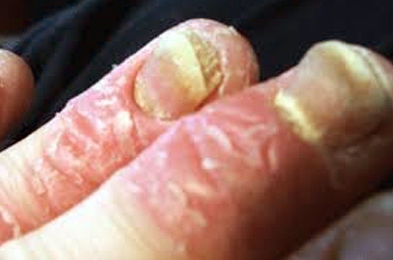
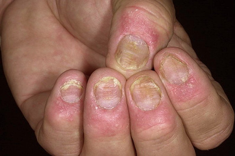
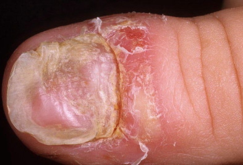
Approximately five percent of people who have nail psoriasis do not have any symptoms that are visible. Although these are not really symptoms of this medical condition they are observed discolorations, which are common, and markings that are commonly found when a person has nail psoriasis which can include:

  • Horizontal lines that run side to side across your nail called “Beau’s lines” that can be visible
  • There may also be white spots that are visible called leukonychia. Besides the white spots leukonychia can also appear as spots or stripes and may cover the entire nail.
  • Having salmon patches or oil spots which can look like blood or oil that has been trapped under your nail.
  • Having a tiny break in your skin that can cause a tiny bleed under your nail that is referred to as a splinter hemorrhage. It may look like a black thin line that runs the length of your nail.
  • Having a congestion of the capillaries under your nail referred to as spotted lunula. It usually appears at the base of your nail as a reddening of the arch there.

You may also have changes in the structure and shape of your nail, which is very common. Some of these changes can include:

  • Pitting of the nail which is quite common
  • The skin under your nail may start to thicken, causing your nail to become lose and separate
  • Crumbling of your nail if this medical condition affects the nail bed

Nail Psoriasis Causes

It is not fully understood what causes a person to have nail psoriasis but what they do know is that heredity does play a part of a person having it. In addition immunologic and environmental factors could contribute to you developing nail psoriasis.

Nail Psoriasis Pictures

nail psoriasis

nail psoriasis 2

nail psoriasis 3

nail psoriasis 4

nail psoriasis 5

nail psoriasis 6

Treatment

At this time there is no known cure for nail psoriasis. When treating nail psoriasis the treatment used focuses on improving the function and appearance of the nails, treating any complicating issues like having an infection, and minimizing the symptoms. When you see your physician for treatment for nail psoriasis the may prescribe ointments and topical creams. If your physician determines that either the whole nail or part of the nail must be removed it can be done through surgery or chemical treatment. To reduce the chances of developing an infection you can trim back the nail to help prevent them from catching on to something and causing them to pull away from your nail bed. When you are caring for the affected nail you must do it very gently. If you do not and use aggressive nail care or grooming it could cause your nail psoriasis to flare up and make the condition worse.

Which treatments are used for nail psoriasis will depend on how severe the condition is. Some of treatments that are used include:

  • If you have a flare-up of general psoriasis that involves your nails it can sometimes be caused by a fungal infection so in this case you would use an anti-fungal ointment or cream.
  • You may also be given oral medications to help suppress the rapid decrease of the cells that are causing your nail psoriasis. When taking these oral medications you will take them in spurts because they could cause you to have other symptoms that are skin related like dry skin and itching.
  • Using ultraviolet light, which can be artificial or natural light, to help ease the production of these extra cells. You should not do this treatment yourself. It should only be done under the supervision of your physician because it is not healthy for you to be exposed to excessive ultraviolet light. Your physician would know which way is best to make sure that it is done effectively and safely
  • Using a variety of ointments and creams that contain corticosteroids and vitamins. Two of the vitamins that should be in the creams are vitamins A and D. These are the two vitamins that are successful in soothing your skin and slowing the growth of new cells. The corticosteroids in the ointments and creams will help to alleviate inflammation. This type of treatment is referred to as topical treatments because they are usually applied directly to your nail. Sometimes you will have a prescription for more than one ointment and cream in order to find the most effective treatment.

If your case of nail psoriasis is severe and you do not treat it this can lead to social and functional problems.

Natural treatment

If you do not want to go to your physician to have this condition treated there are some natural treatments that you can try which can include:

  • Soak your nails in different warm vegetable oils like sunflower oil, olive oil, coconut oil, etc to keep your cuticles and nails moisturized.
  • Use a coal tar bath by putting three capfuls of coal tar bath oil in a container with warm water. How large a container you need will depend on if you are soaking your toenails or fingernails. Soak for thirty minutes and then trim your nails to the point of their attachment. Apply a moisturizer on each nail.
  • Take protein mixtures and vitamins that are for “skin, hair, and nails.”
  • Make sure that when you are doing work that could hurt your nails that you wear protective gloves.
  • Soak your nails in warm water for ten to fifteen minutes to soften them so you can gently file away the thickened parts of your nails with an emery board.

Although this is not really a treatment you can also use artificial nails or nail strengtheners to help protect the fragile nails from damage.

No responses yet

Telangiectasia

Jan 24 2017 Published by under Skin Conditions

What is Telangiectasia?

This is a blood vessel that is most often found in a mucous membrane or in the outer layer of your skin. This blood vessel can be a capillary, arteriole, or small vein. They have dilated and have been left enlarged permanently. It can be found anywhere on or in your body but are most commonly seen on your legs and face. You will usually see them on below your knees and on the surface of your ankles and thighs and around your nose, cheeks, or chin on your face. They can also be seen on the whites of your eyes. Although they are usually harmless there is also a chance that they can occur in your brain and cause some major problems from bleeding. On your legs they are sometimes referred to as spider veins.

Telangiectasia Symptoms

Many times when these lesions develop on people that are healthy they usually do not cause any symptoms. What you might see is that this permanent dilation can cause a reddish discoloration in the surrounding skin. This happens because of the small lesions that develop. When this happens it makes telangiectasia easier to see.

  • When telangiectasia appears on your skin, which is also called vascular ectasias, they will look like tiny lines that range in color from red through blue and purple.
  • They measure between one and three millimeters in width. They are usually somewhat longer than it is wide.
  • They can look unattractive and cause discomfort
  • The formation of these lesions is gradually
  • They can be painful because of the pressure on the venules
  • Itching

Telangiectasia Causes

The exact cause of telangiectasia is not known but according to researchers they believe that it could be because of overexposure to the sun. The reason that researchers think this is because these lesions will usually appear in places that are more exposed to sunlight. Some of the other causes that can possibly cause telangiectasia can include:

  • Pregnancy because this can put a large amount of pressure on your venules
  • Aging because as you get older the blood vessels start to weaken
  • Alcoholism which can affect the flow of blood in your vessels

They can be seen with a number of other diseases like acne rosacea, birthmarks, prolonged use of topical or oral corticosteroids, and long time use of oral contraceptives, several different inherited disorders, and more. It has also been found that telangiectasia can be caused by routine damage to your skin or just appear spontaneously.

Telangiectasia Pictures

telangiectasia

telangiectasia 2Telangiectasia on Face

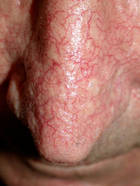
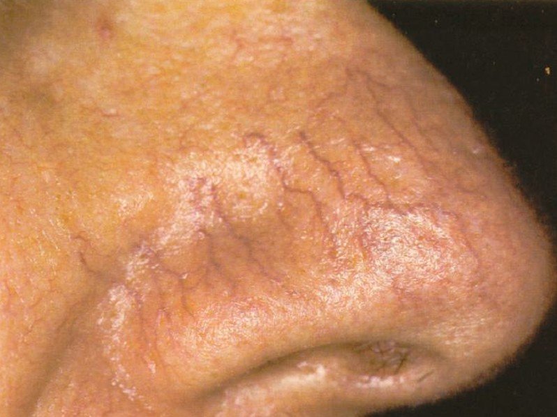
telangiectasia 3Telangiectasia on Nose surface

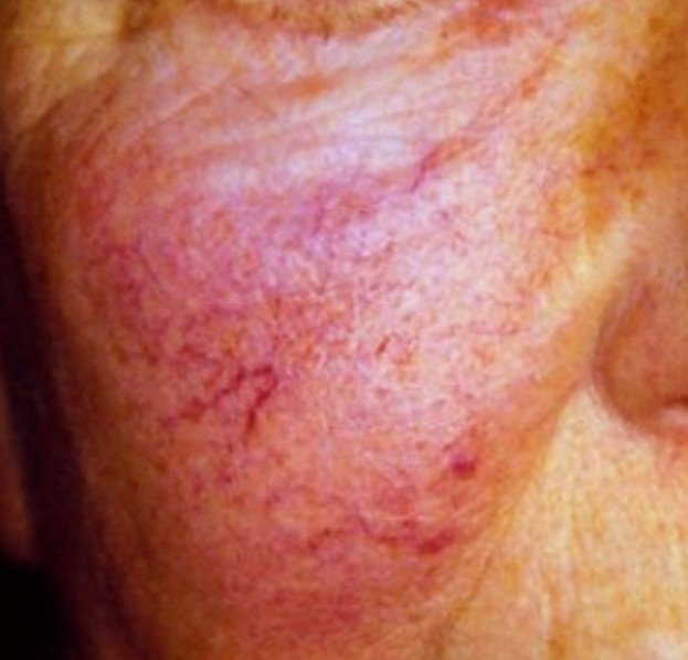
telangiectasia 4Telangiectasia on the right cheek

telangiectasia 5Telangiectasia on the nose

Diagnosis

When seeing your physician or dermatologist they may be able to diagnosis telangiectasia just by the obvious signs of this disease because it can be seen as lines that are red or marks on your skin. Some may do other tests to rule out any underlying diseases that can include:

Sturge-Weber disease

This is a disorder that is rare and can cause nervous system problems and a birthmark that is called port wine stain.

Xeroderma pigmentosa

This is where your eyes and skin are very sensitive to the ultraviolet light. It is considered a rare medical condition.

Hereditary hemorrhagic telangiectasia

This is an inherited disorder of your blood where the blood vessels form abnormally. These are called arteriovenous malformations.

Spider angioma

This is an abnormal collection of blood vessels near the surface of your skin

Some of the diagnostic tests that can be used to see if it is a result of an underlying or not include:

  • CT scans
  • MRI scans
  • X-rays
  • Blood tests
  • Liver function studies

Telangiectasia Treatment

If you notice telangiectasia on your skin it is advisable that you see your physician to rule out the presence of an inherited or underlying condition. There are also treatments that your dermatologist can do if they are detracting from your personal appearance your dermatologist could treat it by electrosurgery, or a pulsed dye laser. If it is not caused by an underlying condition you could cover it up by using makeup.

Other methods to treat telangiectasia can include:

Laser ablation

The laser targets the vessels that have been widened and seals them. With this treatment there is little pain and a short recovery. This the type of treatment used on your face.

Surgery

With surgery the widened vessels can be removed but this treatment it can take you a long time to recover and can be painful.

Sclerotherapy

This is a cosmetic surgery in which the dermatologist injects a sclerosing agent into your veins. The dermatologist uses one injection for every one inch of vein and as a result you may need between five to fifty injections to treat telangiectasia. This is the treatment that is used on your legs.

Because they are not attractive looking and are causing discomfort many decide to have them removed. This is accomplished by causing some type of damage to your blood vessel, forcing the blood vessel to scar or collapse. By doing this it will decrease the appearance of the patterns or red marks on your skin.

No responses yet

Next »

© 2011-2019 MDDK.com - Medical Tips and Advice. All Rights Reserved. Privacy Policy
The health information provided on this web site is for educational purposes only and is not to be used as a substitute for medical advice, diagnosis or treatment.