Diseases General Health Skin Conditions

Archive for September, 2016

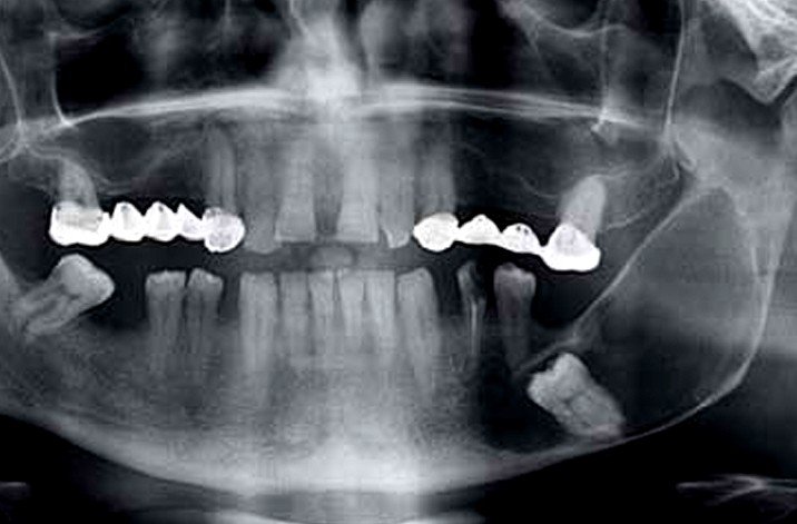
Dentigerous cyst

Sep 07 2016 Published by under Oral Health

What is a Dentigerous cyst?

This is a type of cyst that normally surrounds the crown of your impacted tooth. It is noticed due to the accumulation of fluid that takes place between the layer referred to as reduced enamel epithelium and the enamel surface of a tooth. This will lead to what looks like a cyst. The crown is found within the lumen of the cyst and is almost always seen in permanent teeth. It is rare to see them in deciduous teeth, which are your temporary, or baby, teeth. A dentigerous cyst falls under the category of developmental odontogenic cysts. The dentigerous cyst is believed to account for one-fifth of all jaw cysts found and is also the second most common of all developmental odontogenic cysts.

You will find this type of cyst it is normally associated with the crown of your unerupted or impacted tooth. It is normally seen in the area of the mandibular and maxillary third molars. Sometimes it may be seen in your upper canine teeth, especially if they are impacted. It may also be found in supernumerary teeth, which are the extra teeth you can have in your mouth. You will normally find dentigerous cysts in people during their twenties and thirties with men having more of a chance of developing them than women. They are usually solitary cysts. With other medical syndromes you may see multiple or bilateral cysts. It is also referred to as a follicular cyst and is usually non-cancerous.

Symptoms

A dentigerous cyst does have the ability to become aggressive and if they become larger in size it could lead to bone expansion. This can lead to a visible swelling and then to facial asymmetry, which is a medical condition in which the right side of a person’s face does not match the left side. It can also increase into a size that may displace the adjacent teeth eventually, leading to discomfort and pain that can be bad. With a severe cyst it can take up the whole angle of your mandible, causing your mandible to be hollowed out. This could make your mandible compromised and weak.

If the dentigerous cyst becomes large in size where it does not cause any symptoms this means that it is asymptomatic, which means there are no symptoms. Unless the cyst becomes infected there will be no pain.

Causes

At this time it is not known exactly what leads to this type of fluid accumulation that eventually results in the formation of a cyst. Some feel that the reason that a person develops a dentigerous cyst is due to the presence of teeth that have not erupted, pulp necrosis, and primary tooth trauma. These teeth that have not erupted include your wisdom teeth and upper jaw canines.

Diagnosis

They symptoms of a dentigerous cyst are what play a critical role in the diagnosis of this type of cyst. Normally it will be diagnosed with an x-ray. The area between the tooth and the cyst lining will normally be larger than five millimeters. Discovering a dentigerous cyst is usually by accident when they are doing routine dental x-rays.

Treatment

The only way to take care of a dentigerous cyst is with surgical intervention as there is no medicine that will help to get rid of it. If they remove the entire cystic lining and cyst the chances of recurrence is minimal. To avoid any complications you should have it diagnosed and treated properly.

Surgery

  • Small dentigerous cyst – this would involve the surgical removal of the entire cystic lining, cyst, and the impacted tooth
  • Large dentigerous cyst – if there is a large amount of bone loss the treatment would be marsupialization or surgical drainage may be necessary. Marsupialization means to cut a slit into a cyst to let it drain. It is not a good idea to surgically remove the large cyst because of the risk of fracturing your jaw

Complications

  • Developing an ameloblastoma, which is a rare benign tumor that develops in your jaw near your molars
  • Developing an epidermoid carcinoma, which is a non-small-cell type of lung cancer that develops from the cells that line the inside of your lungs
  • Risk of a large jaw fracture

Dentigerous cyst Pictures

dentigerous cyst

dentigerous cyst pictures

No responses yet

Celiac Plexus Block

Sep 07 2016 Published by under Pain Management

A Celiac plexus block is a medical procedure in which a local anesthetic and medication are injected into the celiac plexus. The celiac plexus is represented by a bundle of nerves, which are located in the center of the abdomen, near the aorta and other blood vessels. The main purpose behind this procedure is to block the pain signals that are transmitted from the territories innervated by the celiac plexus (abdominal area) to the brain.

The celiac plexus block is recommended in patients who have been diagnosed with chronic pancreatitis and chronic abdominal pain, caused by different types of cancer. The procedure is performed with intravenous sedation, as it can be quite painful, due to the fact that the doctor will need to reach the deeper tissues. In case the patient does not respond to the first injection, a further injection is going to be recommended. As the response to the procedure varies from one patient to the other, there are no set numbers of injections recommended. In general, relief from the symptoms is provided after 2-10 injections.

Technique

Before the actual procedure, you will be administered intravenous anesthetics, so that you become relaxed and tolerate the procedure, without any pain or discomfort. You will be lying on your back, while the doctor applies a local anesthetic to the point of entry. Using guided X-raying (fluoroscopy), the doctor will insert a thin needle and deliver the pain medication to the respective site. Among the medication that can be administered, there are: epinephrine, corticosteroids or clonidine. The doctor might also decide to administer alcohol or phenol. The injection can last between ten and thirty minutes to be completed.

Is Celiac plexus block effective?

As it was already mentioned, the celiac plexus block has as main purpose the blockage of pain signals from the abdominal area to the brain. The pain relief provided by this procedure vary from one person to the other. In some patients, the pain relief can last for several weeks, while in others the period of pain relief might extend to a couple of years. The procedure can contribute to a great improvement in the overall quality of life, allowing patients to resume their daily activities and even return to work. The efficiency of this procedure can be increased by repeating the injections, according to the recommendations made by the doctor.

Procedure

The celiac plexus block is a procedure that is done with intravenous sedation, so as to ensure the highest level of tolerance for the patient. In order to identify the exact location of the injection, the doctor will use fluoroscopy or guided X-rays. For the visualization of the injection site to be possible, the doctor will inject radiographic dye first. Then, he/she will administer the medication for pain relief. The procedure lasts for about half an hour, provided there are no complications. In the majority of the cases, the patient is allowed to go home in the same day.

During the procedure, the vital signs of the patient are monitored. The doctor will monitor the activity of the heart, the pulse and the breathing capacity.

How long does it take for a celiac plexus block to work?

As the doctor also injects a local anesthetic, the pain relief is immediate and this effect lasts for a couple of hours. The celiac plexus block remains effective for a couple of weeks to several years. It should be noted that the efficiency of the celiac plexus block increases after each injection.

What to expect after a celiac plexus block?

Immediately after the procedure, you might experience a sensation of warmth at the level of the abdomen and a reduction in the pain intensity (due to the local anesthetic). Temporary side-effects that may appear with the celiac plexus block include: temporary weakness, numbness sensation in the abdominal wall, similar sensations at the level of the leg.

Given the fact that you have been sedated, it is important to have someone waiting to take you home. During the first days after the procedure, the doctor will recommend that you avoid intense physical effort. It is for the best that you stick with the activities that you can tolerate, meaning those that cause no discomfort or pain. Physical therapy might be recommended after the celiac plexus block, promoting a better and a faster recovery.

It is difficult to predict how much the celiac plexus block helps, as each patient has a different response to the administered treatment. The response might be reduced in patients who have suffered from health problems at the level of the abdomen for prolonged periods of time or in those who have been diagnosed with severe cases. The doctor can provide the most accurate information about the expected response, upon the performing of the celiac plexus block.

The doctor will also talk to you about the potential contraindications that this procedure presents. These contraindications include: taking certain medication (blood thinners), suffering from diabetes (severe forms), and the presence of infection at the level of the body (active state). In these situations, the doctor might decide to postpone the celiac plexus block, until it is safe for this procedure to be performed. As for medication, you might be required to stop taking the medication prior to the procedure, so as to reduce the risk of hemorrhage.

In general, the celiac plexus block is a relatively safe procedure. However, for the first couple of days after the procedure, you might feel pain or soreness at the site of injection. In rare cases, the following complications might occur: bleeding, infection, nerve injury, damage to the surrounding blood vessels, collapsed lung. The complications are often the result of misguided applications. As for the other side-effects of this procedure, this might include the hypotension and the change in the bowel movements (diarrhea). Nevertheless, you must remember that these side-effects are temporary, going away on their own.

In conclusion, the celiac plexus block is a recommended procedure to all patients who suffer from chronic abdominal pain, due to different types of cancer or other chronic conditions. It is especially indicated in those who have failed to respond to other types of treatment, improving the overall quality of life.

No responses yet

Monteggia Fracture

Sep 06 2016 Published by under Fractures

Definition

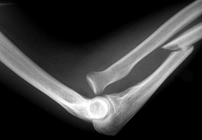
This is an arm fracture in which the joint with the radial head at your elbow becomes dislocated and the ulna, one of the bones in your forearm is broken. When a person has this type of fracture it is normally very evident because it causes extreme pain. Your range of motion will also be limited. It is named after Giovanni Battista Monteggia. He is the one who described this fracture in the early 1800’s. It is rare for an adult to have a Monteggia fracture but is most common in children between the ages of four and ten.

Relevant anatomy

The radial head is a bone that makes up part of your elbow. Your ulna is a bone in your forearm that is broader near your elbow and tapers as it approaches your wrist. It is on the pinky-finger side of your arm near the radius bone, which is the lateral bone of your forearm between your elbow and hand. The radius bone is always aligned with your thumb.

Pictures : How Monteggia Fracture Looks like?

monteggia fracture

Classification

There are four classifications of a Monteggia fracture. What classification they fall into depends on the displacement of the radial head. They are referred to as Bado classifications.

  • Type 1 — fracture of the middle third of your ulna with the anterior, or front, dislocation of your radial head. This accounts for approximately sixty percent of these types of fractures and is referred to as an extension type. This is most common in adults and children.
  • Type 2 — fracture of your middle third of your ulna with posterior, or back, dislocation of your radial head. This accounts for approximately fifteen percent of fractures and is referred to as a flexion type. This is more commonly seen in adults.
  • Type 3 — fracture of the wide portion of your ulna, referred to as the metaphysis with the lateral, or the side, dislocation of your radial head. This accounts for approximately twenty percent of these fractures and are referred to as a lateral type.
  • Type 4 — fracture of the middle third of your ulna and radius with the anterior, or front, dislocation of your radial head. This accounts for approximately five percent of these fractures and is referred to as a combined type.

Symptoms

  • The only general symptoms of having a Monteggia fracture are swelling and pain at the elbow joint
  • You may also have pain in your arm that gets worse every time you move your elbow or wrist
  • When looking at it you may or may not see any obvious dislocation but you may notice that there may be loss of range of motion at the elbow because of the dislocation
  • You may also have swelling in your hand, wrist, and forearm
  • Forearm tenderness
  • Numbness

Causes

A Monteggia fracture can be caused by:

  • A blow to the back of your upper forearm
  • Hyperextension, which is when you move your tendons, muscles, or joints beyond the normal range of motion
  • Falling on an outstretched hand to break your fall. This is referred to as a hyperpronation injury.

Diagnosis

To diagnosis a Monteggia fracture the radiographer will use an x-ray machine to visualize the joints and bones in your arm. They will take several angles so they can tell the full extent of your injury.

Treatment

Although the exact treatment used depends on how severe the fracture is the conservative treatment for a Monteggia fracture is to cast it in order to immobilize the arm so the joint and the ulna have a chance to heal. After your arm has been in a cast for several weeks your physician will order more x-rays to confirm that the healing is even and the bones are knitting together. If they have pulled out of position or not healing the cast will be remove to correct the situation. Unless the fracture appears problematic casting is often sufficient for younger patients. Many times the Monteggia fracture will have to be treated using surgery. How long a cast will stay on depends on the classification of the fracture.

  • Type 1 — three weeks in a cast above your elbow and then another three weeks with a cast on the area below your elbow. This type rarely requires surgery.
  • Type 2 — three weeks in a cast
  • Type 3 — four weeks in a long arm cast
  • Type 4 — three to four weeks for the lower part of your arm past your elbow and three week for the area above your elbow

The casting is done by an orthopedic physician who is trained in the branch of medicine that deals with your musculoskeletal system.

Other treatments can include:

  • Resting your arm by elevating it higher than your heart
  • Cold compresses but you have to be careful not to get the cast wet. Put the ice pack into a plastic bag and wrap in a towel before you put it on the cast so the cast will not get wet
  • Use a shoulder sling
  • Taking either a prescription medication or over-the –counter medication for the pain.

Surgery

If the Monteggia fracture is severe you may need to have surgery. The surgery is done to pin the fractured ulna and to stabilize the joint if necessary. This type of surgery is referred to as open reduction internal fixation. The surgery is done by an orthopedic surgeon, who is a surgical specialist that has received special training working with injuries to the bone, especially this type of fracture. Because it is a very complex surgery you may need several surgeries to correct a Monteggia fracture. After the surgery is finished a cast will normally be applied to give it stability during healing. Depending on the patient, healing time can take six weeks or longer. For children who have a type four classification they may use intramedullary wires or plate fixation to stabilize the fracture.

Complications after surgery

After surgery there could be complications or long range problems such as:

  • Nerve palsy and damage
  • Muscle damage
  • Tendonitis
  • Infection
  • Arthritis
  • Loss of range of motion and stiffness
  • Audible snapping or popping
  • Compartment syndrome, which is an increase in pressure in one of your body’s compartments that contains nerves and muscles.
  • Deformity
  • Chronic pain that is associated with surgical hardware like the plates, pins, and screws

Problems if not healing correctly

If the fracture does not heal correctly you could have one arm that is shorter than the other arm. It can also predispose you to another fracture. A big risk with an open fracture, which means that your skin has been damaged during the injury or the bone has broken through your skin, is necrosis. This is when the tissue and bone die because they are not being supplied with blood or there is an infection.

Management

A Monteggia fracture is often managed with resetting and casting in children. After having surgery there will be a restriction on range of motion on the elbow but that is normally lifted after three months. In three months after the cast is removed the orthopedic physician or surgeon will have you start rehabilitation. Rehabilitation is necessary in order for your muscles to become stronger.

No responses yet

Subclinical Hypothyroidism

Sep 05 2016 Published by under Ear, Nose and Throat

What is Subclinical Hypothyroidism?

The subclinical hypothyroidism can be defined as a medical condition in which the thyroid does not function correctly, the dysfunction being classified as mild. The condition is characterized by normal peripheral thyroid hormone levels, while the thyroid stimulating hormone (TSH) is slightly increased.

According to the specialists in the field, this condition affects approximately 3-8% of the general population. The most important implication is the increased risk of progression to clinical hypothyroidism; studies have shown that 1 in 10 people who are diagnosed with subclinical hypothyroidism is going to be diagnosed with clinical hypothyroidism within a period of three years. This condition is more often encountered in women than men and its prevalence increases as a person ages. Some studies have identified subclinical hypothyroidism as a risk factor for cardiovascular disease but further research is necessary.

Symptoms

Given the fact that the thyroid dysfunction is only mild, there are a lot of people who do not present any symptoms. However, there may be patients experiencing the below-mentioned clinical manifestations:

  • The skin is drier than usual
  • The cognition is affected, with the thinking process becoming slower
  • The memory becomes poorer (both declarative and working memory are impaired)
  • The muscles become weaker and the person is easily fatigued (progressive tiredness)
  • Increased frequency of muscle cramps
  • High sensitivity to cold (patients declare they feel a lot colder than they used to)
  • The voice changes, becoming deep and hoarse
  • The retention of water is possible, with the puffiness around the eyes becoming visible
  • The bowel movements are slowed, with constipation being a frequent complaint
  • Mood swings, anxiety and depression can also appear in these patients
  • In the cases that are closer to the progression to clinical hypothyroidism, the characteristic goiter can appear
  • Patients gain weight
  • The hearing might become impaired as well
  • Possible risk for cardiovascular disease
  • Menorrhagia in women
  • Reduced tendon reflexes (most commonly seen in the knee reflex)
  • Reduced cardiac rhythm is also possible (bradycardia).

Causes

These are the most common causes that can lead to the appearance of subclinical hypothyroidism:

  • Hashimoto’s disease
    • Medical condition that is also known as chronic autoimmune thyroiditis
    • Most often incriminated in the appearance of subclinical hypothyroidism (accounts for 90% of all the cases)
  • Hyperthyroidism treatment
    • The changes characteristic of subclinical hypothyroidism appear in particular in patients who have undergone treatment with radioactive iodine, for the overly-functioning thyroid gland
  • Surgical intervention or anti-thyroid medication
    • Both account for 5-25% of the patients who are diagnosed with subclinical hypothyroidism
  • Medication
    • Lithium
    • Amiodarone
  • Surgical intervention at the level of the neck or head (affecting the thyroid as a complication)
  • Radiation therapy for different forms of cancer
    • The thyroid gland is quite sensible to radiation therapy and its functioning can be very easily affected by this kind of treatment.

Diagnosis

These are the most common solutions available for the diagnosis of subclinical hypothyroidism:

Blood testing

  • TSH hormone levels
    • Repeated measurement should be made due to the circadian fluctuations (the TSH hormone levels are higher at night)
  • Serum FT4
    • Confirm that the condition did not yet progress to clinical hypothyroidism
  • Thyroid antibody testing
    • Identification of the exact cause of the subclinical hypothyroidism – chronic autoimmune thyroiditis
    • Also recommended in patients who have a goiter, other autoimmune conditions or increased levels of TSH

Ultrasound

  • Identification of specific thyroid echo pattern (inhomogeneous)
  • This pattern appears before the circulating thyroid antibodies, so it is considered a useful method for the early detection of autoimmune thyroiditis

Aspiration cytology

  • Most sensitive method used for the diagnosis of autoimmune thyroiditis
  • Last resort, due to the fact that it is considered an invasive method of diagnosis.

Treatment

The patients who would benefit the most from the treatment are the ones who have had high levels of TSH hormone for prolonged periods of time and the blood tests have shown the presence of antithyroid antibodies (increased risk for progression to clinical hypothyroidism). In general, it is considered that treatment should be administered to all patients who have TSH equal or over 10 mIU/L and especially I those who already present some of the clinical features.

In case of pregnant women or those who are planning to conceive, getting treated for this condition is mandatory, so as to reduce the risk of pregnancy complications and prevent the impairment of cognitive functioning in the newborn baby.

In patients who have TSH levels under 10 mIU/L, the doctor will pursue treatment only in those who present specific symptoms, in those who suffer from infertility, have a goiter or antithyroid antibodies identified through the blood testing. The treatment for subclinical hypothyroidism is not indicated in elderly patients who are over 85 years of age.

The standard medication used for the treatment of subclinical hypothyroidism is Levothyroxine. This drug is preferred to other medication, as it has a long half-life (approximately one week) and it offers constant levels of both T3 and T4 in the body, requiring only one single dose per day. The dosage differs according to the age of the patient, being of 50 micrograms for those who are young and of 12.5-25 micrograms for those who are older. In order to establish the correct dosage, the patient will have to be monitored for about six to eight weeks. After that period, the monitoring will be performed every six to twelve weeks.

The main purpose of the medication is to lower and maintain TSH at normal levels (between 1 and 3 mIU/L). Other goals of the treatment include the lowering of bad LDL cholesterol and the improvement of the symptoms experienced by the patients. The contraindications of this treatment include the risk of bone fractures and osteoporosis.

Diet

If you want to improve the symptoms of subclinical hypothyroidism, you will have to eliminate all the refined carbohydrates from your diet. Say goodbye to the products that are made from white flour or to those who contain high quantities of sugar. Instead, search for sources of good carbs, such as the products that are made from or contain whole grains.

You should also eliminate the foods that have a high content of unhealthy, saturated fats. Instead, you should load up on the foods that contains healthy fatty acids, such as fish (salmon, mackerel and sardines), nuts (walnuts) and different types of seeds (flaxseed).

There are certain things that you will have to avoid completely, as they will only increase the TSH levels. These are known as goitrogens and they can do a lot of harm – so, be sure to avoid broccoli, Brussels sprouts, cabbage and kale. You should also eliminate peanuts and soybeans from the diet, for the same purpose as above.

Improve your health by taking dietary supplements, including the following ones: selenium, zinc, vitamin A, vitamin C and vitamins from the B complex (B1, B6, B12 and folic acid).

The Mediterranean style diet is especially recommended for those who are suffering from such health problems. The diet includes plenty of fresh fruits and vegetables, seafood and the extra virgin olive oil that is quite healthy. So, you can definitely consider changing your diet and, soon, you will also see an improvement of your symptoms.

No responses yet

Synovial Fluid

Sep 04 2016 Published by under Bone, Joints and Muscle

Definition: What is Synovial fluid?

The synovial fluid is the one that is found inside the synovial joints; with a viscous consistence, it has the purpose of lubricating the interior of the joint and also of reducing the friction between the joint cartilage the actual bone joint during different types of movement.

Anatomy

The synovial fluid is actually secreted by the synovial membrane, which coats the interior of the synovial joints. What happens is that the synovial fluid coats the surface of the joint cartilage, by forming a thin layer over it. Given the fact that the joint cartilage might present certain irregularities and small cavities, the synovial fluid will cover all of them, filling each empty space inside the joint. In a way, the synovial fluid found inside a joint is actually a reserve for the rest of the joints. When a joint is moved, the synovial fluid is going to be used in order to ensure the lubrication of the respective joint (mechanical squeeze over the surface of the cartilage)

Composition

The composition of the synovial fluid includes: hyaluronic acid, which is secreted by cells that resemble fibroblasts and which are part of the synovial membrane and interstitial fluid, the latter being filtered from the plasma.

It should be noted that the composition of the synovial fluid is 100% sterile, being primarily organized of connective tissue that is vascularized. It is composed from two main types of cells. The first type – type A – refers to the cells that derive from monocytes (blood cells); these cells work to remove the debris that results from the wear and tear process, which occurs normally with aging (these are also known as phagocytic cells). The second type – type B – are the cells that actually produce the synovial fluid. Apart from hyaluronic acid, the synovial fluid also contains other substances, such as lubricin (hence the lubrication properties), proteinases and collagenases.

In healthy people, this is the composition of the synovial fluid: hyaluronic acid, disaccharides and glucosamine. Keep in mind that the hyaluronic acid is produced by the cells of the synovial membrane and that it has different purposes: increased viscosity of the synovial fluid, better elasticity for the joint cartilage and lubrication of the articular surface, commonly found between the synovial membrane and the actual cartilage.

Lubricin is an essential component of the synovial fluid, being secreted by the fibroblasts of the synovial membrane and guaranteeing its lubricating properties. The main purpose of lubricin is to reduce the friction that would otherwise occur between the different cartilaginous surfaces. Recent studies have shown that lubricin might also help with the regulation of the growth process for the synovial cells.

Function of Synovial Fluid

The main function of the synovial fluid is to reduce the friction that can occur between the articular cartilage and the actual bone joint, during movement. In order to reduce the friction, the synovial fluid covers the joint cartilage and lubricates the moving joints.

There is one more function that is essential for the body, meaning the one of shock absorption. The interesting thing is that, the more pressure is applied to a joint, the more viscous the synovial fluid is going to become, thus absorbing all the shocks on the respective joints. This is often noticed at the levels of the diarthrotic joints, with synovial fluid becoming thicker in order to absorb the shocks and protect the joints. When there is no shock to be absorbed, the synovial fluid returns to its normal consistency, resuming its lubrication function. Apart from that, you might be interested to know that the synovial fluid also provides the joints with the necessary oxygen and nutrients. It also removes the carbon dioxide and metabolic waste that results from the activity of the chondrocytes, contributing thus to the health of the respective joints.

Synovial fluid analysis

In analysis the synovial fluid, one will find that, in healthy patients, the levels of glucose are equal to the ones commonly found in the serum. Apart from that, these are the tests that entail the synovial fluid analysis:

  • Mucin clot test
    • Useful to determine the inflammatory type of synovial fluid
    • Acetic acid is added to the synovial fluid collected from the joint
    • If the synovial fluid is healthy, the hyaluronic acid will congeal, forming the mucin clot
    • In case of inflammatory cells, the mucin clot does not form, suggesting the degeneration of the hyaluronic acid
  • Lactate levels
    • Elevated lactate levels – septic arthritis diagnosis (over 250 mg/Dl)
  • Complement factors
    • Decreased levels – rheumatoid or lupus arthritis diagnosis
  • Microscopic analysis
    • Assessment of cell count and crystals
    • Crystals assessed include: corticosteroid crystals, hydroxyapatite, calcium pyrophosphate and monosodium urate
      • Monosodium urate crystals – gout, arthritis caused by gout
      • Calcium pyrophosphate crystals – pseudogout
      • Hydroxyapatite crystals – calcific tendinitis
      • Corticosteroid crystals – after therapeutic injections with corticosteroids

How to increase synovial fluid?

The synovial fluids can be increased by:

  • Eating fish or taking fish oil supplements (Omega 3 and 6 fatty acids)
  • Include soy or soy-based products in your diet, so as to stimulate the production of hyaluronic acid
  • Eat foods that are rich in magnesium, such as: fruits (apple, pear, melon, banana), veggies (tomatoes)
  • Reduce your intake of red meat and also the amount of dairy
  • Avoid high intakes of carbs, including pasta
  • Take supplements that have the following ingredients in their composition: collagen, glucosamine sulfate, chondroitin sulfate, hyaluronan and methyl-sulphonyl-methane.

Clinical significance

The procedure through which the synovial fluid is collected from a joint is known as arthrocentesis or joint aspiration. The doctor will use a special syringe in order to collect the synovial fluid from the respective joint capsule. The procedure can be used with diagnosis purposes, identifying medical problems such as gout, inflammatory changes at the level of a joint (arthritis) or infectious conditions (septic arthritis).

The synovial fluid can be classified into different types, including:

  • Normal – volume <3.5 ml; high viscosity; clear; colorless/straw; <200 WBC/mm3; <25% polys; negative gram stain
  • Non-inflammatory – volume >3.5 ml; high viscosity; clear; straw/yellow; <2000 WBC/mm3; <25% polys; negative gram stain
  • Inflammatory – volume >3.5 ml; low viscosity; cloudy; yellow; 5000-75.000 WBC/mm3; 50-70% polys; negative gram stain
  • Septic – volume >3.5 ml; mixed viscosity; opaque; mixed color; >50.000 WBC/mm3; >70% polys; negative gram stain
  • Hemorrhagic – volume >3.5 ml; low viscosity; mixed clarity; red color; WBC/mm3 and polys similar to blood levels; negative gram stain.

The viscosity of the synovial fluid can be a good indicator of different pathologies. However, there are medical conditions in which the synovial fluid remains normal, such as: arthritis as a result of trauma, arthritis due to age-related degeneration and inflammation of the synovial membrane (condition known as pigmented villonodular synovitis). The viscosity of the synovial fluid can remain within normal consistency or be reduced in patients who have been diagnosed with systemic lupus erythematosus. As for the conditions in which the viscosity of the synovial fluid is always reduced, these are: rheumatic fever, rheumatoid arthritis, gout and other types of arthritis (septic, tubercular).

The non-inflammatory type synovial fluid is encountered in the following medical conditions: amyloidosis, acromegaly, hemochromatosis, sickle cell anemia, arthropathy of neuropathic causes, erythema nodosum, systemic lupus erythematosus, polymyositis, scleroderma, gout or pseudogout (chronic condition), rheumatic fever, trauma and degenerative joint diseases, such as osteoarthritis.

The inflammatory type synovial fluid is associated with the following medical conditions: acute crystal synovitis, Lyme disease, infections caused by bacteria, viruses or fungi; arthritis associated with IBS (spastic colon or inflammatory bowel disease), ankylosing spondylitis, systemic lupus erythematosus, polymyositis, scleroderma, gout or pseudogout (acute condition), acute rheumatic fever, arthritis of different types (psoriatic, rheumatic and rheumatoid).

The septic type synovial fluid is found in patients who suffer from pyogenic bacterial infections and septic arthritis. The hemorrhagic type synovial fluid is found in patients who suffer from: neuropathic arthropathy, Ehlers-Danlos syndrome, scurvy, hemophilia or other coagulopathies, neoplastic growths and trauma.
No responses yet

Exostosis

Sep 02 2016 Published by under Bone, Joints and Muscle

Definition: What is Exostosis?

This is a bony outgrowth that can occur anywhere on your body and is non-cancerous. It is basically the formation of new bone on the surface of another bone. You will mainly notice them on your ribs but you may see larger growths on your shoulders, knees, ankles, hips, elbows, jaw, but never on your skull.

Exostosis Symptoms

Other than it could become painful the symptoms that exostosis might cause would be related to the cause. The pain a person feels from having any form of exostosis can range from mild to debilitating severe. The pain depends on where the exostosis is located, the size, and shape.

  • Surfer’s ear – with this cause, which is considered environmental stress, causes a person to have difficulty with hearing. They may also have pain in their ears caused by the growth
  • Subungual exostosis – with these they are usually painful because of the pressure that is applied to the nail plate and bed. They can also destroy your nail bed
  • Buccal exostosis – this type is painless
  • Retrocalcaneal exostosis – this is a painful bump and you may also have tenderness, swelling, or redness along the bony prominence on the back side of your heel. The pain usually becomes worse when wearing shoes that rub the back of your heel or with any type of activity. There can also be pain at the insertion site of your Achilles tendon on the back of your heel bone
  • Dorsal exostosis – this type is often very painful. With dorsal mid-foot exostosis if you wear the wrong shoes and cause the bump to rub against the top of the shoes you may notice swelling and redness of the bump. With dorsal exostosis of the big toe joint you may experience range of motion limitations, limited extension of the big toe, and pain
  • Metatarsal cuneiform exostosis – pain with direct pressure from your shoes and is described as a dull ache that radiates between your first and second toes.

Causes

An exostosis can be caused by many different things ranging from genetics to environmental stress.

  • Environmental stress – one example of this is if the bone grows into your ear canal and becomes known as surfer’s ear. This is caused by frequent flooding of your ear with cold water
  • Damage to your joints – with this cause the strain on your joints will result in the development of a small deposit of bone near or on the joint.
    Excess calcium buildup.

Sometimes there is no clear environmental cause and it just occurs spontaneously.

Subungual exostosis

This type of exostosis involves your fingers and toes. They usually arise from the dorsal, or back, surface of your distal fingers or toes. This means outermost bone on your toe or finger. You will normally find them growing under the nail, especially your toenail of the big toe. They are less common on your fingers but when they occur there it is usually on your middle and index fingers. The exact cause is not known but they can occur because of some trauma to the toe. It is also thought that having a chronic infection or constant irritation can cause them. They can occur at any age but more cases occur by twenty years of age and are more prominent in females.

Buccal exostosis

This type of exostosis involves your jawbone. In this case your lower jaw will be more prone than your upper jaw. It forms on your outer cheek facing the side of your upper jaw just above your teeth. It could also affect the cheek-facing side of your lower jaw. It is much less common to find it on your lower jaw than on the upper jaw. They usually start to form in early adulthood and over time slowly enlarge

Retrocalcaneal exostosis

This is a bone spur or bump on the back of your heel bone and often involves your Achilles tendon. It is a problem that affects most adults, especially women because of the types of shoes they wear. The shoes that irritate this bony growth the most are pumps or very high heeled shoes. They can also occur because of short or tight Achilles tendon, inflamed pad, bursitis, inflammation, Haglund’s deformity, or Achilles tendonitis.

Dorsal exostosis

This is a distinct bump on the top of your foot and is often referred to as dorsal mid-foot exostosis. Having this type can make wearing some shoes very difficult. If they are open-toed they will usually pose no problems but if you wear enclosed shoes it can cause the bump to compress and rub against the top of your shoe. It can also be caused by rolled-in feet but can also occur in people who have high arches. Injury and trauma to the area can also cause this bony bump. You can also get dorsal exostosis of the big toe joint and is similar to bunions. Dorsal exostosis of the big toe can be cause due to biomechanical factors that can alter the alignment of your foot, tight calf muscles that force your arch to say, or trauma to the joint of the big toe.

Metatarsal cuneiform exostosis

This is often referred to as a saddle bone deformity and is a prominence of a bone that forms on the top of your food above your arch. It can happen because of hyper-mobility in your first metatarsal cuneiform joint, which are three different bones that are side-by-side in your mid-foot and lead to the base of each toe. You usually see this between the ages of twenty-five and sixty and are found equally in women and men.

Osteocartilaginous exostosis

This cause involves a growth of cartilage and bone and is also referred to as osteochondroma. This type normally appears at the ends of your long bones.

Hereditary multiple exostosis (HME)

This is when exostoses randomly occur across your body because of an inherited condition. If you have a family history of HME you should monitor your children as they develop in order to detect early any signs of bony growths. This cause is estimated to affect one in fifty thousand people. Most are unaffected at birth but by the age of twelve they begin to develop multiple ones.

Diagnosis

It can be identified by your physician from an examination, taking a family history, and x-rays.

Treatment

Once your physician has determined the extent of the exostosis and the cause then they will talk with you about treatment options. In some cases the growth may be allowed to remain but it becomes aesthetically displeasing or painful surgery is a treatment option. If surgery is done your physician will talk with you about the cause to see what can be done or modified to help prevent it from recurring. When it is allowed to remain your physician will check it often to identify any signs of complication that could emerge.

  • With osteochondroma, this non-cancerous tumor can be removed or left in place. It all depends on whether it interferes with your quality of life.
  • If the cause of exostosis is surfer’s ear, a surgeon can remove the bone in order to give you more comfort and restore hearing.
  • Subungual exostosis — treatment for this type is surgical excision and is very effective. It is done under local anesthesia and once the growth is removed the underlying bone is scraped clean to prevent it from recurring.
  • Retrocalcaneal exostosis — how it is treated will depend on the severity of the pain. To help with the inflammation of the tendon you may be given a prescription for steroids or an oral NSAID. You may also be given a prescription for an adhesive pain patch or pain gel that you can apply to the back of your heel. Your physician may order heel lifts, which are a wedge that goes under your heel to lift it so the tension on your Achilles tendons decreases. This heel lift can also raise your heel out of your shoe a little so the back of the shoe is not rubbing on the painful spot. For severe pain you may have to have your foot immobilized in a walking boot to decrease the strain and stress on your Achilles tendon. The last option for severe pain is to put your foot in a cast and have you use crutches so you do not put any weight on the foot. Your physician may also have you undergo physical therapy to decrease the inflammation of the tendon and increase the flexibility of your Achilles tendon. If all of these non-surgical methods do not help then you may have to have surgery.
  • Dorsal exostosis — with dorsal mid-foot exostosis you should modify your footwear to avoid compressing the bump. For rolled-in feet and high arches you may need orthotic therapy. Your physician may give you a prescription of anti-inflammatory medication or have you take some over-the-counter anti-inflammatory medication. To help provide relief to the joint and nerves your physician may use injection therapy. Having surgery would be the final treatment if nothing else works or the pain becomes unbearable. With dorsal exostosis of the big toe in order to improve foot function you need to do stretching exercises, correct the biomechanics that caused the problem, and orthotics. Any of these will ease the stress on the big toe joint. Again, if non-surgical treatment does not work then you may need surgery to clean the big toe joint up.
  • Metatarsal cuneiform exostosis — the best treatment is to wear shoes that put no pressure on the bony bump. These can include low cut dress shoes, flip-flops, or sandals. You can also use a tongue pad to create a small area on the underside of the shoe tongue to relieve pressure on the bump. If surgery is needed it will be done in a surgery center or hospital under general anesthetic or sedation and local anesthesia. After surgery you can put weight on the foot but can take approximately four to six weeks before you can return to normal activities.

No responses yet

« Prev

© 2011-2019 MDDK.com - Medical Tips and Advice. All Rights Reserved. Privacy Policy
The health information provided on this web site is for educational purposes only and is not to be used as a substitute for medical advice, diagnosis or treatment.