Diseases General Health Skin Conditions

Archive for January, 2016

Brown Spots on Skin

Jan 30 2016 Published by under Skin Conditions

When the skin is exposed for prolonged periods of time to the sun, the cells in the skin are multiplied. An increased in the number of melanocytes can lead to a darker skin but, in many cases, it causes the appearance of brown spots. These modifications on the skin are also known as age spots, as they often appear in the elder population. However, they are also presented as sun spots or liver spots, depending on the exact cause that has led to their appearance in the first place. Also, the treatment might be different according to the existence of an underlying medical condition.

The brown spots appear most often on the areas where the skin is directly exposed to the sun, such as the face, neck, chest and forearms. The back, the shoulders and, of course, the hands and the feet are often affected. The condition is more severe in patients who have had periods of prolonged exposure to the sun (for tanning or occupational purposes) and in those who have suffered from frequent sunburns. The elderly population is often diagnosed with brown spots on skin and it is seems that there is also a genetic predisposition to developing this condition.

The condition can be aggravated in cases of constant stress and anxiety, by pregnancy, in case of different types of vitamin deficiency and also in case the liver does not function properly. The good news is that these brown spots are harmless for the patient. Most of the patients seek out treatment and they try out different homes remedies because of the aesthetic concern.

What Causes Brown Spots on Skin?

These are the most common causes that can lead to the appearance of brown spots on the skin:

  • Prolonged and constant exposure to ultraviolet
  • Repeated artificial tanning
  • The following are considered as risk factors:
    • Light skin that rather burns than tans
    • Frequent sunburns

Treatment

These are the most common treatments recommended for brown spots on the skin:

Topical applications

Bleaching cream – active substance: hydroquinone

Topical retinoids

These should be used only for reduced periods of time as a treatment, as they can have serious consequences on a person’s overall health

Corticosteroids

Topical corticosteroids of mild potency are recommended in this situation

Broad spectrum sunscreen

As the brown spots appear as a direct result of the prolonged sun damage, it is important to protect yourself from the sun and use sunscreen with high protection factor

Laser therapy/ intense pulsed light therapy

  • Recommended to reduce the number of melanocytes in the skin
  • The surface of the skin is not damaged
  • Several therapy sessions are required before the first results are noticeable
  • The skin might be slightly discolored after the therapy
  • It is recommended that you use a high SPF sunscreen after undergoing laser or intense pulsed light therapy

Cryotherapy

  • Liquid nitrogen is applied to the area with brown spots, freezing the melanocytes
  • The results are noticeable after the skin heals
  • Recommended to be used on single spots or on small clusters of brown spots
  • Potential side-effects include:
    • Temporary skin irritation
    • Scarring (may be permanent)
    • Discoloration

Dermabrasion

  • This is a procedure in which the surface of the skin is practically sanded down with a specialized tool
  • This will allow for a new layer of skin to form and it will take care of the brown spots
  • Potential side-effects include:
    • Treated area is red (temporarily)
    • Scab formation

Chemical peel

  • This is a procedure in which the surface of the skin is practically burned, with the help of a chemical substance
  • After the chemical peel procedure, a new layer of the skin is going to appear and the brown spots will no longer be present
  • Several sessions of chemical peeling are required before the results become noticeable
  • It is recommended that you wear a high SPF sunscreen after you have undergone a chemical peel
  • Potential risks include:
    • Temporary skin irritation
    • Discoloration

Home remedies

These are the most common home remedies you can try out for brown spots on the skin:

Lemon juice

  • Direct application to the brown spots on the skin
  • Applications of half an hour
  • Cold water rinse after the application
  • Daily repetitions until an improvement in skin appearance is noticed
  • For sensitive skin, use water to dilute the fresh lemon juice

Buttermilk

  • Application to the brown spots with the help of a cotton ball
  • Applications of several minutes
  • Cold water rinse after the application
  • Can be combined with lemon juice for better results
  • Daily repetitions are recommended until the first results are noticeable

Castor oil

  • Recommended because of its healing properties
  • Cotton ball applications
  • Applications lasts several hours
  • Cold water rinse after the application
  • Daily applications (morning and night)
  • Other types of oil you can use for the treatment of brown spots: vitamin E oil, coconut oil, olive oil, almond oil

Apple cider vinegar

  • Mix with water, honey or orange juice
  • Application lasts several minutes
  • Cold water rinse after the application
  • Daily applications are recommended

Aloe Vera

  • Recommended because of its healing properties
  • It can also be used to rejuvenate the skin after having undergone different therapies for brown spots
  • Fresh Aloe Vera is indicated to be used through direct application
  • Half an hour application
  • Cold water rinse after the application
  • Daily repetition of applications

Sandalwood

  • Recommended because of its abilities to reduce excessive pigmentation
  • It can be either used as a powder or as oil

Horseradish

  • Direct applications to the skin as paste
  • Alternative: mix with cider vinegar

Papaya

  • Direct applications of fresh grated papaya
  • Rinse and repeat daily

Yogurt

  • Direct applications on the brown spots
  • Cold water rinse

Onion juice

  • Direct application of onion slices on the brown spots
  • Daily repetition

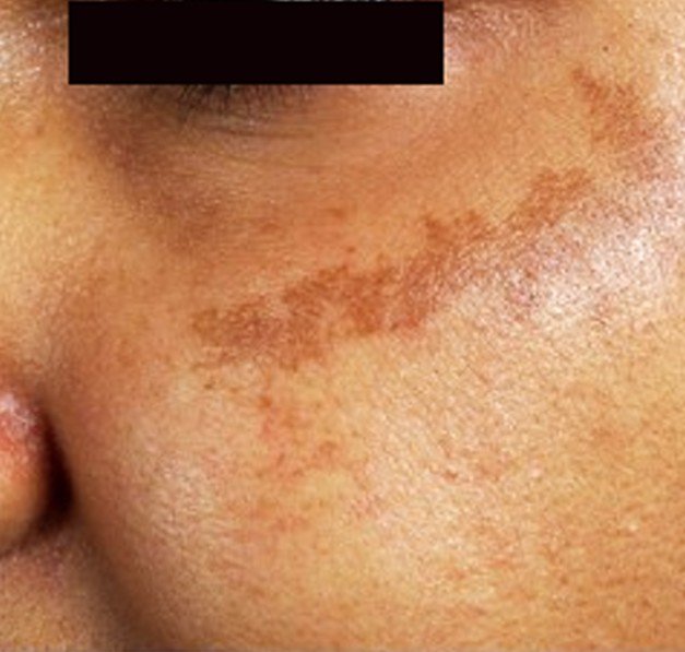
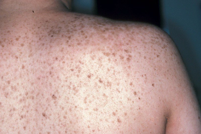
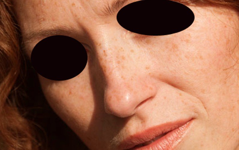
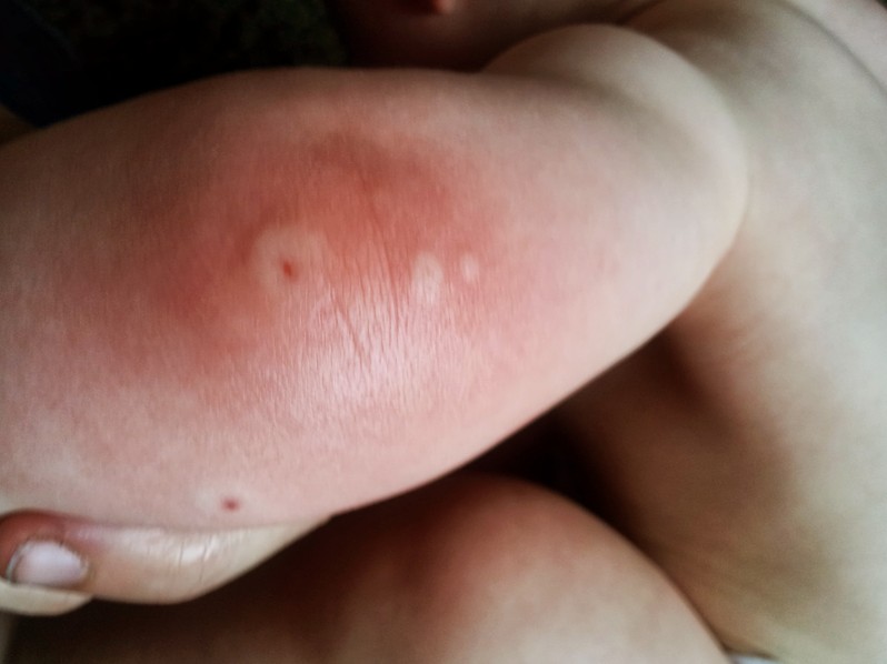
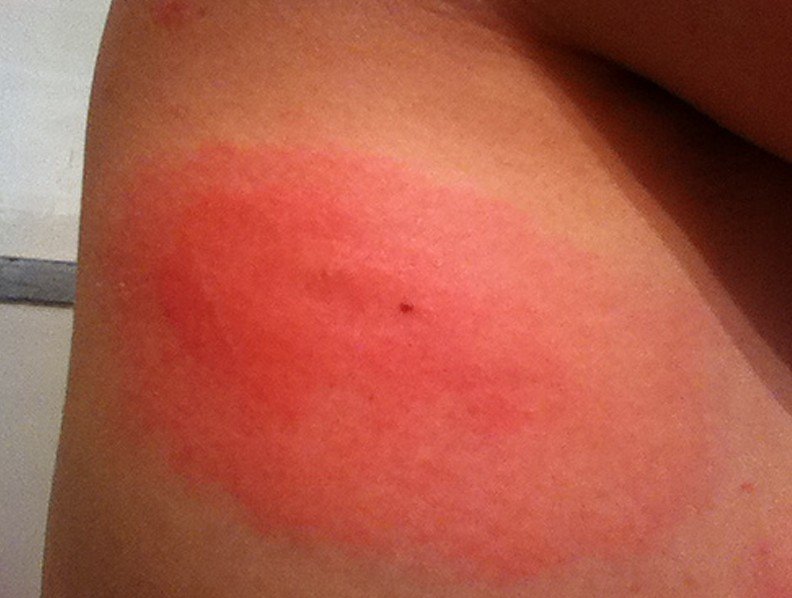
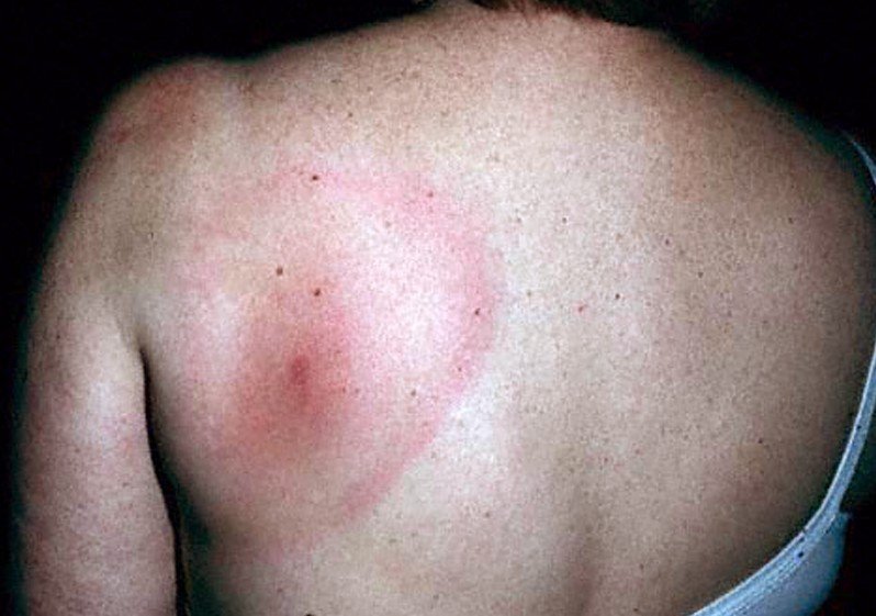
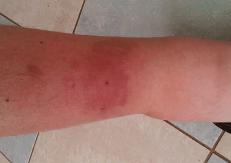
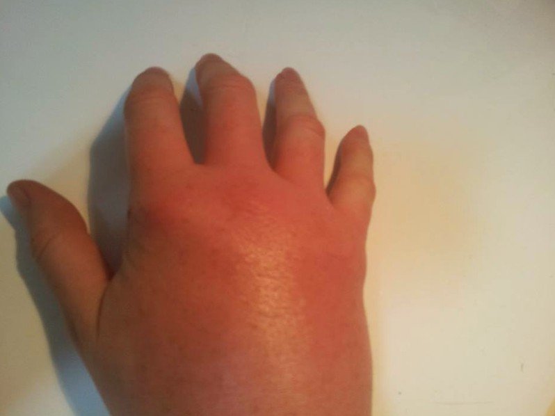
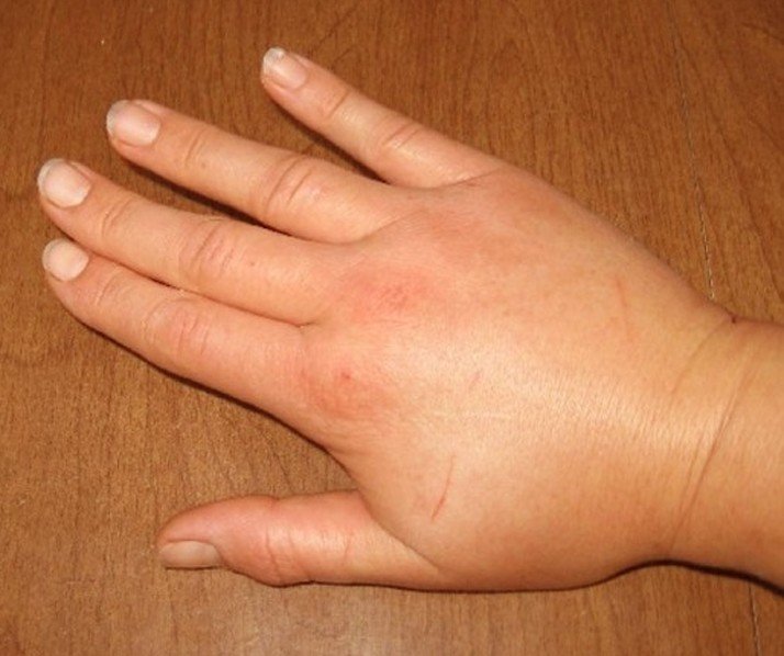
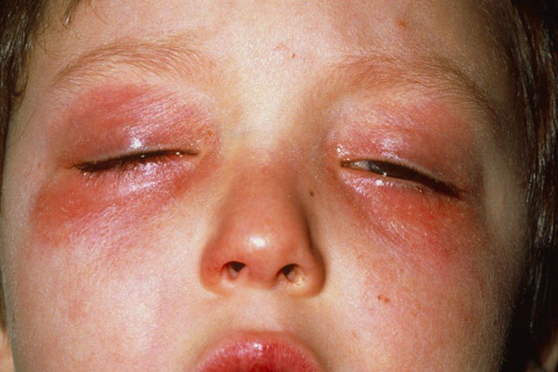
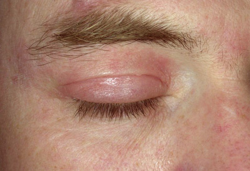
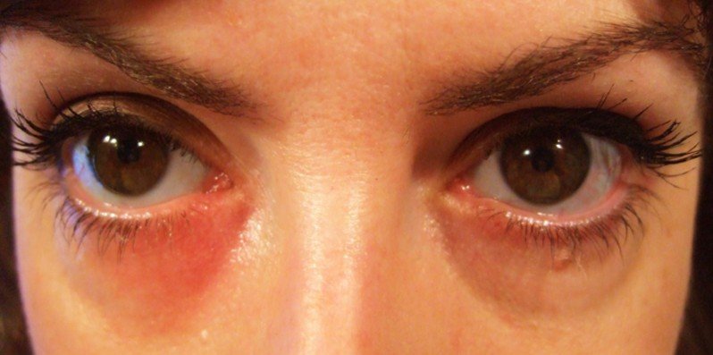
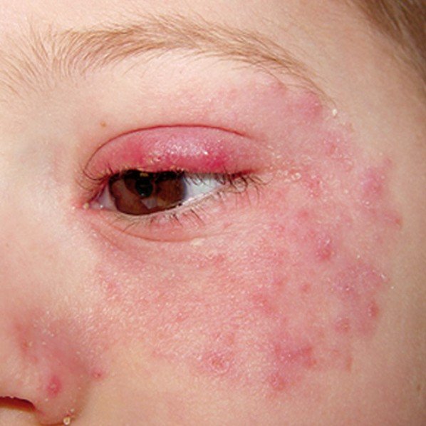
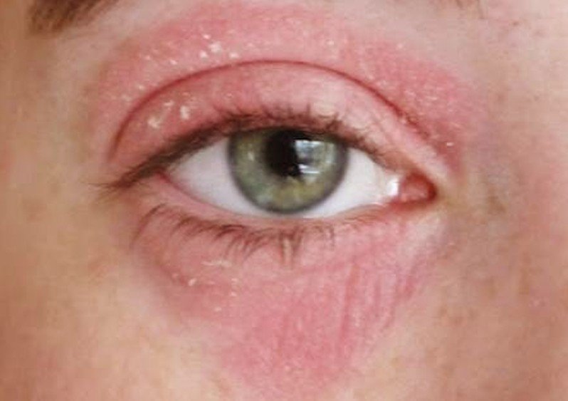
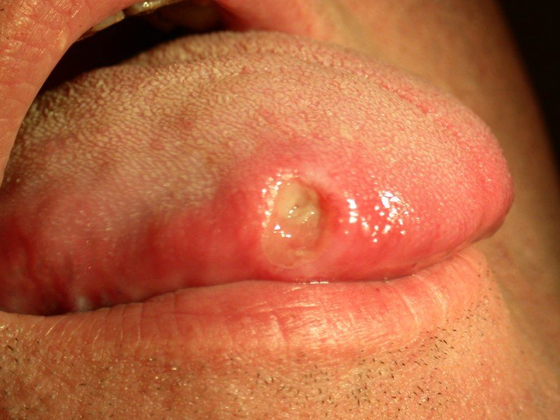
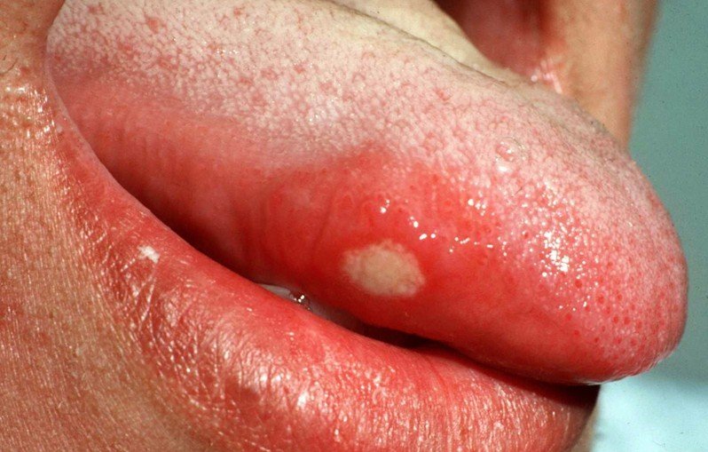
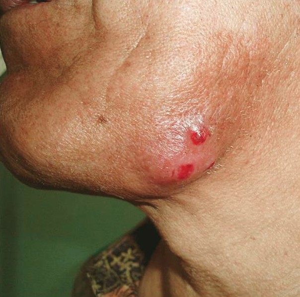
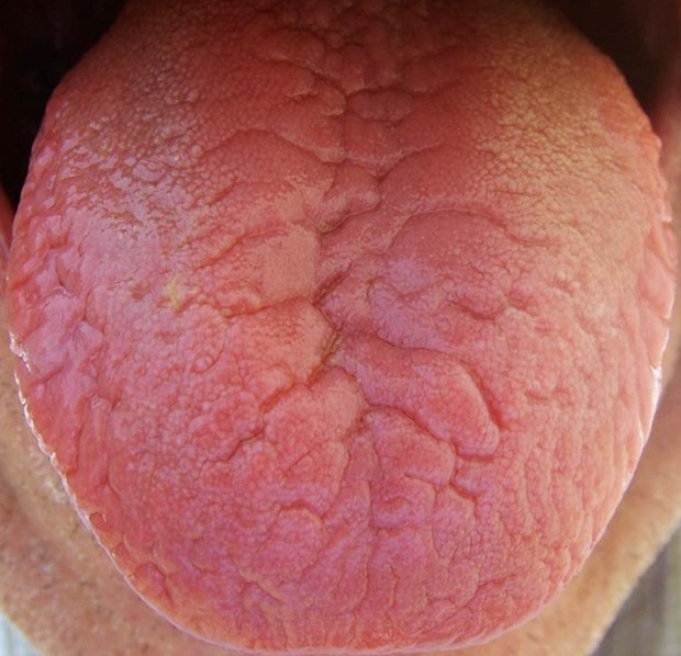
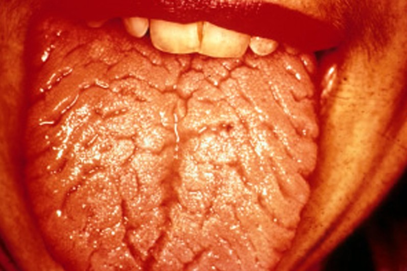
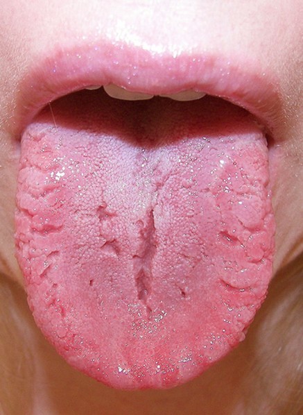
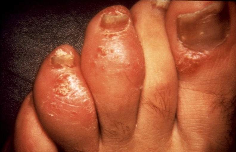
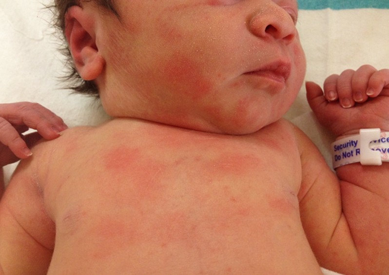
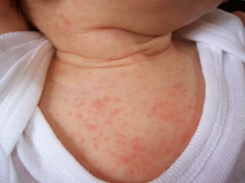
Pictures of Brown Spots on Skin

Collection of photos, pictures of brown spots on skin…

brown spots on skin

brown spots on skin pictures

brown spots on skin pictures 2

brown spots on skin pictures 3

brown spots on skin pictures 4

brown spots on skin pictures 5

brown spots on skin pictures 6

brown spots on skin pictures 7

brown spots on skin pictures 8

No responses yet

Bee Sting Swelling

Jan 28 2016 Published by under General Health

When bees sting, they can trigger an allergic reaction. However, in many cases, the normal reaction from a sting is confused with an allergic reaction. Also, it is important to note that the severity of the reaction can vary from one person to the other. The person might experience a normal reaction to the bee sting, in which the swelling is strictly confined at the site of the sting and accompanied by local pain and erythema. An extended, yet normal reaction to the bee sting, means that the bee sting swelling has gone beyond the original site. For example, if a person was stung on the hand, then the entire arm might swell up.

It is important to make the difference between a normal reaction to a bee sting and the one that is actually an allergic reaction. If a person has an anaphylactic reaction to the bee sting, then the bee sting swelling might be extensive and the person might also experience systemic symptoms. Among these symptoms, you will find any of the following: difficult breathing, swelling of the face and throat, difficulty swallowing, wheezing, extreme agitation and anxiety, rapid heartbeat and vascular collapse. Emergency medical treatment with adrenaline is required in case of severe allergic reactions to bee stings.

How long does Bee sting swelling last?

Depending on the severity of the reaction to the sting, it may take somewhere from two to five days for the affected area to heal. During this period, it is recommended to have an exemplary hygiene, so as to prevent secondary infections.

Treatment

These are the most common recommendations and courses of treatment in case of bee sting swelling:

  • If the bee sting has occurred on the fingers, it is important to immediately remove any rings, before the swelling becomes worse. The same goes for bracelets or any other tight fitting jewelry that you might be wearing.
  • Avoid leaving the sting needle in the skin, as this will only deliver more venom and it will cause the swelling to accentuate. It is recommended that the sting needle is removed within 30 seconds of the sting.
  • Avoid scratching the area of the sting, as it will make the swelling and the itchiness worse. Also, by constantly scratching, you will break down the protective barrier of the skin and you will expose yourself to secondary infections.
  • The area where the stung has occurred needs to be washed with warm water and antiseptic soap, in order to prevent secondary infections.
  • No specific antivenom exists that can be injected in order to counteract the effects of the bee venom.
  • Sting area is recommended to be covered with a protective bandage until it heals.
  • Anti-histamines are recommended not only for the bee sting swelling but also for the associated itchiness and rash
    • Oral administration
    • Not recommended in children or pregnant women
  • Anti-inflammatory medication is also recommended for bee sting swelling
    • Acetaminophen
    • Ibuprofen
    • Aspirin – not recommended for people who are under 18 years of age
  • Severe allergic reactions accompanied by swelling are treated with epinephrine or adrenaline
    • Self-injections – most people who know that they are allergic to bee stings carry with them an epinephrine self-injector or they also wear a medical alert bracelet. This identifies the allergy to the bee stings and will help people get you to the emergency room in case it is necessary. Also, emergency allergy kits can be prescribed by the doctor.
    • Administration by medical personnel
    • Additional treatment: Intravenous fluids, Oxygen, Intravenous antihistamines and cortisone and Beta agonist (to  improve the breathing capacity)
    • Injections are recommended to be administered in the outer muscle of the thigh; they should not be injected into the buttocks or in a vein.
    • The injections should not be made into the hands or feet, as they can cause serious tissue damage.
    • More than one adrenaline injection might be necessary, depending on the severity of the allergic reaction.

Topical applications

  • Corticosteroids – hydrocortisone : These are recommended to reduce the bee sting swelling
  • Calamine lotion – This is indicated as it has a soothing effect in the inflamed skin. There is also a special calamine lotion that contains analgesic substances
  • Antibiotic cream – Recommended to prevent secondary infections that might arise in the sting area
  • Benzocaine sticks – It is recommended that the benzocaine stick is rubbed against the sting area. Guarantee a short period of relief from the symptoms of the bee sting

Home remedies for Bee sting swelling

These are the most common home remedies you can consider for bee sting swelling:

  • Cold applications
    • Ice pack – these should not be applied directly on the skin, but rather wrapped in something.
    • Cold water compress
    • Be careful when it comes to how long you leave the cold applications on the skin; this is especially valid for ice packs, as prolonged application to the skin can cause circulatory problems.
  • Elevating the arm/leg (if the swelling has occurred in those areas)
  • Anti-itchiness natural remedy
    • Mix baking soda with warm water and apply it to the affected area on the skin.
    • Mix baking soda with vinegar and meat tenderizer. The latter is recommended to be thrown into the mix because it contains an active substance (papain) that can actually break down the toxins of the bee venom.
    • Honey applications – these are recommended because honey can relieve the itchiness but also because of its antibacterial and antiseptic properties. It can be useful to prevent secondary infections.
  • A less common home remedy is to rub a wet aspirin tablet to the sting area
  • Toothpaste application
    • This helps with the itchiness commonly associated with a bee sting
  • Apply roll-on deodorant to the sting area
    • Not always recommended, as it may help with the symptoms in the sting area but have a negative effect on the surrounding area
  • Application of raw potato
    • This can help with the swelling, causing it to subside
    • Allows for faster healing of the lesion

Pictures

Collection of pictures of bee sting swelling…

bee sting swelling pictures

bee sting swelling pictures 2

bee sting swelling pictures 3

bee sting swelling pictures 4

bee sting swelling pictures 5

bee sting swelling pictures 6

bee sting swelling pictures 7

bee sting swelling

No responses yet

Rash Around Eyes

Jan 26 2016 Published by under Eye Health

Having a rash around your eyes can be a difficult thing to handle, as it can cause a lot of discomfort. In the majority of the cases, the rash is accompanied by pain and it makes vision difficult. For women, there is also the cosmetic concern, as the rash around eyes can be difficult to conceal. There are many reasons that can lead to the appearance of a rash around the eyes, so it might be for the best to consult a dermatologist. The reason why you need to turn to a specialist is that the root cause of the rash has to be addressed for the condition to be cured.

Only a specialized dermatologist can recommend a treatment that works. Below, in this article you will find the potential causes that can lead to a rash around the eyes, including allergies, taking certain medication, infection, using the wrong skin care products, hormonal imbalances and poor ocular health. You will also find the treatment guidelines for this medical condition, so that you know what to expect. Just make sure you consult a dermatologist, so that you get the proper treatment for the root cause of your skin problem.

Causes of Rash Around Eyes

These are the potential causes that can lead to the appearance of a rash around eyes:

  • Atopic dermatitis
  • Allergies: Hay fever, Chlorine allergies
  • Contact dermatitis
  • Infection: Lyme disease
  • Blepharitis
  • Conjunctivitis
  • Skin care products
  • Medication

How to treat Rash Around Eyes?

These are the most common causes of treatment for the rash around eyes:

Corticosteroids

  • Topical applications – these are recommended to reduce the itching and inflammation or redness commonly associated with dermatitis. Low potency corticosteroids are indicated as the first course of treatment.
  • Oral or injected corticosteroids – these are recommended in cases of severe allergies, accompanied by intense itchiness. However, they are not recommended as treatment for prolonged periods of time, as they can have serious consequences on a person’s health.

Antibiotics

  • Recommended in cases of bacterial agents responsible for the rash around the eyes
  • Broad spectrum antibiotics are usually recommended
  • The patient is instructed to take the antibiotic for the entire duration of time that is recommended; otherwise the infectious agent (bacteria) will develop increased resistance to the antibiotic.
  • In cases of severe bacterial infections, such as Lyme disease, intravenous antibiotics are the recommended course of treatment. Less severe cases might also benefit from oral treatment with antibiotics but this depends from one patient to the other. Insect repellents might also be recommended as prevention for Lyme disease.

Antihistamines

  • Oral antihistamines are indicated for severe itchiness.

Immunomodulators

  • These are recommended in order to reduce the inflammation normally associated with allergic reactions.
  • Two of the most recommended Immunomodulators are tacrolimus and pimecrolimus.

Topical creams

  • These contain active ingredients that have a soothing effect for the skin, such as calamine.
  • They are recommended to reduce the inflammation and irritation of the skin.
  • They are highly effective when it comes to reducing the intensity of the itchiness.
  • Once applied, the patient is advised to refrain from scratching the skin, otherwise the topical cream might not have the desired effect.

Cool compresses

  • Applying a cold compress to your eyes will help reduce the inflammation and it will calm the irritated skin.
  • A cold compress can also be of help in case you experience a burning sensation.

Emollient cream

  • A good cream to moisturize the skin will promote a faster healing process
  • It will keep the skin properly hydrated and it will prevent it from becoming excessively dry
  • Be sure to use an emollient cream that is made from natural ingredients; otherwise, you could end up making the rash around the eyes even worse.

Avoiding the allergen that has caused the rash in the first place is 100% recommended. In case the allergen in question comes from expired makeup, it is indicated that the area is thoroughly washed with clean water.

Avoiding the environment that is causing allergies, such as botanical gardens (lots of flowers and pollen), farms (hay) and even swimming pools that are treated with chlorine can help reduce the risk for the rash to reappear.

Natural remedies

  • Applications of cucumber slices on the eyes have been shown to also reduce the rash around the eyes. Patients have declared the burning sensation to have subsided after the application of cucumber slices in the eye area.

In conclusion, you have to understand that a very important part is the prevention. If you know that you are predisposed to allergies, then you need to avoid potential allergens and keep the area around the eyes as clean as possible. For the situations when the rash occurs, be sure to follow the treatment guidelines that were presented above.

Pictures of Rash Around Eyes

Collection of images, photos and pictures of Rash Around Eyes…

rash around eyes

rash around eyes pictures

rash around eyes pictures 2

rash around eyes pictures 3

rash around eyes pictures 4

rash around eyes pictures 5

No responses yet

Ecthyma Gangrenosum

Jan 22 2016 Published by under Skin Conditions

Ecthyma gangrenosum is a medical condition in which the skin becomes infected with pseudomonas aeruginosa. The people who are most at risk of developing this skin condition are those who are critically ill or those whose immune system is severely compromised. In many of these patients, this condition is a sign of generalized infection in the body (with the same pathological agent). Ecthyma gangrenosum is mainly characterized by the appearance of lesions in the skin in the form of vesicles or pustules. These tend to be hemorrhagic and they quickly progress to necrotic ulcers.

This condition occurs when the skin no longer represents a defensive barrier and the bacteria easily disseminate into the body. What happens is that pseudomonas aeruginosa enters the body and affects the arteries and veins in the skin, causing their walls to be inflamed and quickly leading to necrosis. The affectation of the blood vessels leads to secondary necrosis in the other layers of the skin, contributing to the aggravation of the disease. This condition has a high mortality rate, especially if emergency medical treatment is not administered. There is no prevalence whereas the gender is concerned and, when it comes to age, it seems that children and the elderly are the most affected (as their immune system does not function properly).

Symptoms of Ecthyma Gangrenosum

These are the most common symptoms of ecthyma gangrenosum:

  • Prodromal symptoms (precede the appearance of lesions on the skin: Diarrhea (in children) and Fever
  • First lesions are macules and pustules
  • These lesions are painless and they have a red surrounding border
  • As the lesions progress, they form a hemorrhagic center (bulla)
  • The lesions evolve into necrotic ulcers
  • The transformation from the initial lesion to the necrotic ulcer can undergo in a matter of hours
  • Single or multiple lesions are present on the skin
  • Areas that are most affected: anal, genital and axillary
  • Any other part of the body can be affected, especially if there is a generalized infection with pseudomonas aeruginosa
  • Other areas that can be affected include:
    • Gluteal region
    • Perineal region
    • Upper or inferior limb extremities
    • Trunk
    • Face – In rare cases – bilateral periorbital lesions
  • Lesions heal with the formation of scars

What are the Causes of Ecthyma Gangrenosum?

These are the most common causes that lead to the appearance of ecthyma gangrenosum:

  • Infection with pseudomonas aeruginosa
  • Other bacterial/fungal infections
  • Gram positive bacteria
    • Staphylococcus aureus
    • Streptococcus pyogenes
  • Gram negative bacteria
    • Escherichia coli
    • Klebsiella pneumoniae
    • Neisseria gonorrhoeae
    • Aeromonas hydrophila
    • Yersinia pestis
  • Fungal infection
    • Candida albicans
    • Aspergillus fumigatus
    • Fusarium solani
  • Infection with the herpes simplex virus

Diagnosis

These are the most common methods used for the diagnosis of ecthyma gangrenosum:

  • Gram stain
    • A sample is taken from the center of the lesion
  • Blood culture
    • Blood is drawn before starting the treatment with antibiotics
    • The best period for a blood culture is when the patient runs a high fever (sign of powerful infection in the body, the bacteria is rapidly disseminated)
  • Skin biopsy
    • This is performed for differential diagnosis, so as to rule other possible causes that leads to the appearance of this medical condition
    • Vascular necrosis is identified through this method
  • Tissue culture
    • This is performed in order to clearly identify the bacteria, fungi or virus causing the infection on the skin
    • Includes the sensitivity tests
  • Urine culture
    • Identify rests of infectious agents
  • Differential diagnosis
    • Cutaneous anthrax
    • Aspergillosis
    • Ecthyma
    • Pyoderma gangrenosum
    • Septic emboli
  • Imaging (MRI) can also be used for the diagnosis of ecthyma gangrenosum

Treatment

These are the most common courses of treatment undertaken for ecthyma gangrenosum:

Antibiotic therapy

  • Antibiotics need to be administered rapidly and in correspondence with the bacterial agent causing the infection
  • The most recommended antibiotics are:
    • Penicillin that works against pseudomonas aeruginosa
    • Aminoglycosides
    • Fluoroquinolones
    • Cephalosporin (third generation)
  • In order to obtain the best results, two antibiotics can be recommended at the same time

Surgical debridement

  • This is recommended in cases when the patients do not respond to the antibiotic therapy
  • The procedure is necessary to stop the spreading of the necrotic ulcers

Other treatments include:

  • Administration of granulocyte-macrophage colony-stimulating factor – Aids with the recovery process
  • In case of fungal infection
    • Anti-fungal treatment
    • Systemic medication against the most common fungi
  • The treatment is only administered after the result of the blood cultures arrives. It is important to know the kind of pathogen that is causing the infection in order to administer the right treatment.
  • Poor prognosis is considered to be in the following cases:
    • Multiple necrotic ulcers present on the skin, spreading continuously
    • Delayed treatment with antibiotics
    • High count of bacteria in the blood
    • Generalized sepsis with pseudomonas aeruginosa

Ecthyma Gangrenosum Pictures

Here are some of the photos, pictures of Ecthyma gangrenosum…

No responses yet

Aphthous Ulcer

Jan 21 2016 Published by under Oral Health

The aphthous ulcer is a common condition that is also known as recurrent aphthous stomatitis in the medical world. The condition is pretty common in children and teenagers, this being the period of debut as well. The ulcers appear inside the mouth, causing a lot of discomfort and sometimes pain. Patients diagnosed with aphthous ulcer often present a family history and the condition usually goes away on its own somewhere around young adulthood. However, if the mouth ulcers persist and there is no spontaneous disappearance, there may be a sign of a systemic illness. Common conditions associated with the aphthous ulcer include the Behçet syndrome, diseases of the gastrointestinal tract, autoinflammatory conditions and HIV/AIDS.

The lesions that appear inside the mouth are benign and they appear in healthy individuals. At the moment, there is no cure for the disease and the treatments that are available are aimed at improving the symptoms. Pain is one of the main objective treatments but medical specialists are also trying to reduce the amount of time that is necessary for healing and also to reduce the number of episodes. Identifying the triggering factors that might lead to the appearance of the aphthous ulcer is also a recommended course to follow.

Aphthous Ulcer Symptoms

These are the most common symptoms of the aphthous ulcer:

  • Lesions inside the mouth, affecting the mucous membranes
  • These lesions are also known as aphthae, hence the name of the condition
  • Lesions tend to come and go, being triggered by a set of factors (the triggering factors will be discussed under causes)
  • From one episode to the other, the lesions heal completely, without leaving any sign
  • A common lesion can last up to ten days
  • On average, a person experiences from three to six episodes per year
  • The hard palate and the dorsum of the tongue are not affected by the lesions, in the majority of the cases
  • The patient can experience discomfort and even pain
  • Usual activities, such as drinking and eating can be impaired. Speaking can be affected as well, in more severe cases.
  • Weight loss appears in severe cases, when the patient does not eat because of the prolonged and extensive lesions inside the mouth
  • No systemic symptoms accompany the disease
  • Before the appearance of the actual lesions, the patient might experience a burning or stinging sensation
  • The pain experienced in the region can be aggravated by food or liquid intake, especially if they are acid
  • The pain is at its highest level immediately after the ulcer has formed
  • As the lesion heals, the pain subsides
  • Chronic cases have new lesions appear before the older ones heal, the chewing process is constantly affected and the pain is constant as well. These are the cases where the weight loss occurs.

Causes of Aphthous Ulcer

The exact cause that leads to the appearance of the aphthous ulcer has yet to be identified. From the research made so far, specialists know that this condition is not caused by an infectious agent and it is not transmitted through sexual contact. The modifications that appear inside the mouth are believed to actually be a response of the immune system to a harmful stimulus. No association has been made so far with autoimmune diseases. However, it seems that the immune system plays an important role in the appearance of this condition. Associations with other systemic conditions have been made.

The persons who are diagnosed with aphthous ulcer usually have problems with immunity, their defensive protective barrier does not function properly and they have an increased sensitivity to developing such lesions. It also seems that there is a genetic predisposition to this condition, as a lot of the patients diagnosed with aphthous ulcer have a family history. Stress is believed to be a triggering factor, because a lot of the persons who are stressed tend to constantly bite their lip or cheek and thus they breakdown the protective barrier. Also, if the thickness of the mucosal barrier is decreased through, for example, local trauma, that increases the risk for the appearance of the ulcer.

Treatment for Aphthous Ulcer

These are the most common courses of symptomatic treatment and recommendations for aphthous ulcer:

Topical treatments

  • Covering agents – these are recommended for the reduction of pain
    • Orabase
    • Triamcinolone
  • Anti-inflammatory medication – reduces both pain and inflammation
    • Benzydamine hydrochloride – in the form of mouthwash/spray
    • Amlexanox (paste)
    • Lidocaine (viscous)
    • Hyaluronan (active agent – diclofenac)
  • Antiseptics – reduces healing time and prevents secondary infections
    • Doxycycline
    • Tetracycline
    • Triclosan
  • Topical corticosteroids – reduce inflammation in the affected area (depending on the severity of the condition, administration can range from mild to medium potency)
    • Hydrocortisone cream (mild)
    • Dexamethasone (moderate)
  • Oral corticosteroids – these are recommended only in severe cases, because of the potential side-effects
    • Prednisolone
  • Surgical excision in rare cases

Home remedies

These are the most common home remedies recommended for the aphthous ulcer:

  • Mouth rinses – Mix warm water with: baking soda, goldenseal powder and elm powder
  • Topical application of milk of magnesia (directly on the ulcer)
  • Paste application – mix baking soda with a small quantity of water
  • Try to chew ice chips in the area near the lesions. They will act as numbing agents, reducing the pain and discomfort.
  • Use special toothpaste recommended for such medical problems and a soft toothbrush.
  • Apply propolis tincture
  • Use licorice extract to reduce the healing time for the lesions
  • Alum powder topical application
  • Probiotics to replenish useful bacteria inside the mouth
  • Avoid eating spicy, acidic or irritating foods
  • Avoid drinking acid beverages
  • Saltwater solution with sodium bicarbonate
  • Hydrogen peroxide solution

As you can see for yourself, the treatment for the symptoms of aphthous ulcer consists of both medication and home remedies. Be sure to try out everything that has been suggested in this article and see which one works better for your own situation. Make the necessary diet changes and you will definitely see an improvement whereas the actual lesions are concerned. These are necessary for preventive purposes as well.

Aphthous ulcer Pictures

Collection of photos, images and pictures of Aphthous ulcer…

aphthous ulcer

aphthous ulcer pictures

aphthous ulcer pictures 2

aphthous ulcer pictures 3

aphthous ulcer pictures 4

aphthous ulcer pictures 5

aphthous ulcer pictures 6

aphthous ulcer pictures 7

aphthous ulcer pictures 8

No responses yet

Actinomycosis

Jan 19 2016 Published by under Diseases and Conditions

Actinomycosis is a polymicrobial infection that occurs extremely rare in humans and more often in animals. The infectious bacterial disease, when it affects humans, can range from sub-acute to chronic. It has different clinical forms, including the one affecting the cervical and facial area, the thoracic and abdominal form. In women, there is a pelvic form that can be developed. The fact that this is a polymicrobial infection means that there are actually several bacterial species causing the problem (usually somewhere between five and ten).

The bacterial species that commonly cause this infection are part of the regular flora of the oral cavity; they are also found in other parts of the body, including the gastrointestinal tract and the genital tract of females. However, in this area, their percentage is reduced. What happens is that these bacteria cannot breach the skin if the mucous membrane is whole; however, if the mucous membrane is not whole anymore because of various reasons, then the bacteria can enter the deeper tissues of the body and cause the polymicrobial infection. This is a severe infection, in which the body develops a powerful inflammatory response and a wide range of clinical features are present.

Actinomycosis Symptoms

These are the symptoms of actinomycosis, depending on the actual form of the disease:

Cervico-facial actinomycosis

  • This condition is also known as a lumpy jaw
  • In a lot of cases, this form is not accompanied by pain
  • Pain might arise along with the inflammation of the soft tissues from the submandibular or peri-mandibular region
  • Abscess formation with formation of sinus tracts and pus discharge
  • Pus contains sulfur granules
  • The condition can enter into remission and re-occur after a period of time
  • The skin in the affected area is discolored
  • Nodular lesions in the angle of the jaw
  • No swollen lymph nodes
  • The patient can also present fever
  • The mastication might be impaired, as the muscles responsible for the process are affected as well.

Thoracic actinomycosis

  • Cough (dry or productive)
  • Sputum with blood
  • Difficult breathing
  • Pain in the chest
  • Fever
  • Weight loss
  • Abnormal sounds in the chest
  • Sinus tracts can form on the chest as well, with pus discharge.

Abdominal actinomycosis

  • Fever (low in intensity)
  • Weight loss
  • Overall weakness
  • Bowel movement is affected
  • Abdominal discomfort
  • Nausea
  • Vomiting
  • Palpable mass in the abdomen
  • Sinus tracts are formed in the abdomen, with pus discharge.

Pelvic actinomycosis

  • Abdominal discomfort (lower region)
  • Abnormal bleeding from the vagina
  • Discharge from vagina
  • Palpable mass in the lower abdomen.

What Causes Actinomycosis?

Actinomycosis is a polymicrobial infectious disease. Below, you will find the bacterial species incriminated in the appearance of the disease:

  • Actinomycetales, family Actinomycetaceae, genus Actinomyces:
    • Actinomyces israelii
    • Actinomyces gerencseriae
    • Actinomyces turicensis
    • Actinomyces radingae
    • Actinomyces europaeus
    • Other Actinomyces: naeslundii, odontolyticus, viscosus, meyeri
  • Propionibacterium
    • Propionibacterium propionicum
  • Actinobaculum
  • Bifidobacterium
  • Other bacterial agents that accompany the original bacteria:
    • Actinobacillus actinomycetemcomitans
    • Peptostreptococcus
    • Prevotella
    • Fusobacterium
    • Bacteroides
    • Staphylococcus
    • Streptococcus
    • Enterobacteriaceae.

Diagnosis

These are the most common methods used for the diagnosis of actinomycosis:

Complete blood count

  • Anemia
  • Leukocytosis
  • Erythrocyte sedimentation rate is increased
  • C-reactive protein is increased as well

Chemistry levels

  • Normal in most cases
  • Hepatic actinomycosis: Increased alkaline phosphatase

Bacteria culture

  • A sample of the bacterial culture is necessary for the confirmation of the diagnosis
  • The sample can be taken from the following sources:
    • Sinus tract with pus discharge
    • Deep tissue (through aspiration)
    • Other possibilities: swap in the oral cavity, sputum sample, urine specimen.
  • Gram-stained smear is also used for the confirmation of the diagnosis
  • Nucleic acid probes
  • Polymerase chain reaction

Examination of sulfur granules under the microscope

Papanicolaou test is recommended in case of suspicion of pelvic actinomycosis. However, this is a not a test used for diagnosis.

X-rays

  • Recommended in case of pulmonary affectation
  • Used for the diagnosis of thoracic actinomycosis
  • The X-ray can reveal a mass-like lesion
  • This lesion can affect the pleura (lining of the lungs and anterior thoracic wall), the thoracic vertebrae, the ribs or the sternum

Computer tomography

  • CT can detect an infiltrative mass, in different regions in the body

Ultrasound-guided fine needle aspiration

  • This is used for the biopsy necessary for the confirmation of diagnosis

Surgical exploration

  • Thoracotomy
  • Open lung biopsy
  • Exploratory laparotomy.

Actinomycosis Treatment

These are the most common methods of treatment used for actinomycosis:

Antibiotics

  • This the number one course of treatment
  • Penicillin G is often effective in treating the polymicrobial infection
    • Administration of high doses
    • Treatment is required to be followed for prolonged periods of time (up to one year)
    • The patients diagnosed with the cervico-form of actinomycosis can take the antibiotic treatment for shorter periods of time (six months)
  • Minimum risk of the patient developing resistance of the treatment with antibiotics
  • Given the fact that this is a polymicrobial infection, it might be necessary to add a secondary treatment to the course of treatment. This happens when one of the bacterial species is resistant to the first course of treatment.
  • Other antibiotics that can be administered include:
    • Metronidazole
    • Aminoglycosides
    • Aztreonam
    • Co-trimoxazole
    • Other penicillins: methicillin, oxacillin, cloxacillin
    • Cephalexin
    • Fluoroquinolones: ciprofloxacin, levofloxacin, moxifloxacin.
  • In more severe cases, intravenous antibiotics will be administered through a peripherally inserted central catheter.
  • The response to the given antibiotic therapy is monitored to the usage of imagining methods, such as the CT and MRI.

Surgery

  • Incision and drainage of skin abscesses
  • Sinus tract excision
  • Removal of fibrotic lesions
  • Cleaning of infected areas through surgical methods
  • Decompressive surgery in case of pelvic actinomycosis affecting the uterus
  • Emergency medical treatment in case of peritonitis (this appears in patients diagnosed with abdominal actinomycosis)

Actinomycosis Pictures

Images, Photos and Pictures collection of Actinomycosis…

actinomycosis

actinomycosis pictures

actinomycosis pictures 2

actinomycosis pictures 3

actinomycosis pictures 4

No responses yet

Fissured tongue

Jan 18 2016 Published by under Oral Health

Fissured tongue is a benign condition in which deep grooves or fissures appear on the lateral sides and back of the tongue. This condition is also known as scrotal tongue, lingua plicata, plicated tongue or furrowed tongue. It is painless in most cases and it is sometimes accompanied by a burning sensation. The fissured tongue affects 2-5% of the general population and it is more frequent in males than females. Even though it may be diagnosed in childhood, it is more frequent in older people and it tends to accentuate with age.

Some studies have suggested that patients diagnosed with fissured tongue have a genetic predisposition. Most of the cases of fissured tongue are discovered through routine oral examination or by accidental findings. The condition is frequently associated with the geographic tongue (benign migratory glossitis) and it is also often encountered in Melkersson-Rosenthal syndrome and Down syndrome. No biopsy is required for the diagnosis of the fissured tongue; the clinical appearance is often times enough for the confirmation of the diagnosis.

Symptoms of Fissured tongue

These are the most common symptoms of the fissured tongue:

  • Deep grooves or fissures present on the lateral and back side of the tongue
  • Might be just one groove or several in number
  • These grooves can have a depth of 2-6 mm
  • Large central furrow is present in some patients, with small fissures extending from the central one (perpendicularly)
  • The grooves can also appear on the dorso-lateral portion of the tongue
  • Burning sensation – this appears especially when the patient drinks or eats certain foods (cold beverages, spicy foods)
  • Soreness
  • Pain
  • Redness of the tongue
  • Difficult eating and swallowing (especially if the tongue is inflamed)
  • No other part of the body is affected other than the tongue
  • The grooves can connect with each other; the tongue will appear as being split into lobes or sections
  • Debris can build up in these fissures, causing upsetting symptoms
  • Symptoms are worse in case the patient also has a geographic tongue

Causes

The exact cause that leads to the appearance of the fissured tongue has yet to be identified. Below, you will find the theories that have been suggested so far:

  • Genetic predisposition – the fissured tongue has been encountered in several members of the same family.
  • Age – the condition is more frequent in older people and it also accentuated with age.
  • Environmental factors are believed to be triggers for the fissured tongue.

Association with other medical conditions:

  • Geographic tongue – this is a condition in which the surface of the tongue has a map-like appearance.
  • Melkersson-Rosenthal syndrome – this is a rare neurological disorder, in which the patient suffers from facial paralysis, swelling in the face (including lips) and tongue fissures.
  • Down’s syndrome – this is a genetic disorder, in which there is an extra chromosome causing a wide range of medical problems, including fissured tongue. Almost all children diagnosed with Down’s syndrome have fissured tongue.
  • Pustular psoriasis – this is an uncommon form of psoriasis, in which raised bumps, filled with pus (white blood cells) appear on the skin.
  • Orofacial granulomatosis – condition in which the soft tissues in the oral area are swollen, without any pain being associated with the disease.
  • Acromegaly – this is a condition in which the anterior pituitary gland produces excessive quantities of growth hormone.
  • Sjögren’s syndrome – this is a chronic auto-immune disease in which the white blood cells practically destroy the exocrine glands (these are the lacrimal and salivary glands). The mouth is excessively dry and the tongue becomes more prone to fissures.

Other causes include:

  • Tongue inflammation
  • Poor nutrition
  • Anemia or other hematologic abnormalities
  • Oral yeast infection (caused by candida fungus)
  • Vitamin B deficiency
  • Electrolytic imbalance
  • Oral cavity or tongue injuries (especially in people who bite their tongue or those who grind their teeth)
  • Excessive intake of alcohol
  • Constant and prolonged tobacco chewing
  • Reaction to certain types of medication

Treatment for Fissured tongue

These are the most common courses of treatment undertaken for fissured tongue:

  • Mechanical tongue cleansing:
    • Recommended as part of the oral hygiene routine
    • Indicated in case of debris accumulated in the tongue fissures, causing halitosis (bad breath)
    • The regular brushing is recommended so as to remove debris accumulation, reduce irritation and also the unpleasant odor in the exhaled breath
    • The cleansing of the tongue can be performed with the toothbrush (most of them have a special part on the back of the head for such purposes) or with specific cleaning devices for the tongue that can be found in most pharmacies
  • Antibiotics and topical creams are recommended in cases of secondary infections or trauma to the tongue
  • Good oral hygiene
  • Drinking a lot of water – this is recommended to prevent excessive dryness of the mouth (if the tongue is hydrated, then there is a higher risk for fissures to appear)
  • Natural remedies – gargling with baking soda can help with the symptoms. Chewing spearmint gum can also help in some cases but there are patients who describe more irritation after doing that

In Chinese medicine, the tongue is a map that shows the entire health of the body. It is believed the deep fissures in the tongue are signs of chronic conditions. The deeper a fissure, the more chronic the condition actually is. It is believed that fissured tongue appears when a person does not drink enough water and when there is a lot of stress. Chinese specialists recommend the treatment of hormonal imbalances and guarantee an improvement of the fissured tongue.

Fissured tongue Pictures

Photos, Images and Pictures of Fissured tongue…

fissured tongue

fissured tongue pictures

fissured tongue pictures 2

fissured tongue pictures 3

fissured tongue pictures 4

fissured tongue pictures 5

Pain relief

In case there are a lot of grooves on the surface of the tongue and debris builds up in those fissures, the tongue might become irritated and painful. In such cases, apart from the mechanical cleansing of the tongue, you might be recommended anti-inflammatory medication in order to obtain pain relief. These drugs are especially recommended when there are fissures in the tongue and there is a lot of inflammation associated with the condition. Pain relief can also be obtained with the help of natural remedies as well (baking soda).

No responses yet

Dactylitis

Jan 13 2016 Published by under Diseases and Conditions

Dactylitis is a medical condition in which a digit (finger or toe) becomes inflamed, taking the shape of a sausage and causing a lot of pain. The name of the condition comes from the Greek word ‘daktylos’, meaning finger. However, in the medical literature, it is known that both the finger and the toe can be affected. The diagnosis can be made with the help of a dactylitis score sheet (finger/toe involvement, circumference of the involved digit, contralateral digit, tenderness).

Dactylitis Symptoms

These are the most common symptoms of dactylitis:

  • Inflammation of the finger or toe – the severity of the inflammation depends on the underlying pathology can leads to the appearance of this condition but also from one patient to the other
  • Fingers and toes can be affected at the same time
  • Painful swelling commonly appears along the flexor tendons (the patient is unable to flex his or her finger)
  • Diffuse inflammation of the digital soft tissue (pseudosynovitis)
  • Sausage-shape or puffy-like appearance of the inflamed digit
  • The inflammation can extend to the other digits of the palm (suggesting involvement of digital and palmo-carpal synovial sheaths)
  • If a finger is involved in the inflammatory process and its synovial sheath is communicating with the ulnar bursa, then there is a very good chance for the swelling and pain to extend to the palm of the hand
  • The inflammation of the thumb and the fifth finger can lead to the teno-synovial inflammation of both the radial and ulnar bursa (according to the synovial sheath territory)
  • Joint synovitis can also be present, accompanied by tenosynovitis and inflammation of the soft tissues
  • Involvement of connective tissue has also been encountered in some patients
  • Intense pain – the pain can be so intense that the patient will refrain from doing a lot of usual activities, avoiding using the affected toes or fingers
  • Tenderness – this can evaluated with the tenderness test or the response of squeezing. The scores are as follows:
    • 0 – no tenderness identified through squeezing the affected digit
    • 1 – tenderness
    • 2 – tenderness and wincing through squeezing
    • 3 – extreme tenderness and immediate withdrawal when the affected toe or finger is squeezed
  • The digits are warm to the touch – this is because there is a lot of blood in the area but the circulation is blocked. Also, the warmth can be a sign of bacterial infection in the area.
  • Symptoms last for one to four weeks
  • Sudden onset
  • If the toes are affected, the child will refuse to bear weight. Older patients can be affected as well, refusing to walk on the affected toes.
  • If the fingers are affected, the patient will refrain from using his or her hands as he or she would normally have (for example, a child will refuse to pick up toys or hold a drinking water bottle or cup)
  • Systemic symptoms include: fever, increased number of white blood cells and mild anemia. These appear most often in bacterial infections and in children.

What are the Causes of Dactylitis?

The main cause behind the appearance of dactylitis is the blocked blood circulation. Another potential cause of dactylitis is the flexor tenosynovitis, without the distension of the joint capsule. The following medical conditions have been associated with dactylitis:

Tuberculosis

  • Dactylitis is a rare manifestation of this condition
  • It can affect the short and tubular bones in the hands/feet
  • Radiologic sign – spina ventosa (particular lesion)

Syphilis

  • Dactylitis can be one of the symptoms of congenital syphilis
  • Similar to the dactylitis in tuberculosis, the difference being represented by the bilateral and symmetrical involvement

Sarcoidosis

  • Dactylitis affects a small percentage of the patients diagnosed with this medical condition
  • Associated with other types of lesions (nodules, plaques)
  • The digits swell to take a sausage-like appearance
  • The involvement is bilateral

Bacterial infection

  • The anterior fat pad of the distal portion of the finger is infected and swollen
  • Type of dactylitis that commonly affects children
  • Most common bacteria involved in this type of dactylitis are: Streptococcus A beta-hemolytic and Staphylococcus aureus

Rheumatologic disease (spondyloarthritis, psoriatic arthritis)

  • Dactylitis affects a third of the patients who are diagnosed with a rheumatologic disease
  • The toes in the feet are the ones that swell and take the form of a sausage

Sickle cell disease

  • This condition affects children under four years of age (most often children between six and nine months)
  • Dactylitis is often the first symptom
  • The inflammation of the digits is used for confirmation of diagnosis
  • May be confused with other disorders (acute osteomyelitis, cellulitis, leukemia, rheumatic fever)

Treatment

These are the most common courses of treatment undertaken for dactylitis:

  • Anti-inflammatory medication – this is recommended in order to alleviate the upsetting symptoms, such as pain and inflammation. Not only will this treatment bring the necessary pain relief, but it will also improve the range of mobility in the affected finger/toe and it will allow for the bearing of weight on the affected toes. The most recommended anti-inflammatory drugs are ibuprofen and acetaminophen.
  • In severe cases, emergency medical treatment might be recommended. Going to the emergency room is recommended if the pain is not relieved by the usually administered anti-inflammatory medication, if inflammation occurs in other parts of the body other than the finger or toes and if the patient runs a high fever. The treatment in the emergency room will constitute of more powerful anti-inflammatory medication and intravenous fluids to maintain proper hydration. Reducing the fever will also be an immediate objective of treatment.
  • Antibiotics are recommended in cases of bacterial infections, caused either by streptococcus or staphylococcus. It is important that the treatment is taken for the recommended period of time; otherwise, the bacteria might develop resistance of the administered antibiotic and the treatment will no longer provide the desired results.
  • Proper hydration can guarantee a faster recovery from this condition. This is especially recommended for children, as they can get dehydrated easily.
  • In many patients, the swelling and inflammation subside on their own, without any medical or surgical treatment.

Pictures of Dactylitis

List of pictures collection of Dactylitis…

dactylitis

dactylitis pictures

dactylitis pictures 2

dactylitis pictures 3

dactylitis pictures 4

dactylitis pictures 5

No responses yet

Erythema Toxicum

Jan 13 2016 Published by under Skin Conditions

What is Erythema Toxicum?

Erythema toxicum is a skin condition that affects only newborns in the early neonatal period and which is characterized by a typical rash. This skin condition is benign, self-limited and, often times, does not present any other symptoms than the said rash. Affecting half of all the healthy newborns, erythema toxicum will manifest itself a few hours after the birth or the first day of life. The condition can last for several days and it can cause great worries for the parents, even though there are no associated illnesses or severe symptoms caused by it. Sometimes, the condition is presented as flea-bite dermatitis, as the lesions present on the skin resemble the ones caused by flea bites very much. However, this is not the case.

This skin condition is also known as erythema toxicum neonatorum, urticaria neonatorum or toxic erythema of the newborn. It is more encountered in babies who were delivered full term rather than those who were premature. As for the gender, there are more male newborns diagnosed with erythema toxicum rather than female newborns. Also, this condition is more often encountered in babies who weigh more at birth and in those who are on artificial diet with powdered milk. It is very important to remember that this condition does not occur outside the neonatal period. Also, it might be harder to identify and diagnose in babies who are dark-skinned.

What does Erythema toxicum look like?

erythema toxicum
The rash characteristic of erythema toxicum constitutes of small papules that appear on the skin. These papules can either be yellow or white in color, the surrounding skin being red. Their size and number varies and they can appear mostly on the face and trunk. Sometimes, they affect the upper arms and thighs and only in very rare cases, the soles of the feet. In some newborns, there are also pustules or vesicles present on the skin.

Symptoms

Most of the cases of erythema toxicum are asymptomatic. There is no fever or swollen lymph nodes associated with this condition. The skin rash is the sole modification identified in the patient. The rash can suffer from a rapid change, as it can appear and disappear in various parts of the body, within a few hours or days. The bumps on the skin become flattened when pressure is applied to the skin. No systemic involvement is noticed in patients diagnosed with erythema toxicum. This condition can appear in newborns of maximum two weeks old. The majority of the cases appear after two days from the birth. The skin rash is generally noticed on dependent areas and it has the tendency to spread in a centripetal direction. All of the lesions are surrounded by a red halo, as it was already mentioned.

Causes of Erythema Toxicum

The exact cause that leads to the appearance of erythema toxicum has yet to be identified. However, several theories have been suggested:

  • Allergies – this is because there are eosinophils present within the skin lesions
  • Normal non-specific inflammatory response of the neonate
  • Increased viscosity of the neonatal skin leads an allergic response (involving eosinophils)
  • Reaction to maternal lymphocytes
  • Response of the immune system to the hair follicle commensal microbes
  • Inflammatory response
  • No infectious agent involved
  • No connection to the type of feeding (natural or artificial)
  • Increased sensitivity to laundry detergents (in bed sheets) or even clothing

Diagnosis

The diagnosis is made through the routine checkup after birth. The rash is characteristic for erythema toxicum and it is easily recognized by the medical specialists. If there are systemic symptoms, then further investigations will be required, as the rash might be caused by another medical condition. Differential diagnosis can be made with herpes simplex virus infection, impetigo, infection with listeria, neonatal sepsis and varicella. Other conditions for differential diagnosis are: folliculitis, congenital cutaneous candidiasis, cytomegalovirus infection, transient pustular melanosis, infantile acropustulosis, miliaria rubra, eosinophilic pustulosis, incontinentia pigmenti, Omenn syndrome and self-healing histiocytosis.

The microscopic analysis of the skin lesion will reveal increased numbers of eosinophils at the site of the lesion. Also, the blood count reveals increased numbers of eosinophils in the circulating blood. Further investigations are made not to confirm the diagnosis but to exclude other potential harmful and more serious conditions of the skin. Among these investigations, the following can be included in the workup: Gram or Wright stain (reveals eosinophils in the skin lesions; if there are neutrophils, then the skin lesions are caused by an infectious agent), fluorescent antibody testing (these are done for suspicion of infection with herpes simplex virus or varicella virus), test for fungal infection (with potassium hydroxide preparation) and blood cultures (for different pathogens, including Streptococcus, Listeria or E. coli). Skin biopsy is recommended only in the situation when other methods of diagnosis have failed.

Treatment

No treatment is required for erythema toxicum, as the condition goes away on its own. No pharmacological treatment has been recommended for this particular medical condition. Also, there are no prevention methods that can be used in this case. Parents are recommended to continue with the normal skin care and not make any changes when it comes to the actual routine or the products being used.

Even though the skin rash does not look great, it does not cause any discomfort to the baby. Parents are recommended to avoid washing the baby excessively, as this can lead to dry skin and complicate the condition. Also, they are advised to refrain from squeezing or breaking the pustules, as this will only expose the baby to different infectious agents.

No topical treatments are recommended for this particular skin rash; during this period, it is recommended to use normal baby moisturizers that are allergen-free and made from natural ingredients. It is important to understand that, when the rash heals, the skin is left normal, without any modifications. Only in rare cases, after pustular lesions, the skin can present erythema that resembles the one from urticaria or some mild desquamation in the affected areas. If the baby has dry skin or other modifications, they are not caused by erythema toxicum but most probably by other co-existing skin conditions. You will need to address a pediatric dermatologist in order to identify the exact cause of the skin problem.

How long does erythema toxicum last?

This is a self-limited condition that normally disappears on its own in a couple of weeks. If the condition does not go away in that period or it seems to be complicated by another medical condition, then the recommendation is to visit a specialized doctor. However, there are no complications that can be caused by this condition. Recent studies have tried to find a connection with atopic disease because of the increased number of eosinophils; however, no study has managed to confirm yet this link. The prognosis for erythema toxicum is excellent, as there are no complications or prolonged treatments to be administered. Only in very rare occasions, the rash can re-appear but the recurrences are mild in intensity. Recurrences can appear until the baby is six weeks old. Also, because there is no infectious agent involved in the appearance of erythema toxicum, it is important to highlight that this is not a contagious condition of the skin. There is no risk to be transmitted to other healthy newborns.

Erythema toxicum Pictures

Photos, Images and Pictures collection of Erythema toxicum…

erythema toxicum pictures

erythema toxicum pictures 2

erythema toxicum pictures 3

erythema toxicum pictures 4

No responses yet

Vesicular Rash

Jan 12 2016 Published by under Skin Conditions

The vesicular rash is quite common and it can have a wide range of causes. If you have noticed that a vesicular rash has appeared on your skin, it is for the best not to attempt to handle it by yourself. You should make an appointment and visit the doctor, so as to get an accurate diagnosis. Once you know the exact cause of the vesicular rash, you can seek out adequate treatment and perhaps try out several home remedies.

What does a Vesicular Rash look like?

vesicular rash

Picture of Vesicular Rash On back side

Characteristic for the vesicular rash is the presence of vesicles at the level of the skin. These can have different sizes, ranging from small to moderate and, in rare situations, even large (blister/bulla). As for the shape, these are always round, being transparent and filled with liquid. Depending on the cause that has led to the appearance of the vesicular rash, the liquid contained in the vesicles can have different colors, such as white or yellow. It is also possible that the liquid is clear or, in the situation that it is mixed with blood, it has a red or pink-like color.

The vesicular rash is generally characterized by the presence of multiple lesions, with redness and inflammation surrounding them. In the majority of the cases, the vesicular rash is extremely itchy. In case of excessive scratching, the vesicles can break, with the liquid coming out of them and forming crusts at the level of the skin. It is highly important not to break these vesicles by yourself, as you expose the skin to bacteria and other potentially harmful pathogens, increasing the risk for infection.

Causes of Vesicular Rash

These are the most common causes that can lead to the appearance of the vesicular rash:

  • Allergic reaction
  • Dermatitis
  • Eczema
  • Contact dermatitis
  • Cold sore
  • Autoimmune conditions (bullous pemphigoid)
  • Viral infection (chickenpox, varicella, shingles, herpes)
  • Skin conditions (porphyria cutanea tarda)
  • Streptococcal or staphylococcal bacterial infection (impetigo).

From the causes that have been listed above, you have probably understood that the vesicular rash can be caused by both minor and serious conditions. Many people develop this kind of rash when they come into contact with potential allergens, so they require specific testing in order to know how to protect themselves in the future. On the other hand, the vesicular rash can also appear as one of the symptoms of more serious medical problems, requiring more aggressive treatment of the underlying condition.

Diagnosis

These are the methods used for the diagnosis of the vesicular rash:

  • Medical history of the patient
    • History of allergies (what are the suspected allergens)
    • Similar symptoms in the past
    • Current and past treatments (the rash can appear as a side-effect of medication)
    • Other symptoms or pre-existing medical conditions
    • Family medical history (inherited conditions)
  • Physical examination
    • Analysis of vesicular rash with the dermoscope
    • Sample testing (biopsy) – the doctor might take a small sample of the vesicle fluid or even a tissue sample, in order to confirm the diagnosis
    • Allergy testing – reveal the potential allergens (prophylactic purpose).

Pictures of Vesicular Rash

vesicular rash pictures

vesicular rash pictures 2

vesicular rash pictures 3

vesicular rash pictures 4

Treatment for Vesicular Rash

From what has been said so far, you can probably understand that the treatment of the vesicular rash is very much dependent on the cause that has led to its appearance in the first place. Anti-inflammatory medication can be administered in order to deal with the symptoms. In case the problem is caused by an allergic reaction, topical or oral antihistamines can be administered, in order to reduce both the inflammation and the itchiness. For more severe allergies, corticosteroids can be administered – topical or oral – but only for short periods of time, due to the negative side-effects they may have on the overall health.

In case of a bacterial infection, the doctor will recommend antibiotic treatment, with oral or topical administration. In the situation that you are given oral antibiotics, make sure to complete the entire course, so as to reduce the resistance of the bacteria to the treatment. Also, ask the doctor for probiotic supplements, as it is a known fact that antibiotics can have a negative impact on the healthy intestinal flora. For viral infections, antiviral medication can be administered, with the purpose of eliminating the virus responsible for the infection from the body. If the vesicular rash is caused by an autoimmune disorder, such as is the case of bullous pemphigoid, the standard treatment is represented by corticosteroids.

Prognosis

Given the fact that, in the majority of the cases the vesicular rash appears as the result of an allergic reaction, the prognosis is excellent. With the adequate treatment, the vesicular rash will disappear and the patient can go back to a normal existence. In the situation that the problem is caused by a viral infection, it is important to mention that the virus can remain latent in the body. Whenever the immune system is going to be weakened for various reasons, it is highly likely that the virus is going to become activated, causing a new infection.

Prevention of Vesicular Rash

If you suffer from allergies in general, it is quite simple to prevent the vesicular rash. You need to get yourself tested and identify all the potentially harmful allergens. In this way, you can avoid them and also ensure the necessary protection against them. As for the bacterial or viral infections, it is recommended to avoid sharing objects such as glasses or forks with other people (high risk of pathogen transmission).

When does the vesicular rash represent a medical emergency?

In some situations, it is possible that the vesicular rash is part of an emergency, with the person experiencing the symptoms of a severe allergic reaction. If the person has difficulties breathing, experiences wheezing and cyanosis, this means that a severe allergic reaction has occurred and emergency medical assistance can make the difference between life and death. An emergency shot of adrenaline has to be administered, in order for the person not to suffocate, due to the throat closing up. Many allergic persons carry such shots with them in case of an allergic reaction, so you need to search for such a shot first and foremost.

No responses yet

Next »

© 2011-2019 MDDK.com - Medical Tips and Advice. All Rights Reserved. Privacy Policy
The health information provided on this web site is for educational purposes only and is not to be used as a substitute for medical advice, diagnosis or treatment.