Diseases General Health Skin Conditions

Archive for January, 2016

Cold Urticaria

Jan 09 2016 Published by under Skin Conditions

What is Cold Urticaria?

Cold urticaria is a skin reaction to the cold temperatures, in which red and itchy bumps appear on the skin. The intensity of the symptoms depends on the actual stimulus (cold weather, cold object etc.) but also from one patient to the other, ranging from minor to severe reactions. This condition is most often encountered in young people, aged between 18 and 25 but it can also appear in children (as young as five years of age).

If cold urticaria develops for a period of more than six weeks, then it is diagnosed as a chronic condition. This allergy to the cold can persist throughout the entire life but it has no predictable course. It can be inherited (familial cold auto-inflammatory syndrome) or acquired. Also, there are different types of cold urticaria to be presented: primary (this appears when the patients come in contact with rain or cold objects, ice cubes included), secondary (this is rare, the patient presents serum abnormalities and association with purpura or Raynaud’s phenomenon), reflex (this is caused by the entire cooling of the body) and familial (this is the inherited one and the symptoms appear after exposure to temperatures below 22 degrees Celsius).

How common is Cold urticaria?

Cold urticaria is one of the most common conditions in which hives appear on the skin. However, it is said that this is a rare condition, affecting one in 1,00,000 people.

Symptoms

These are the most common symptoms of cold urticaria:

  • Red and itchy bumps (hives) on the skin
  • The hives are temporary and they appear on the areas that were exposed to the cold
  • If the person goes from cold to warm, the skin reaction will aggravate
  • Hands become swollen if a person holds a cold object
  • Lips and throat become swollen if a person drinks cold beverages or eats cold foods
  • In severe cases, the patient can present anaphylactic shock, with the following symptoms: syncope, rapid pulse, vascular collapse, airways closing in, tongue and lips swelling (difficult or impossible breathing). This requires immediate medical attention.
  • Symptoms appear after immediate exposure to cold
  • The skin is sensitive to temperatures below 4 degrees Celsius
  • In some cases, the skin reaction is present to warmer temperatures
  • Wind, rain and excessive humidity increase the risks for this condition
  • Generalized skin reaction can appear after full body immersion in cold water
  • Patients also describe a burning sensation along with the lesions on the skin

Causes of Cold Urticaria

These are the most common causes that lead to the appearance of cold urticaria:

  • Sensitive skin cells (inherited condition, viral infection or other associated illnesses)
  • Allergic reaction – the cold temperatures lead to the release of histamine from the mast cells
  • Risk factors – age (children, young adults), infection (pneumonia), underlying pathologies (hepatitis, cancer), genetic (inherited condition)
  • Idiopathic (unknown cause)
  • General exposure to cold temperatures or cold water
  • Hematologic conditions
  • Underlying pathologies that have been associated with cold urticaria: cryoglobulinemia, chronic lymphocytic leukemia, lymphosarcoma, varicella, viral hepatitis, infectious mononucleosis.

Treatment

Cold urticaria cannot be cured but there is symptomatic treatment that you can follow. These are the most recommended treatments and indications for cold urticaria:

  • Anti-histamines – these block the histamine release from the mast cells and they help with the itchiness and inflammation:
    • Fexofenadine
    • Desloratadine
    • Ebastine (H1 anti-histaminic)
  • Topical anti-histamine creams are also recommended but they are not as effective as oral treatments
  • Other successful treatments:
    • Leukotriene antagonists – these act against leukotrienes, which are compounds of the immune system (commonly recommended in allergies)
    • Ciclosporin – this is an immunosuppressant recommended commonly for organ transplants but it has shown promising results in patients with cold urticaria as well.
    • Systemic corticosteroids – these are recommended only for severe cases of cold urticaria and the treatment should be administered only for short periods of time, as it can have serious side-effects. Also, the treatment should be withdrawn gradually, as the patients can experience serious withdrawal symptoms when the treatment is stopped all of a sudden.
    • Dapsone – this is an antibacterial drug that can help in cases of secondary infections.
    • Oral antibiotics – these have helped patients with cold urticaria and bacterial infections.
    • Danazol – this is actually a synthetic hormone.
  • Doxepin – this is a drug commonly recommended for the treatment of depression and anxiety but it has shown promising results in treating the symptoms of cold urticaria.
  • Omalizumab – this is a drug commonly recommended for the treatment of asthma but it has helped with the treatment of cold urticaria symptoms in patients who have not responded to other types of treatment.
  • Patients are recommended to take with them an epinephrine auto-injector that can be used in case of severe anaphylactic reactions. Or, you can carry one of those medic alert bracelets.
  • Avoid sudden temperature changes. Do not stay a lot of time in the cold and then enter all of a sudden into a warm room. Undress gradually and avoid putting your hands on a warm radiator or near a fire. This will only make the condition worse and it will accentuate the hives.
  • Protect your skin from the cold. When going outside in cold temperature, be sure to protect the extremities by wearing gloves, scarf around the neck, warm socks and something on your head.
  • If you are going to swim in cold water, then it is for the best that you wear an insulating wetsuit.
  • Avoid drinking cold beverages and eating cold foods to protect your lips and throat.
  • Avoid rooms with air conditioning (low temperatures).
  • Desensitization of the skin – gradual and slow exposure to the cold, it can increase tolerance (for example, by taking regulate cold showers).
  • Watch out for the factors that can aggravate the allergy to the cold, such as: excessive dryness of the skin, fabrics of the clothes that you wear (wool is a common culprit), laundry detergent or fabric softener used for the washing of the clothes and skin care products (most often, the soap you use leads to dry skin). By eliminating these aggravating factors, you can reduce the intensity of the cold urticaria.

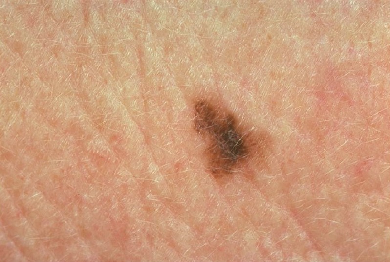
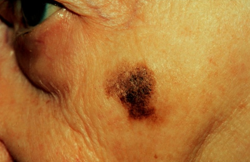
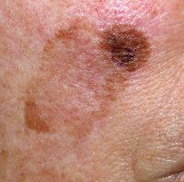
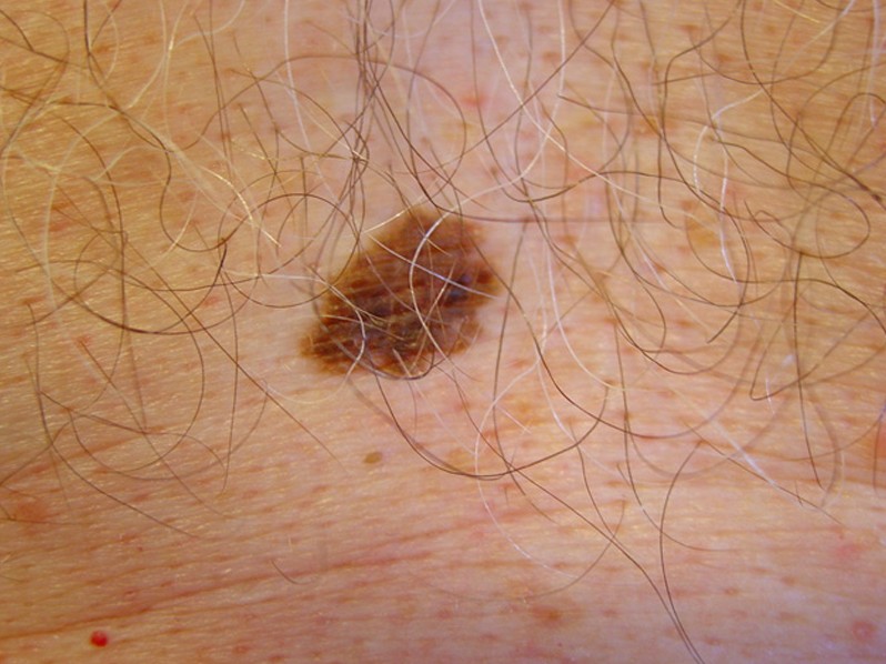
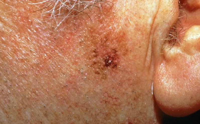
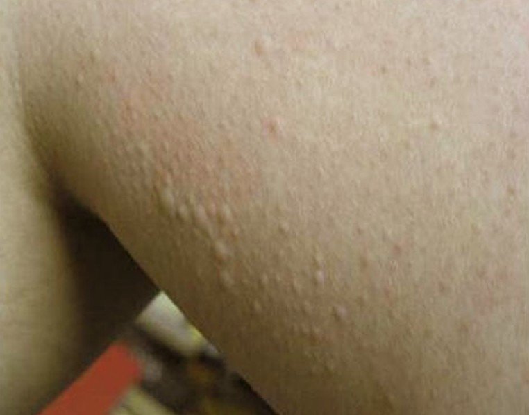
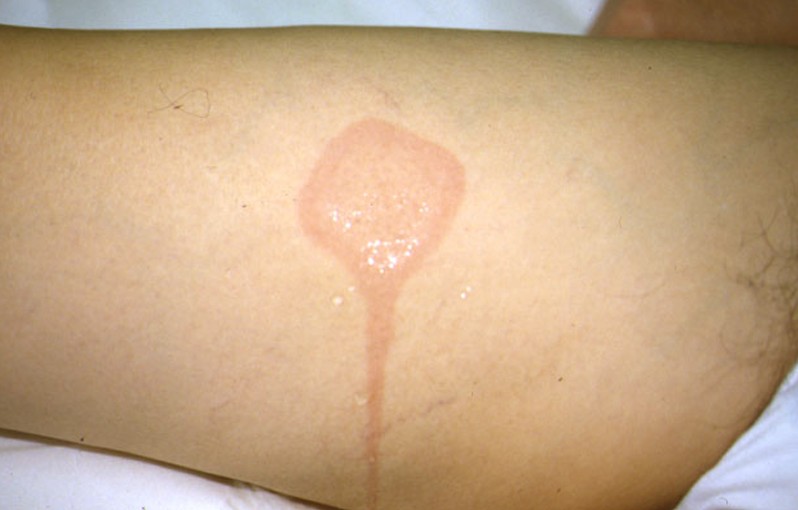
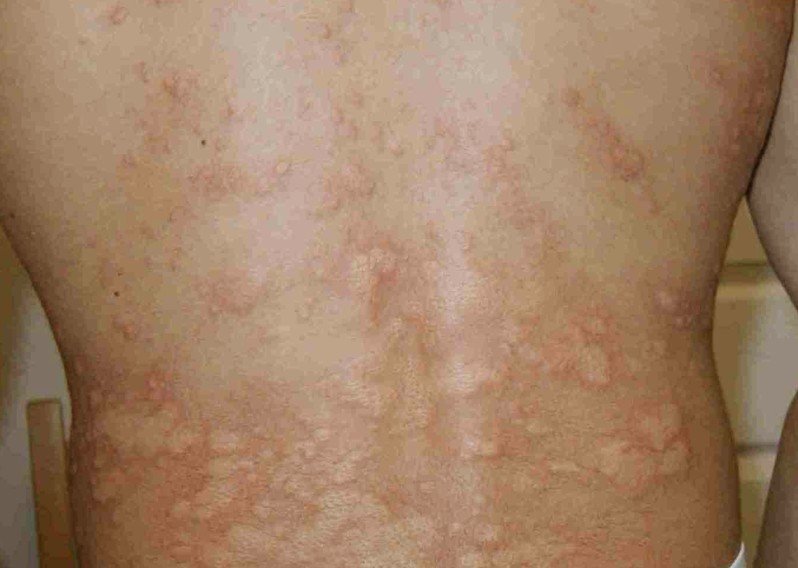
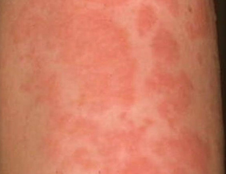
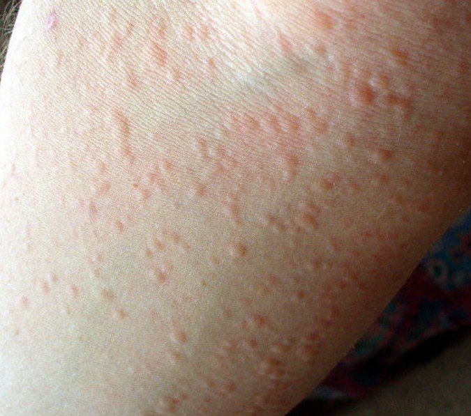
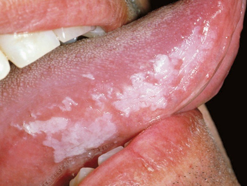
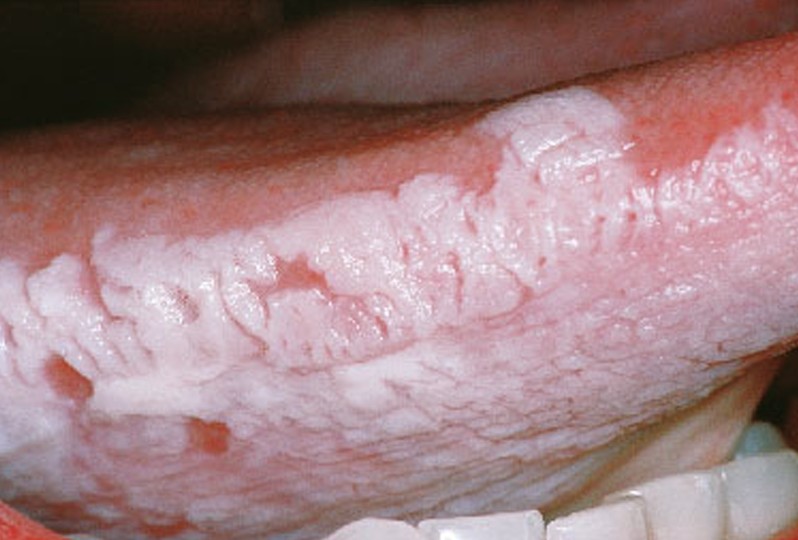
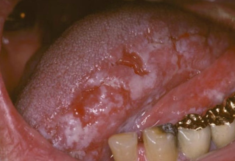
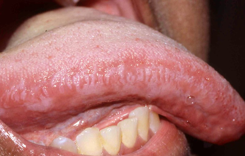
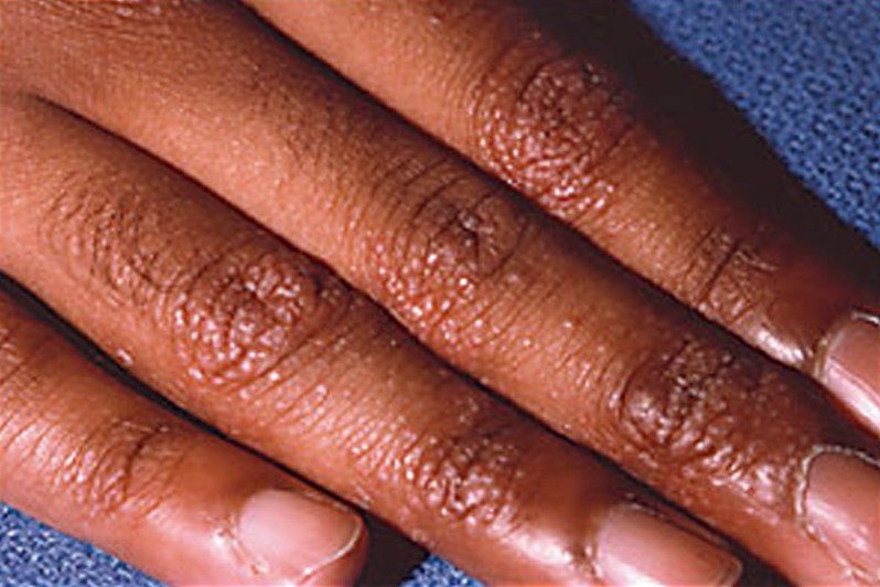
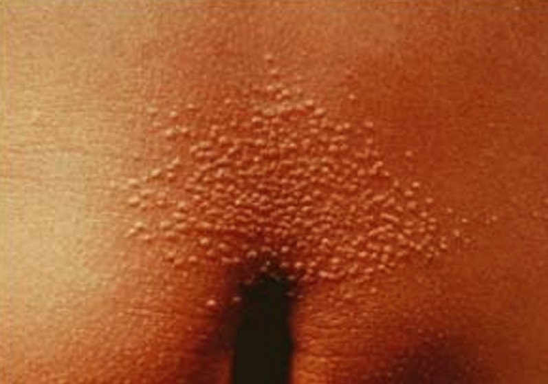
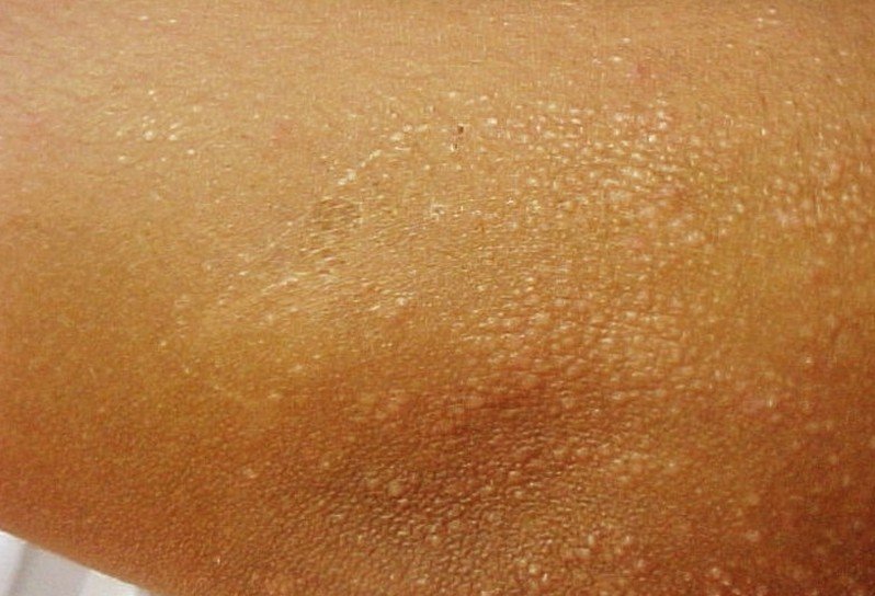
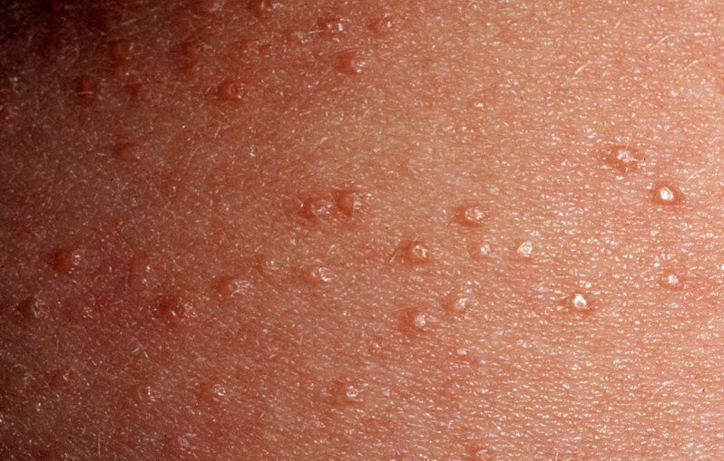
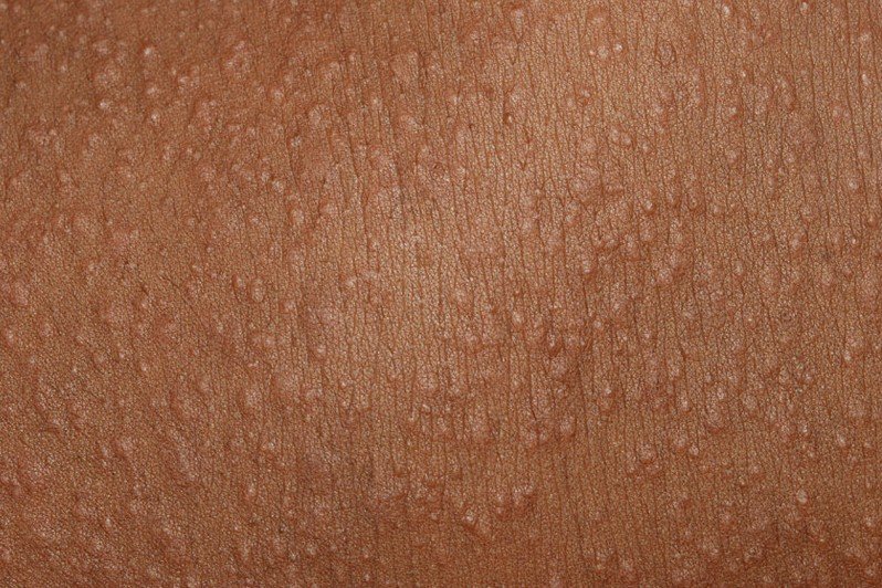
Cold Urticaria Pictures

Collection of images, photos and pictures of the skin condition Cold Urticaria…

cold urticaria

cold urticaria pictures

cold urticaria pictures 2

cold urticaria pictures 3

cold urticaria pictures 4

cold urticaria pictures 5

No responses yet

Oral Hairy Leukoplakia

Jan 08 2016 Published by under Oral Health

Oral hairy leukoplakia is a disease of the oral mucosa that was first described in 1984. It is strongly associated with the HIV infection; this is why it is also known as HIV-associated hairy leukoplakia. This condition is primarily characterized by the appearance of a white patch on the sides of the tongue; this patch has a hairy-like appearance, hence the name. The lesion cannot be scraped off but it is benign.

The main culprit behind the diagnosis of oral hairy leukoplakia is the infection with the Epstein-Barr virus. Once infected, this virus stays inside the body for the entire lifetime and it becomes activated when the immune system is weakened. This is the reason why there is such a strong association between HIV/AIDS and this medical condition. In many cases, the appearance of the white patch on the tongue is a clear sign that the treatment administered against the HIV virus does not provide the desired results. It may also be the first sign that suggests an infection with the HIV virus or the first sign of an infection with another virus (however, it generally suggests that the immune system has been weakened).

Oral hairy leukoplakia appears in HIV negative patients as well, especially those that have a compromised immune system due to other types of infection or various medical conditions (for example, in patients who have undergone organ transplants or who have received chemotherapy for different types of cancer). Even though its name is similar to the one of hairy tongue, these two should not be confused. Oral hairy leukoplakia and hairy tongue are two separate medical conditions, with different symptoms and treatments.

What does Oral Hairy Leukoplakia look like?

oral hairy leukoplakia
The primary lesion in this medical condition is a white patch on the sides of the tongue. The patch can appear on one side or on both sides of the tongue. The hairy like appearance is characteristic for this condition.

Symptoms of Oral Hairy Leukoplakia

These are the most common symptoms of oral hairy leukoplakia:

  • White patch on the lateral borders of the tongue
  • Often asymptomatic (no pain)
  • Daily change of appearance
  • Spontaneous appearance and disappearance of the lesions
  • Unilateral or bilateral involvement, not necessarily symmetric
  • The lesions are continuous or discontinuous along the tongue lateral borders
  • The lesions cannot be scraped off, as they are adherent (scraping will only remove the superficial layers)
  • Can also affect the dorsal or ventral surface of the tongue and the buccal mucosa or the gums
  • In rare occasions, it can affect the soft palate, the pharynx or the esophagus
  • Lesions can become thickened or hardened in some areas
  • The lesions look similar to the ones that appear in cases of candida infections (oral thrush), the difference being that they cannot be scraped off
  • Lesions can be:
    • Small, flat and smooth (this type of lesion is characteristic for the appearance on the ventral surface of the tongue, the buccal mucosa or gums)
    • Hairy appearance (prominent folds)
    • Red prominent lesions – these are a sign of potential malignancy (pre-cancerous lesions) and they appear quite rarely
  • Mild pain
  • Abnormal sensations in the tongue
  • Taste alteration
  • No associated inflammation or redness
  • Psychological impact (cosmetic concern)

What are the Causes of Oral Hairy Leukoplakia?

These are the most common causes that lead to the appearance of oral hairy leukoplakia:

  • The white patch appears because of the increased keratin production and the hyperplasia of the epithelium
  • Infection with the Epstein-Barr virus – the virus enters a person’s body and will remain there for the entire life, waiting for the weakening of the immune system in order to be reactivated.
  • Frequent association with HIV/AIDS
  • General immune-suppression (viral infections)
  • Association with organ transplants and treatments for cancer and especially leukemia (chemotherapy, immunosuppressive medication)
  • Association with Behçet syndrome and ulcerative colitis
  • Appears in rare cases in otherwise healthy people, with a competent immune system
  • Chewing tobacco, drinking excessive quantities of alcohol and smoking are considered risk factors (especially in men who have already received a diagnosis of HIV infection)

Treatment

The lesions in oral hairy leukoplakia are benign, so they do not require any particular treatment. However, these are the most common courses of treatment in case of associated symptoms:

  • Antiviral drugs – the administration of this medication in high doses leads to the rapid disappearance of the lesion but the white patch will re-appear just as fast the moment the treatment is discontinued or the immune system becomes more and more compromised. The most often administered antiviral drugs are:
    • Acyclovir
    • Desciclovir
    • Ganciclovir
    • Foscarnet
    • Valacyclovir/Famciclovir – new drugs, higher oral bioavailability, can be taken less often
  • Topical treatments – these provide temporary remission as well. The most commonly recommended topical treatments are:
    • Podophyllum resin (cytotoxic agent) – the treatment has the following adverse effects: pain in the local area of application, discomfort and taste alteration
    • Retinoids
  • Antiretroviral drugs – these guarantee the most promising results when it comes to the regression of oral hairy leukoplakia lesions. The recommended choice for anti-retroviral medication is:
    • Zidovudine
  • Ablative therapy – this treatment is recommended only in case of small lesions.
  • Cryotherapy – even though it has shown promising results for the treatment of oral hairy leukoplakia, this is not a common course of treatment.
  • Topical applications with gentian violet have also shown to improve the lesions in this medical condition.
  • Treatment of secondary infections (Candida) should be provided as well. This should be done with the proper anti-fungal medication.
  • In rare cases, the surgical removal of the lesions is recommended. However, this is not often the primary choice of treatment and it may be done only on large patches.
  • Ongoing medical treatment is necessary to prevent the lesions from occurring. It is recommended that you go to the doctor’s office for follow-up every three months.
  • This condition can be prevented by reducing the alcohol and tobacco intake.
  • The treatment for the HIV infection should be re-considered and a new treatment plan should be devised, as the presence of oral hairy leukoplakia is often a sign that the actual treatment is not working.

Oral Hairy Leukoplakia Pictures

Here are some of the pictures collection of the medical condition Oral Hairy Leukoplakia…

oral hairy leukoplakia pictures

oral hairy leukoplakia pictures 2

oral hairy leukoplakia pictures 3

oral hairy leukoplakia pictures 4

No responses yet

Lichen Nitidus

Jan 05 2016 Published by under Skin Conditions

What is Lichen Nitidus?

Lichen nitidus is a rare, inflammatory skin condition, in which skin-colored, micropapules that glisten appear on the skin. At the moment, it is known that, in this disease, the skin cells present an abnormal inflammatory activity but the exact cause of the inflammation has yet to be identified. Lichen nitidus affects predominantly children and young adults and it rarely causes upsetting symptoms. Once believed to be a variant of lichen planus, now it is found in the medical literature as a skin condition on its own.

Perhaps the most important things that one should understand about lichen nitidus is that the flat-topped papules, be they skin-colored, red or brown, they do not present an increased risk for skin cancer to develop. At the same time, these papules that appear on the skin are not infectious, so you might want to remember that this is not an infectious skin condition. And once a disease is not infectious, it is not contagious either, so you do not have to worry about it being transmitted to another person.

Parents might become frightened when they will see these papules on the skin of their children but it is important to remember this is an inflammatory skin condition. Even though it may look distressing, it is not life-threatening and certainly it does not represent a sign for more serious illnesses.

Lichen Nitidus Symptoms

These are the most common symptoms of lichen nitidus:

  • The appearance of small, skin-colored bumps or papules on the skin surface; primarily on the trunk, arms, legs and genital area but they can appear on any skin surface. Keep in mind that the appearance on palms or soles is rare, and so is the one on mucous membranes or fingernails
  • If the fingernails are affected, then they may ridge, split or have linear striations on them
  • If the inside of the mouth is affected, the the patient might have the following features:
    • Flat papules on the inside surface of the cheek
    • White plaques on tongue and hard palate
  • Papules can heal in a certain surface area and appear on another
  • Severe itchiness can appear in rare cases
  • Lesions might present an occasional linear pattern or they can gather into clusters, forming plaques
  • Tiny blisters or vesicles can appear in rare cases
  • Rarely, the pustules can present a central depression

Lichen Nitidus Causes

The exact cause of lichen nitidus has yet to be identified. At the moment, it is known that skin cells present an abnormal inflammatory activity, controlled by the white blood cells that are also known as lymphocytes.

However, there are certain risk factors that the medical literature presents, as lichen nitidus may be associated with the following diseases:

  • Lichen planus – as it was already said, at some point, it was believed that lichen nitidus is a variant of lichen planus. However, today, these are two separate entities and it seems that the existence of lichen planus is one of the risk factors involved in the appearance of lichen nitidus
  • Atopic dermatitis – this is also an inflammatory skin condition, believed to favor the appearance of the skin-colored micropapules from lichen nitidus
  • Crohn’s disease – this is also an inflammatory condition, affecting the interior lining of the digestive tract. As lichen nitidus can affect the mucous membranes from inside the body, you can certainly understand the connection between the two
  • Juvenile rheumatoid arthritis – another inflammatory disease, affecting the joints of the body and predisposing to lichen nitidus
  • Tuberculosis – infectious disease suppressing the immune system and favoring the appearance of other medical problems, including this inflammatory skin condition
  • HIV infection – it has to do with a suppressed immune system as well
  • Down syndrome – people with this genetic disorder present an increased risk for various skin conditions, including this one.

Lichen Nitidus Pictures

Collection of images and pictures of the skin condition Lichen Nitidus…

lichen nitidus

lichen nitidus pictures

lichen nitidus pictures 2

lichen nitidus pictures 3

lichen nitidus pictures 4

Treatment

In the majority of the cases, lichen nitidus goes away on its own, without any treatment. There are no scars or changes to the skin color after the lesions have healed. However, there are certain cases in which treatment might be required. These are the most common courses of treatment undertaken by specialized doctors:

  • Corticosteroids – these can be administered orally or topically, helping in the reduction of inflammation. However, long term treatments with corticosteroids are not recommended, as they can have serious side-effects and the withdrawal process is quite complicated
  • Retinoids – these have the same precaution, as they can irritate the skin and have other serious side-effects as well
  • Antihistamines – these are recommended in the situation where there is itchiness reported
  • Phototherapy – UVA and UVB light exposure can help with the faster healing of the papules
  • Topical calcineurin inhibitors – tacrolimus or pimecrolimus
  • Ciclosporins – for more severe cases
  • Antibiotics – these are recommended for situations in which there is an underlying infectious condition, such as tuberculosis.

Precautions that you need to take in order to guarantee fast healing:

  • Avoid humid and warm environments
  • Do not rub the micropapules with a towel when drying off, as this will only contribute to irritating the skin and slowing the actual healing process
  • Eliminate sugary foods and wheat from your diet.

Alternative treatments for lichen nitidus include:

  • Borax (sodium borate)
  • Tea tree oil
  • Lavender oil
  • Coconut oil
  • Comfrey root
  • Baking soda

It is important to understand that the natural remedies presented above do not represent a treatment per say for the disease, as this can go away on its own most of the times. They are, however, recommended for patients who are experiencing upsetting symptoms, including inflammation and itching. Plus, it goes without saying that, as opposed to regular medication, these are natural remedies and they do not have any side-effects. They will work strictly on the symptoms you have and bring that much-needed relief you were looking for. Be sure to contact your doctor in case the symptoms do not subside, as you might need medical treatment as well.

No responses yet

Levoscoliosis

Jan 03 2016 Published by under Bone, Joints and Muscle

What is Levoscoliosis?

Levoscoliosis is a medical condition in which the spine becomes curved to the left side of the body. The symptoms will be identical for the general type of the scoliosis; however, there is going to be one main concern, as the heart is located on the left side as well. These concerns are related to the fact that the left-side curvature is going to press on the ribcage, leading to deformation and possible health problems for the lungs and heart. The most common segment of the vertebral column affected by levoscoliosis is the lumbar spine. When encountered in the thoracic spine for example, it was a consequence of a spinal tumor.

Symptoms of Levoscoliosis

These are the most common symptoms of levoscoliosis:

  • Back pain
  • Postural changes – these are noticed after the pain, that has lasted for several months, decreases
  • The shoulders are at different heights
  • The ribcage appears deformed
  • The shoulder blade on the left side is protruding
  • The anterior superior iliac spines on each side are at different heights
  • The muscles on the left side contract, causing spasms to appear
  • One leg may appear shorter than the other
  • The body may appear as tilting on one side

Levoscoliosis Causes

levoscoliosis pictures
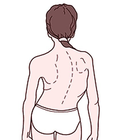
Levoscoliosis can appear under the following circumstances:

  • Idiopathic – the cause is unknown and the person appears to be healthy otherwise
  • Congenital – the bones of the spine have formed abnormally
  • Neuromuscular – there is no control of the the nerves that go into the muscles supporting the spine (for example, in cerebral palsy or muscular dystrophy)
  • Degenerative – in case of degenerated, herniated vertebral discs
  • Puberty – because of hormonal changes, the spine tends to grow faster than the muscles. Plus, the muscles are weakened and levoscoliosis can easily appear.
  • Poor posture – this is not a cause per say but rather a predisposing factor. The one thing you should remember about scoliosis is that the poor posture in which the body finds itself does not hurt but rather when you try to straighten it.

Treatment

The treatment of levoscoliosis depends extensively on the age of the patient, whether the growth period has ended or not and also on the curvature angle. Let’s see the most common courses of treatment that are undertaken:

Physical therapy

In case of levoscoliosis, the physical therapist will perform an initial assessment in order to determine how advanced is the curvature to the left side and use additional investigations (MRI) for confirmation. The PT program will consist of passive and active exercises that are meant to strengthen the muscles on the weakened side. Plus, there will be exercises done on the fixed stairs and plenty of mirror corrective postures to discover.

Braces

Depending on the age of the patient and the curvature angle, the doctor might either recommend a hard or a soft brace. Today, soft braces are widely preferred, as they are more comfortable to wear, while delivering a number of advantages. They have shown to reduce the amount of pain experienced by the patient and also to reduce the progression of the curve. Not only are they recommended for the initial correction but they can also help stabilize the vertebral column, preventing from left side curvature from advancing. Plus, the muscles on the weakened side are stimulated.

The FED method

This is also known as the dynamical and tridimensional therapy of scoliosis. Practically, the spine deviation is treated through the 3D fixation of the vertebral column in elongation with corrective adjustable and derotating pressure. This method originates from Spain and it has treated successfully numerous cases of scoliosis so far, including the one that is the subject here.

Scoliosis surgery

When it comes to surgery for levoscoliosis, this is only recommended in case of the curvature spine angle goes over 45 degrees. Even then, the surgery might not be able to reduce it completely and additional physical therapy will be required in order to reduce the angle even further. Physical therapy will be performed for the rest of the patient’s life, in order to prevent another surgery from happening.

No responses yet

Lentigo Maligna

Jan 02 2016 Published by under Skin Conditions

What is Lentigo Maligna?

This is a skin condition, also known as Hutchinson’s melanotic freckle or melanoma in situ. Basically, in parts of the skin that were damaged by the sun, these malignant cells appear without any invasive growth. The progression of the skin condition is very slow and it may remain in the non-invasive form for years.

Lentigo maligna is commonly encountered in people who have entered their 9th decade, the most affected areas being the ones that received constant sun exposure, such as the face (especially nose and cheeks) and the forearms. In this form, the malignant cells are confined to the initial tissue and their growth is slow but it can be make the transition to genuine melanoma. That is known as lentigo maligna melanoma and it is characterized by the malignant cells transforming into bumpy surfaces.

What does lentigo maligna look like?

lentigo maligna

Initially, blue or black stains appear on the skin. As this condition appears in the elderly, it should come as no surprise that the skin is thin. In many patients, the skin becomes discolored in certain areas and this areas expand slowly with the passing of time. The resemblances in the initial period is of freckles or brown spots, also known as lentigines. The affected areas become more obvious in time, having the possibility to grow to even as several centimeters over the course of decades.

In order to recognize lentigo maligna, one has to follow what is known as the ABCDE rule. A stands for asymmetry, B for irregularity of the border, C for variation of color, D for large diameter and E for evolving. According to this rule, lentigo maligna has a large size, an irregular border, color variation (brown, blue, pink, red or white) and a smooth surface to the touch (this is because the skin becomes thinner due to aging).

Symptoms of Lentigo Maligna

These are the most common symptoms of lentigo maligna:

  • Discolored skin surfaces that may expand with the passing of time
  • Growths on sun-exposed skin – nose, cheeks, ears, neck, forearms
  • Tingling or itching can occur
  • If the skin is hard and lumpy to the touch, bleeding, oozing or crusting, then it has probably already migrated to lentigo maligna melanoma

Causes

The exact cause why the skin cells become malignant has yet to be identified but it seems that a genetic mutation might be involved.

Prolonged and constant sun exposure is the main trigger factor incriminated, especially during the young years of one’s life. This is the reason why having a fair skin and living in a country where there are lots of sunny days become two other potential triggering factors. It goes without saying that the constant exposure to sources of artificial tanning can increase the risk for lentigo melanoma to appear as well.

Having a fair skin is a potential risk because this is the type of skin that is easily damaged by the sun. These are the people who rather burn than tan, having light-colored hair (blonde or red) and light-colored eyes (blue or green). They also have freckles and, as it was already mentioned, lentigo melanoma is also known as the melanotic freckle. It goes without saying that this is a skin condition less common in people with darker skins.

As sun exposure is considered to be the main triggering factor, it seems that having periodic severe sunburn episodes is also a favoring factor. If those episodes took place during childhood and they were severe enough for blisters to form on the skin, then that skin was already hurt and predisposed to lentigo melanoma in older years. Last, but not the least, it seems that people with numerous moles on their bodies and especially hairy birthmarks present an increased risk for this skin condition, as there are those who suffer from immune-deficiency.

Diagnosis

The ABDCE rule is often used for the diagnosis of lentigo melanoma but this is only related to the initial assessment. Often time, a specialist doctor will prefer to see the affected tissue under a microscope. The whole area will be removed surgically and then it will be sent to a specialized laboratory for further examination. In cases of areas that have grown too extensively, then only a sample will be taken, which is known as a biopsy. This is extremely important, as it can determine whether the malignant cells have breached the border and what course of treatment should be taken.

In most of the situations, the surgical removal of the entire affected area is contraindicated, as the initial identity of the lesion cannot be so easily pinpointed. The type of biopsy performed is then the one where the doctor will take samples from multiple points. The samples taken might show different things and it will help the doctor to locate the initial identity of the lesion, thus being able to excise it completely.

Prognosis

First of all, you should understand that lentigo maligna is not dangerous, as it evolves slowly over the years but the malignant cells stay within a certain border. However, if they make a turn for melanoma, then the prognosis changes. The prognosis depends on whether the lesions can be removed surgically or not, the efficiency of other treatments and also on the appearance of new, modified lesions. The moment the diagnosis of lentigo maligna melanoma has been made, the prognosis depends on the thickness of the tumor.

Treatment

These are the most common courses of treatment for lentigo maligna:

  • Surgical removal of affected area, followed by additional skin grafting
  • In cases where the surgery cannot be performed because the affected area is too large, there are other treatment options available:
    • Radiotherapy
    • Cryotherapy
    • Topical creams – the results of these creams are still in the research phase.

When it comes to having lentigo maligna, prevention is the key, in order to prevent it from becoming lentigo maligna melanoma. These are a few key recommendations:

  • Go to the doctor the moment a mole has changed its form, color, growth or if it has started to bleed or ooze.
  • Protect yourself from the sun using creams with high SPF factors and wearing appropriate clothing during hot summer days.
    Do not use artificial tanning.

Survival rate

The survival rate for lentigo maligna is normal, as this is a slowly progressing disease. Co-existing factors might affect the rate of survival, especially if the patient will have to undergo prolonged surgery for extensive removal of the affected area. In the situation that lentigo maligna turns into a melanoma, then the survival rate for those over 4 mm is of 50% during a period of ten years.

Once again, the survival rate depends on how the lesions have spread and whether they have suffered modifications. The survival rate can be definitely influenced by the overall health of the patient, as it a well know fact that older patients can have various chronic medical problems.

As a patient, you should not wait to go to the doctor until it is too late. The treatment and the survival rate are both influenced by the actual time when you go to the doctor, as the progression from lentigo maligna to melanoma can happen without any notice.

Lentigo Maligna Pictures

Collection of pics, photos and pictures of the skin condition Lentigo Maligna…

lentigo maligna pictures

lentigo maligna pictures 2

lentigo maligna pictures 3

lentigo maligna pictures 4

lentigo maligna pictures 5

lentigo maligna pictures 6

No responses yet

« Prev

© 2011-2019 MDDK.com - Medical Tips and Advice. All Rights Reserved. Privacy Policy
The health information provided on this web site is for educational purposes only and is not to be used as a substitute for medical advice, diagnosis or treatment.