Sigmoid Volvulus
Definition
Sigmoid volvulus is a medical condition, in which the sigmoid colon practically twists on the sigmoid mesocolon. These changes frequently lead to the large bowel obstruction. It is estimated that, out of all large bowel obstructions, approximately 5% are due to the sigmoid volvulus. This condition is more often encountered in the elderly population, being one of the most common types of volvulus that can occur at the level of the gastrointestinal tract. If the conditions is left untreated, serious complications – such as venous infarction, perforation and fecal peritonitis – can occur.
According to the most recent research, the sigmoid volvulus is more often encountered in populations from South America, Africa, Asia and Eastern Europe. On the other hand, it is rarely encountered in developed countries, such as Japan, Australia, USA or the UK. While this condition is often encountered in adults, it is quite rare in children or infants. The same studies have shown that men are more predisposed to developing sigmoid volvulus than women.
Symptoms of Sigmoid Volvulus
These are the most common symptoms associated with the appearance of sigmoid volvulus:
- Constipation
- Bloating (obvious distension of the abdomen)
- Nausea
- Vomiting – appears at a later stage, when the distension of the abdomen is already severe
- Abdominal pain – sudden, colicky and located in the lower part of the abdomen; in some patients, only some slight abdominal discomfort might be present (insidious form)
- Failure to pass either flatus or stool
- The patient might present a history of recurrent mild attacks – however, these have been relieved, as the patient has managed to pass a large quantity of flatus or stool
- The examination of the abdomen can reveal the distention of the abdomen, as well as a palpable mass
- In the situation of complications, such as the colonic perforation, the patient can present systemic symptoms (fever, shock)
- The rectal ampulla is empty upon physical examination
The onset of the sigmoid volvulus can be acute or chronic. Complications such as the perforation of the colon and peritonitis can occur, especially if the diagnosis is delayed and the necessary treatment measures are not taken.
Causes
These are the most common causes that can lead to the appearance of the sigmoid volvulus:
- Chronic constipation
- Excessive or prolonged usage of laxatives
- A diet that is too rich in fiber (geographic prevalence – Africa)
- Chagas disease (geographical prevalence as well – Africa)
There are certain predisposing factors that can increase the risk for sigmoid volvulus, such as having a megacolon or suffering from an excessively mobile colon.
The sigmoid volvulus can be found in association with the following:
- Chronic neurological conditions
- Parkinson’s disease
- Multiple sclerosis
- Pseudobulbar palsy
- Treatment for psychiatric conditions that are chronic, such as schizophrenia.
Diagnosis
The diagnosis of sigmoid volvulus can be made based on the following investigations:
- X-ray
- The abdominal X-ray can identify the site of the problem with specific signs, such as the coffee bean sign or the absence of rectal gas
- Key radiologic feature – double-loop obstruction (present in 50% of all the patients)
- Fluoroscopy/ barium enema examination
- Water-soluble contrast enema – reveals the bird beak sign
- Rarely performed nowadays
- Not recommended in the situation that a gangrenous bowel is suspected, in the situation of pneumoperitoneum or peritonitis
- CT
- Identifies the closed-loop at the level of the sigmoid colon
- Whirl sign present – the mesenteric vessels are twisted
- Beak sign present – this appears only if rectal contrast has been administered
- Recommended for the identification of the etiology and also to see the actual site of the obstruction (suggestive of other pathologies)
- Least invasive imaging techniques that can be used for the assessment of complications, such as ischemia
- MRI
- Recommended for the assessment of large-bowel obstruction (not specifically for sigmoid volvulus)
- Ultrasonography
- Can be used for the diagnosis of large-bowel obstruction and, thus of sigmoid volvulus
- Not the most faithful investigation whereas the correct diagnosis is concerned
- Magnetic resonance angiography
- Recommended in case of suspected complications, such as mesenteric ischemia
- Sigmoidoscopy
- This procedure is indicated for the suspicion of an ileosigmoid knot
The differential diagnosis can be made with the following conditions: large bowel obstruction of other etiologies, caecal volvulus and colonic pseudo-obstruction.
Sigmoid Volvulus Pictures

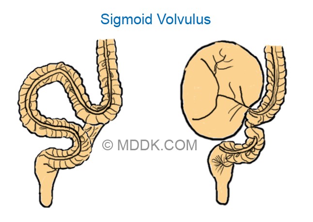
Sigmoid Volvulus Picture 1 : In the left side image, you can see the unattached loop of bowel getting twisted resulting in the obstruction of bowel lumen which you can see on the right image.

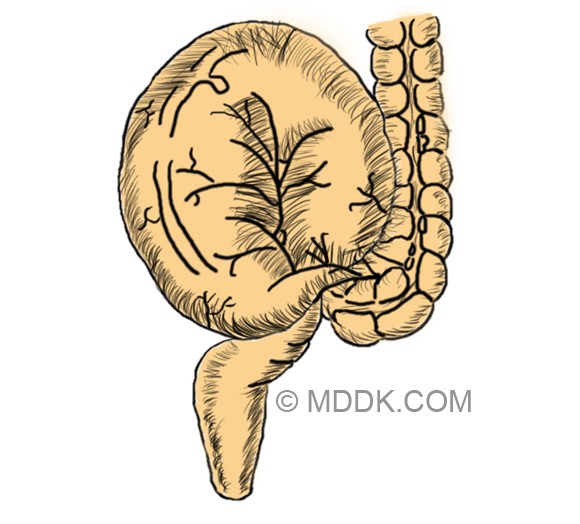
Picture 2 : Obstructed bowel lumen
Treatment for Sigmoid Volvulus
The main approach for the treatment of sigmoid volvulus is the insertion of a rectal tube. The decompression of the sigmoid volvulus can be guided through endoscopy or fluoroscopy. It is important to understand that the acute sigmoid volvulus is a medical emergency and that it should be treated as such. The risk of complications is quite high with the acute onset of the sigmoid volvulus. While non-operative decompression is successful in treating the majority of the patients, the surgical intervention is more recommended, as it can reduce the risk of recurrence.
For the decompression procedure, the patient is placed in a left lateral position. The decompression and untwisting of the sigmoid colon are performed with the help of a sigmoidoscope and a flatus tube. Basically, it is the flatus tube that will solve the problems, with a rush of liquid feces and flatus actually relieving the obstruction. Given the immediate results of this procedure, it should come as no surprise that the patient will experience immediate relief from the previous symptoms.
The flatus tube will remain in position for approximately one day, so as to maintain the decompression and reduce the risk for decompression. Another purpose of the flatus tube is to give the vascular supply to the bowel wall the necessary time to recover. After the intervention, it is essential to monitor the patient. In the situation that the patient suffers from constant pain in the abdomen or blood appears in the stool, a second surgery might be required (these symptoms suggest that ischemia has occurred at the level of the GI tract).
In the situation that the patient suffers from recurrent volvulus episodes, the surgical intervention is going to be recommended. The procedure refers to the resection of the redundant part of the sigmoid colon, being also indicated in the situation that tube decompression has failed or in those who have suffered from bowel ischemia.
Prognosis
The prognosis is influenced by the appearance of complications, such as bowel ischemia. The mortality rate for sigmoid volvulus varies between 20 and 25%, according to how much time has passed between the diagnosis and the actual treatment intervention. The earlier the diagnosis is made on the abdominal X-ray, the better the prognosis will be for this condition.