Diseases General Health Skin Conditions

Archive for February, 2017

Prurigo Nodularis

Feb 18 2017 Published by under Diseases and Conditions

What is Prurigo Nodularis?

Prurigo nodularis is an inflammatory skin condition characterized by the onset of itchy papules in the extremities. The itch and scratch cycle in prurigo nodularis results in the development of more papules.

Prurigo nodularis is often exhibited as multiple eruptions of excoriated papules on the surfaces of the limbs resulting from the incessant scratching. The more scratching made the more lumps will develop.

Prurigo nodularis is also known by another name such as Hyde prurigo nodularis and Picker’s nodules. The disease was first described in 1909 by Hyde and Montgomery describing the disease as itchy nodules on the extensor surface of the lower extremities. The disease was first described among middle aged women.

Prurigo nodularis is the most severe form of prurigo. This skin condition on the other hand is rather benign. Prurigo nodularis can affect the quality of life as the extent of pruritus and nodules can be debilitating and bothersome. The disease does not increase the chances of mortality except in the presence of underlying condition where the onset of prurigo nodularis is a secondary disease. There are cases where prurigo nodularis is being linked with a malignancy such as Hodgkin disease where prurigo nodularis is the manifestation of the primary disease. In modern day medicine, prurigo nodularis is regarded as the most common dermatosis in HIV patients where the incidence is deemed as a sign of severe immunosuppression.

The incidence of prurigo nodularis is found to be common among middle aged individuals and older adults although it can arise in any age group. No racial predilection has been recognized and the disease is deemed to occur in people worldwide. The frequency of the disease between sexes has not been fully recognized although there have been reports where prurigo nodularis is found to be prevalent among women.

Prurigo Nodularis Symptoms

Prurigo nodularis is common among middle aged individuals and older adults although children may also get affected. It initially develops as a small and an itchy red bump on the extensor surface of the limbs. Prurigo nodularis causes an intense itching that provokes a vigorous scratching to relieve the itchiness. Scratching of the nodules however only intensifies the symptom of itchiness and only brings in more nodules.

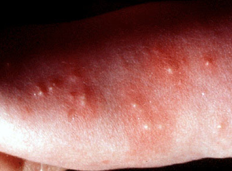
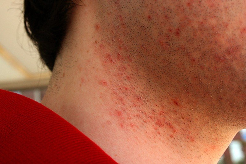
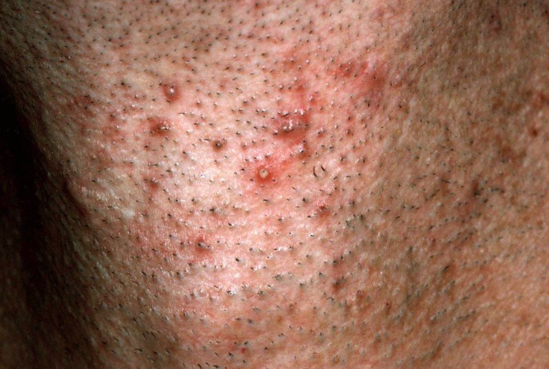
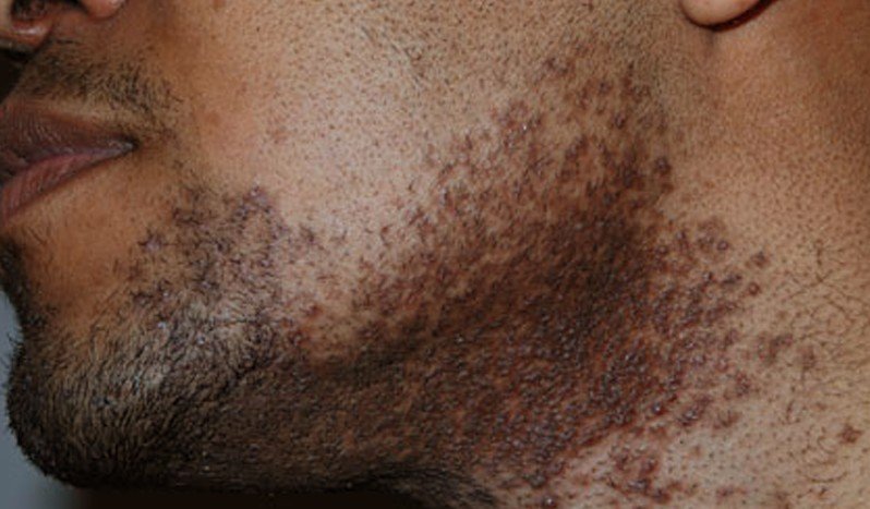
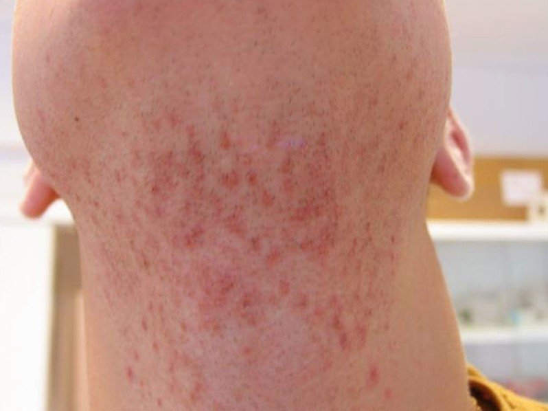
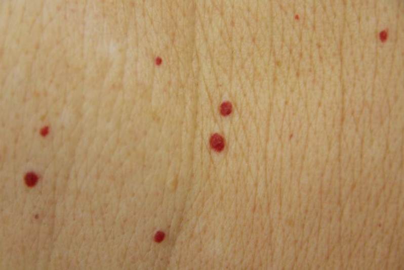
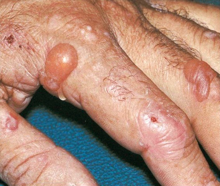
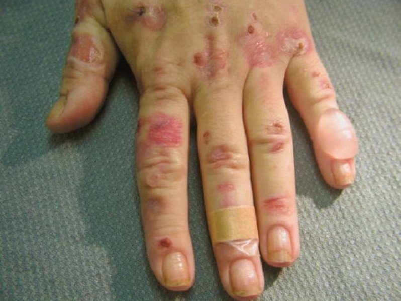
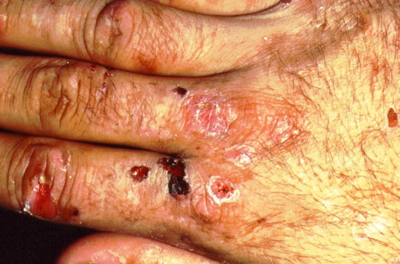
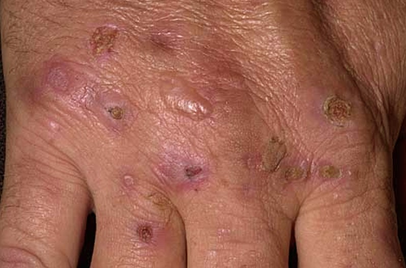
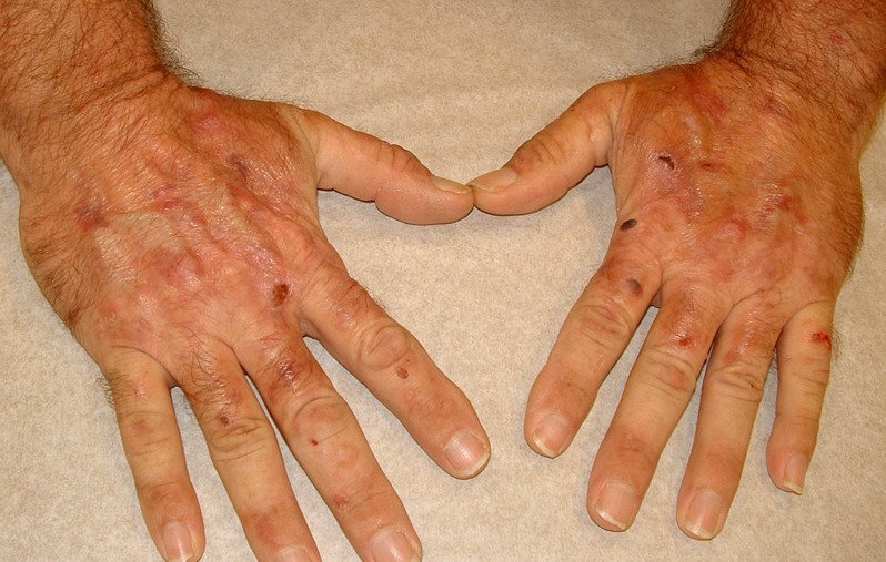
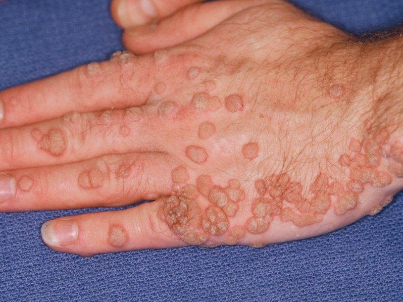
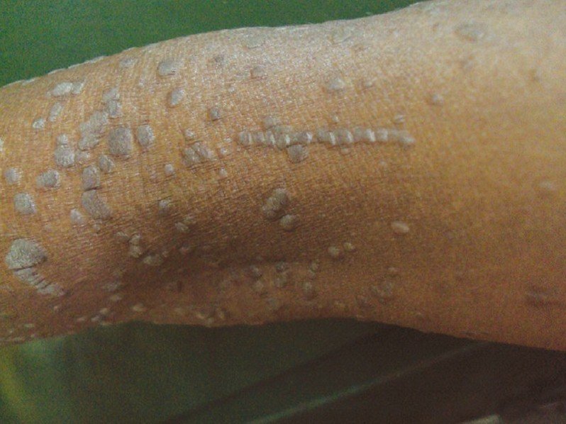
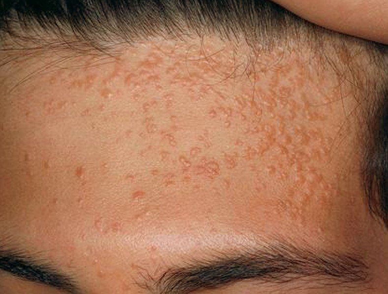
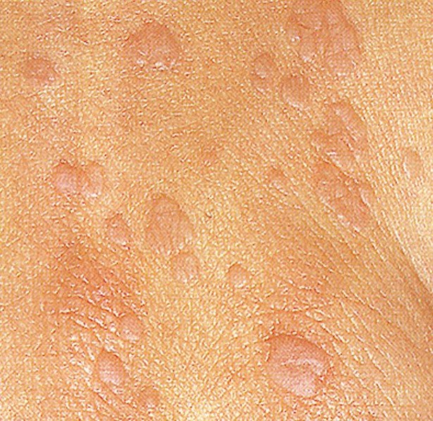
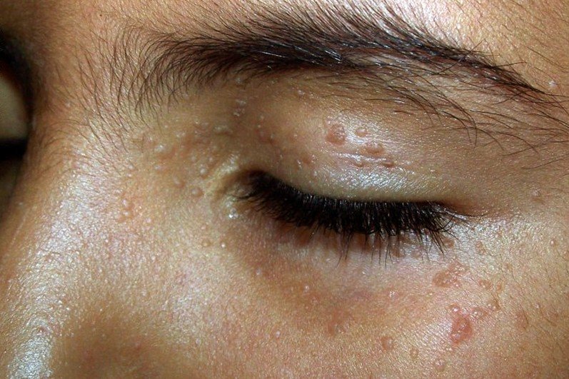
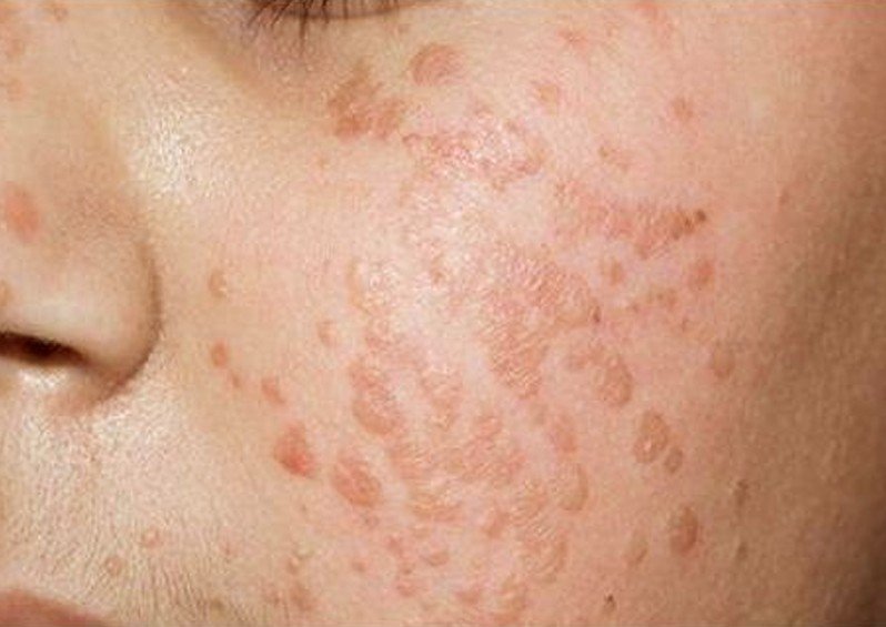
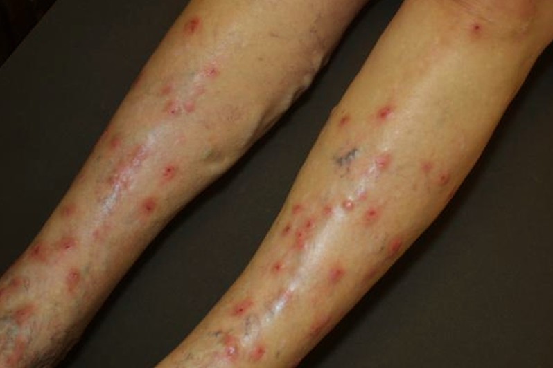
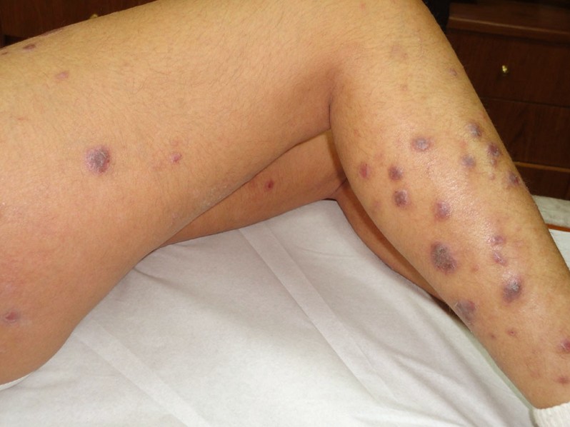
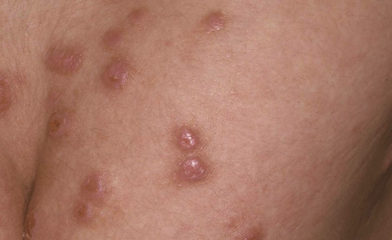
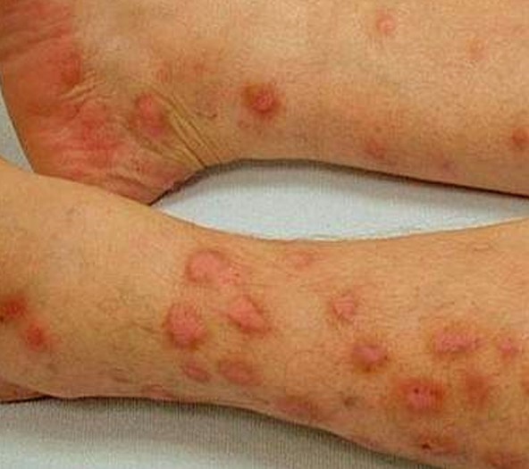
The individual size of the nodule is about 1 cm to 3 cm in diameter and initially develops in the arms or legs although the nodules may develop in any part of the body. The nodules are discrete and appearing in hundred or two hundreds approximately and often appear in clusters and are symmetrically distributed. Excoriation of nodules is evident with a top that is flat and umbilicated or may have a crusted top. The crusty or scaly presentation of the nodules is due to the incessant scratching that brought the nodules to break off and sore. The color of the nodules may be purplish, reddish or brownish and the surface of the nodules is rough. The face and the palms can also get affected with nodules. The neck, upper arms and the shoulders can also be seen with nodules of prurigo nodularis with the cape of the neck as the most obvious area.

Itchiness is one of the hallmarks of prurigo nodularis along with the development of the nodules. The frequency of itchiness is so intense that it can distract the daily activities and can even disturb the sleep particularly at night time. The itchiness can also provoke a non-stop scratching to alleviate the itchiness although scratching can only make the condition worse. It only adds growth of new nodules while scratching can also cause the nodules to break off leading to sore that will cause the nodules to bleed. When the skin surface breaks off, it is highly potential for infection.

Long standing pruritus is the common complaint of patients with prurigo nodularis. The intensity of the pruritus causes the affected individual seems anxious, restless and worried. The disease usually lingers for a long time that it may cause depression and stress to the patient. Scratching does not provide relief, but only result in formation of new nodules whereas the old nodules do no resolve spontaneously. The old nodules will only leave a scar which can be darkly or lightly pigmented.

Prurigo Nodularis Causes

The exact etiology of prurigo nodularis remains unidentified. The pathophysiology of whether the lump or the itchiness occurred first remains vague as well. The exact cause of the inflammation and the formation of lumps and the amplification in the size of the nerve of the skin all remain unclear.

While the exact cause of prurigo nodularis remains unclear, several factors or conditions are considered to predispose the incidence of prurigo nodularis.

Atopic dermatitis

It is among the considered trigger of prurigo nodularis. Several numbers of patients reported to have a family history of atopic dermatitis prior to the development of prurigo nodularis.

Insect bites

Insect bite is considered a trigger of prurigo nodularis. The reaction to insect bites is believed to have perpetuated the itchiness and nodule formation.

Other conditions been linked to the onset of prurigo nodularis include the following:

  • Immunodeficiency disorders including HIV
  • Iron deficiency anemia
  • Gluten enteropathy
  • Malignancies
  • Psychiatric illnesses
  • Liver and renal dysfunction

Prurigo Nodularis Treatment

Prurigo nodularis is an inflammatory skin condition that is rather difficult to treat. It is considered as a resistant skin condition that the success of treatment is either mild or moderate. The challenge of controlling the disease is often done with a combination of medications.

The primary aim of treatment is to stop the patient from scratching which proves to be difficult on the part of the patient. Scratching only worsen the condition that it is necessary to alleviate the pruritus through medical means and not from scratching.

Treatments used to alleviate the itchiness and to reduce the inflammation and the lesions include the following:

  • Antihistamines
  • Corticosteroid
  • Capsaicin cream
  • Topical anesthetics

For more severe prurigo nodularis and for systemic treatments, the following are considered:

  • Anticonvulsants
  • Phototherapy
  • Thalidomide
  • Systemic retinoids
  • Oral steroid
  • Tricyclic antidepressant

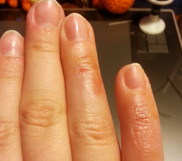
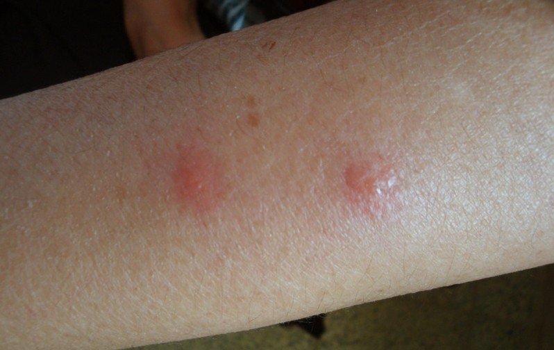
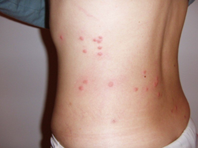
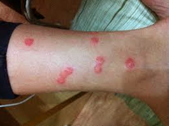
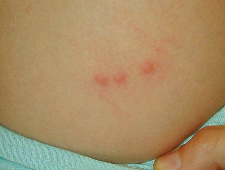
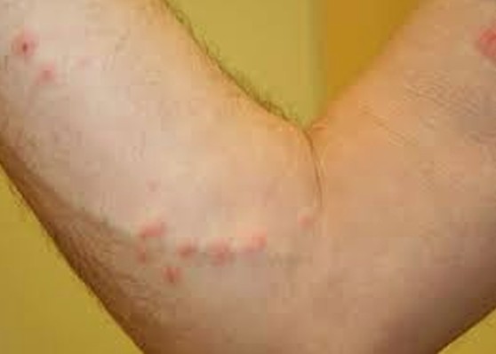
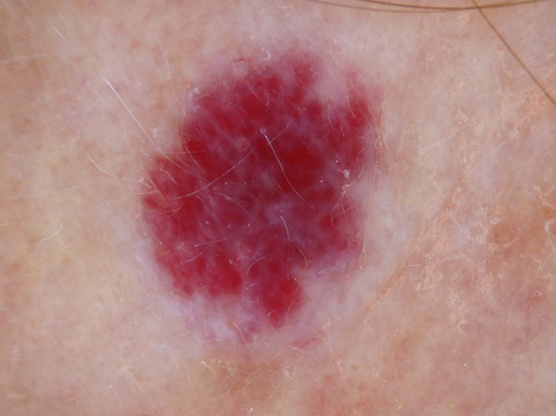
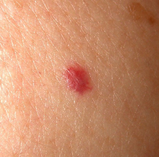
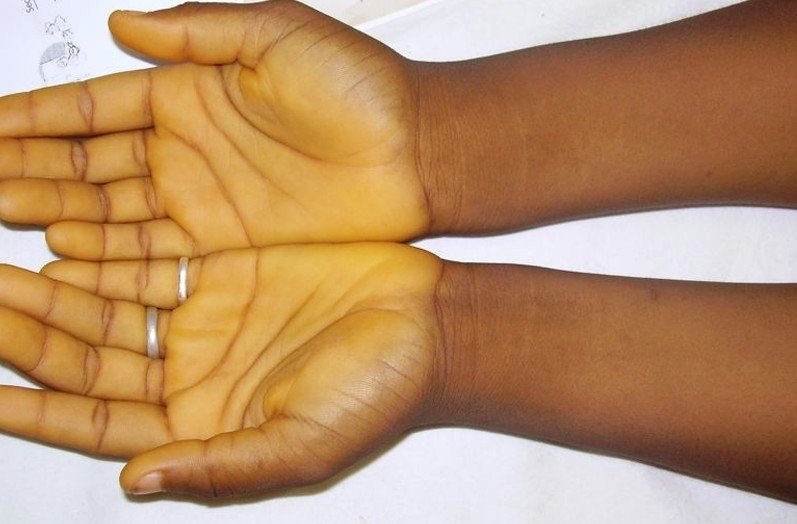
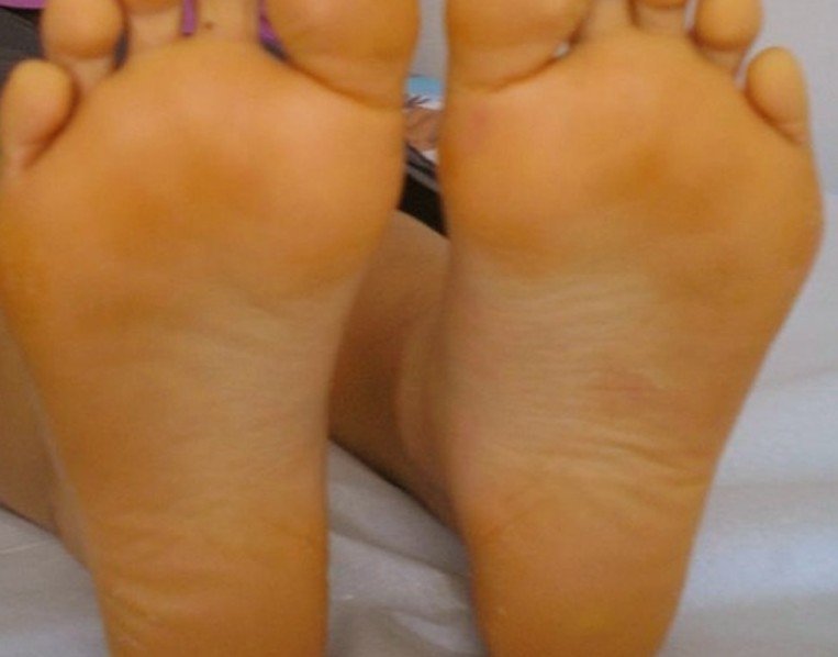
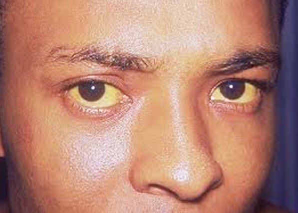
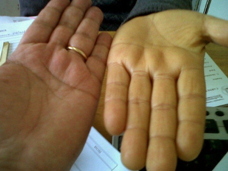
Prurigo Nodularis Pictures

Collection of photos, images and pictures of the health condition Prurigo Nodularis…

prurigo nodularis

prurigo nodularis 2

prurigo nodularis 3

prurigo nodularis 4

prurigo nodularis 5

prurigo nodularis 6

prurigo nodularis 7

Is Prurigo Nodularis contagious?

Prurigo nodularis is not a contagious disease. It is an inflammatory skin condition with an unknown cause. The condition is believed to be from a repetitive trauma to the skin such as the mechanism of an itch – scratch cycle that resulted in the development of itchy nodules. No harmful pathogens are considered to cause and spread this inflammatory skin condition.

No responses yet

Steam Burns

Feb 12 2017 Published by under Skin Conditions

Steam burns are ugly experiences everyone would love to avoid because of the pain and the mental torture associated with them. You may not understand the importance of what we are trying to say here until you are exposed to steam injury or your beautiful skin is destroyed by steam.

Steam burns can potentially damage a high degree of skin, muscles or tissues than you would naturally think is possible. Though they are very painful and have the tendency to leave some scars behind, these injuries are treatable and the scars can be minimized with professional management.

Why do steam burns hurt a lot?

steam burns

There is a release of heat (in form of energy) when there is condensation of steam to water and then the water is cooled to the skin temperature. Latent heat of vaporization is released in the process which results in more injury or damage to the skin. The level of energy released in the process results in deep penetrating burns which is why steam burns hurt a lot.

Latent heat of vaporization is the amount of heat energy that is needed to change the phase or state of matter from liquid to gaseous state. The liquid absorbs the energy but does not change the temperature. This latent heat is released by the gas when the gas condenses and becomes a liquid before it can cool below its boiling temperature. At sea level, water’s latent heat of vaporization is 2250 J/g. A great deal of that heat is absorbed into your skin if you place your hand into a jet of steam as the heat is being released into the atmosphere. The water still remains at 100⁰C as the steam condenses into your skin, penetrating deep into your tissues and causing damage to the cells. That is why steam burns from kettle can be very painful and sometimes serious.

First Aid for Steam burns

There are some steps you can easily take in order to minimize the damage caused by steam burns, especially before the arrival of a medical doctor or before you are taken to the hospital. Here is the First Aid management for steam burns.

Stop the burning

  • Stop the contact with the steam immediately
  • Tear or cut around any cloth that is sticking on the skin

Cool the burn

  • Place the burned area under a running tap of cool water and allow the water to run for about 10 to 15 minutes. This is to completely cool the temperature and prevent further penetration of the heat
  • If there is no running water, use cool compresses to douse the area
  • Never cool burns with ice water, as ice itself is capable of causing burns. It can also lower body temperature and cause shock.

Protect burn

  • The burn should be covered with a sterile, non-adhesive bandage or a clean cloth
  • The following should not be applied to the burn – egg, butter, salt, ointments or any other liquids
  • Do not attempt to break the blister
  • Do not breathe or cough on the burn

Treat pain

  • Relieve pain with over-the-counter drugs such as naproxen (Aleve), Ibuprofen (Motrin), or Acetaminophen (Tylenol).
  • Do not give anything by mouth, especially if there are severe burns and pain

Give emotional support

Calm the patient down and reassure him/her. Let the patient know that you are there to help him/her. This will reduce the level of panic and ensure compliance to treatment.

Consult the doctor

You may need to see the doctor if:

  • The pain and redness refuse to stop after several hours
  • The pain is getting worse
  • You need to take anti-tetanus injection or booster dose
  • There are signs of infection such as increased pain, swelling, redness, offensive discharge, fever, etc.

Steam Burns vs Fire Burns

Steam burns are occasionally misunderstood with fire burns or burns resulting from dry heat. As the name implies, steam burns are caused by exposure to steam or boiling water while fire burn comes from dry source of heat. It is usually easier to sustain steam burns on wrist, fingers, face, lips, etc. because these parts are easily exposed to sources of steam.

Steam may sometimes not be hotter than the fire. Nevertheless, when the skin gets in contact with steam, the pain is felt faster than when it is  with dry fire. The reason is because when the steam comes in contact with your skin, it tries to transfer its own heat to your skin (the skin being the new source of fuel) and you feel the heat within a matter of few seconds.

The moment the heat is transferred to your skin, the temperature increases momentarily and exceeds that of the hot steam. That’s the reason why you easily feel the heat from a boiling kettle more than that of Bunsen burner or candle.

How to Treat Steam burns

The treatment for steam burns is usually done on a First Aid basis. Other treatments may be carried out in the hospital. Having talked about the first aid management earlier, let’s quickly look at what can be done at the hospital.

Hospital admission

Admit the patient if the burns are extensive; otherwise the burns should be treated as an out-patient case.

Barrier-nursing

The patient should be nursed under insect-proof net to prevent flies and other insects from contaminating the surface of the burns (if there are areas with open wounds).

Wound dressing

Burns are usually dressed daily using Normal Saline solution to clean the surface before applying layers of sterile dressing. This helps to remove dead cells and exudates from the wound surface. But there are two basic types of wound dressing you can apply to the steam burns

  • Low Adherent dressing: These are readily available and are quite cheap. Their main work is to help maintain a moist wound bed while allowing exudates to pass through into a secondary dressing. They come in the form of tulles (cloth that is open weave and soaked in chlorhexidine or soft paraffin), perforated films of plastic, or textiles. They can also be smeared with silver sulfadiazine, nadifloxacin or other materials. They are used to cover the wound to prevent infection and aid quick granulation/healing.
  • Semi-permeable Films: They are made of plastic sheets of polyurethane that are sterile and coated with hypoallergenic acrylic adhesive. They are most of the times used as transparent cover for wounds. They do not allow the passage of fluid and bacteria but allow free circulation of air into the burns. Some of the types of semi-permeable films you can use for daily dressing of steam burns include Hydrogels, Hydrocolloids and Alginates.
  • You can also apply biological dressing like collagen dressing, which is quite safe, aids granulation faster, is hypoallergenic, and non-pyrogenic. They come in the form of powder or gel.

Antibiotics treatment

  • Broad spectrum antibiotics injection or capsules can be administered to prevent infection of the burns.
  • Anti-tetanus serum or tetanus toxoid should be administered to prevent infection by tetanus bacterium

Control of pain

Control pain with over-the-counter drugs such as Indomethacin, Ibuprofen, paracetamol, Diclofenac, Piroxicam, etc.

How to Heal steam burns quickly

Your steam burns will heal quickly if you do the following:

  • Prevent infection from setting in: You can do this by protecting the wound with a dressing. Antibiotics prophylaxis also helps in preventing infection. When there is no infection, wounds heal faster
  • Avoid further injury: You must not injure the wound further. Avoid scratching its surface or hitting the burn on hard surfaces
  • Stick to wound dressing as advised: Do not break the blister if there is any. This will help prevent the wound from being exposed to microbes
  • Eat balanced diets: this will improve your body’s ability to generate granulation tissues to heal the burn quickly.
  • Take a lot of Vitamin C: Ascorbic acid is also known as the healing vitamin because of its ability to heal wounds. Take enough of it daily. At least 300mg daily should really help out.

Home Treatment

There are a lot of things you can actually do at home to help take care of your steam burns, reduce the pain associated with it and aid healing. Some of them include:

  • Honey: Honey is known to aid wound healing faster than a lot of the orthodox medications out there. Some health surveys have shown that honey takes an average of 18 – 16 days to heal burns compared to Silver Sulfadiazine which takes about 32 – 68 days.
  • Clean the wound surface of the burns with normal solution or sterile water and apply honey. Do this daily and watch your wound heal fast. However, it must first be established that the wound is not infected and that the patient is not diabetic. This is to prevent bacteria from thriving on the honey to cause more problems.
  • Egg white: They help in controlling the pain that is associated with burns. Apply the egg white to the surface of the burns (without an open wound) and allow it to stay as long as it can. When it dries up, pain is likely to return. In that case, reapply the egg white again to keep the burn area cool.
  • Whole milk: Soak a piece of clean non-fluffy cloth or gauze in cool, whole milk and apply it gently over the burn for about 10 to 15 minutes. Apply fresh one as the milk warms up on the wound.
  • Vinegar: Diluted vinegar is equally helpful in handling steam burns at home. It helps to relieve pain and relax the tissues. Soak a cloth in vinegar that has been diluted with cool water and apply it to the burn area. Repeat when necessary.
  • Onion juice: This has been in use by chefs for centuries for the home treatment of burns. Cut a fresh onion in half, squeeze out the juice and apply it to the burns to minimize pain and reduce blister formation. Freshly cut onions have better soothing properties.
  • Cool water: Have you suffered steam burns from kettle or your cooking pot? Consider using cool water to reduce the burning effect of the steam and prevent further damage to skin and underlying tissues. Put the part under running water and allow it to stay up to 10 to 15 minutes to cool the part.
  • Aloe Vera: This soothing plant also aids burn healing. For first-degree burns, apply the fresh Aloe Vera or Aloe Vera gel to the area immediately after cooling with water or treating with vinegar. But for second-degree burns, you need to wait for the wound to start healing before applying Aloe Vera or Aloe Vera gel. It will help in reducing pain and aid healing of the tissues. Before using Aloe Vera, be sure you are not on blood thinners and that you have no heart problems.
  • Lavender essential Oil: Applying lavender essential oil to the skin immediately after the burns also helps in minimizing the pain, promoting healing and reducing scar formation. You can pour the lavender on a piece of cloth and apply to the skin if the surface involved is big.

Does steam burns leave scar?

Steam burns usually leave some level of scar. But this is highly dependent on the extent of the injury caused by the steam. In cases of first-degree burns without major blisters, the wound will heal without any scar.

If there is an open wound with tissue damage, the possibility of forming a scar is high. Second-degree burns will most likely leave a scar behind. For example, if you had steam burns from kettle that you placed your arm too close to the spout, the burn is going to be much. This would leave a scar. But if it is something you moved your arm across but over a longer distance, the scar may not form.

Conclusion

Steam burns on wrist, fingers, face, lips or any other part of the body can be quite painful and distressing. But with the remedies we have listed in this post, you have several options to help take care of your steam burns.
If you try out our suggestions and they fail to work for you, then you need to see the doctor, especially if there are signs that the burns might be infected. The home remedies are quite helpful and need to try them too.

No responses yet

Papular Urticaria

Feb 11 2017 Published by under Diseases and Conditions

What is Papular Urticaria?

Papular urticaria is a self-limited disorder that often occurs in children. It is characterized by the onset of chronic papules as a result of hypersensitivity to insect bites. This skin condition is not a true urticaria as papular urticaria often persists for several days to several weeks unlike the true urticaria which only lasts for several hours.

Papular urticaria is common during the summer months particularly in places where there are a lot of insects. It is common in the skin areas that are often exposed or are not usually covered with clothing.

Papular urticaria is not a contagious disease although the eruption is distressing as it is often itchy and may leave persistent scars especially if the papules are scratched deeply. The potential for infection is relatively high as intense itching may cause children and adults to scratch the lesions incessantly thereby breaking the lesion causing entry for harmful pathogens.

Children between the ages of 2 and 7 years are often affected particularly during the summer time when children are involved in outdoor activities and are exposed to different insects outdoors. The population of the insects often increases during the summer time that papular urticaria is common. Children also have weaker resistance compared to adults that papular urticaria is highly possible. Papular urticaria may also occur in adults but because adults have more tolerance, the incidence of papular urticaria is rather seldom.

Papular urticaria can affect both boys and girls without racial predilection. It can affect people globally, although the incidence is more common in places with hot climates and with increased populations of insects. The incidence is more common during the hot summer months and resolves during the winter or cold season.

Papular Urticaria Symptoms

The lesions in papular urticaria appear like crops of bug bites although the eruption is more likely to originate from a single bug bite only. The eruptions are likely to emerge, vanish and re-emerge from one spot to another. The crops of eruption of lesions are usually triggered by a single bite or few bites from an insect. The lesions usually linger for a couple of days or more before they disappear then spontaneously recur.

Pruritus or itching is the initial manifestation of papular urticaria. The itching is so intense that scratching is inevitable to get relief from the itching. Incessant scratching will lead to crusting of the lesion which will only make it more susceptible to infection. It will also make the lesion sore and provoke old lesion from cropping up.

The lesion of papular urticaria is approximately 0.2 to 2 cm in diameter and is often reddish in color. The lesion usually starts as clusters of lesions. The development is usually found on the face, forearms and legs and other areas of the skin that are usually not covered and are often exposed. The lesion, however, may also occur in other parts of the body, including those that are covered. The lesion may also be blisters filled with fluid and a diameter of about 1 cm.

The distribution of the lesions is somewhat symmetrical which may be localized in the area of the insect bite although the lesions may spread to other parts of the body. The onset of lesions usually occurs right after the symptom of itching. The lesion is elevated and which may be small or large in size and lingers for several days. It emerges, vanishes and emerges again unexpectedly and elsewhere in the body. The lesions are also firm and reddish-brown in color and often develop at the site of the old bite from the insect.

Papular Urticaria Causes

Papular urticaria is thought to be an allergic reaction from the environment and from insect bites. Most cases of papular urticaria are from insect bites although not all papular urticaria originated from insect bites. There are cases of papular urticaria in the absence of pets and the source of the lesion is rather difficult to identify.

The initial bites of the insects are long gone before the onset of papular urticaria is recognized and this makes it rather difficult for doctors to diagnose and isolate papular urticaria from other skin conditions with similar presentation. One similar disorder is prurigo simplex or chronic papular urticaria which has an unknown cause and is considered as another form of atopic dermatitis.

Papular urticaria is common during the summer months and during autumn. The lesions usually clear up during the winter or cold months. Papular urticaria is also common in places with an infestation of insects.

The onset of papular urticaria is believed to be as hypersensitivity reaction of the body from insect bites. Various insects are being linked to papular urticaria although it is difficult to pinpoint the exact culprit as the bite is usually gone before papular urticaria is identified.

Insects from cats and dogs and other domesticated animals are the most implicated in papular urticaria. The different insects considered are the following:

  • Bed bugs
  • Mites
  • Mosquitoes
  • Gnats
  • Fleas
  • Moths
  • Caterpillar

Pyemotes ventricosus or straw itch mite is a small parasite that lives in other small insects. This insect is so small that they are hardly seen by the naked eye. They basically feed on the body fluid of their insect host. This insect is particularly implicated in papular urticaria evident in the cases of papular urticaria among farmers, packer, bakers and indoor workers.

Papular Urticaria Treatment

Papular urticaria is generally not a serious disorder although it can be very annoying. The disorder tends to clear on its own within several days to several months but can spontaneously recur.

The primary aim for treatment of papular urticaria is the relief of itchiness to prevent incessant scratching that might cause infection. Several treatments are available to address the symptom of itchiness and these are:

  • Topical steroid such as cream is the recommended treatment that is immediately applied on the affected skin and as soon as the lesion develops.
  • Topical soothing agent such as calamine lotion is beneficial in relieving the symptom of itchiness.
  • Oral antihistamine is found to be more beneficial in reducing the symptom of itchiness and in reducing the development of the lesions as well.
  • Antiseptic cream or ointment is often prescribed to prevent secondary infection from happening due to incessant scratching from intense itching.

Papular Urticaria Pictures

Collection of images, pics, photos and pictures of Papular Urticaria…

papular urticaria

papular urticaria 2

papular urticaria 3

papular urticaria 4

papular urticaria 5

papular urticaria 6

No responses yet

Pseudofolliculitis Barbae

Feb 09 2017 Published by under Diseases and Conditions

What is Pseudofolliculitis Barbae?

Pseudofolliculitis barbae also known as shaving bumps or razor bumps is a condition characterized by the development of papules and pustules resulting from an inflammatory reaction against a foreign body.

Pseudofolliculitis barbae is a skin condition known to exist commonly in curly haired individual particularly dark skinned individuals. The inflammatory reaction against foreign body is the consequence of shaving. It is a type of folliculitis that developed from a shaved hair that grows and curves back into the skin. Overtime, the papules or pustules that developed will later become a keloidal scarring which appears to be hard bumps in the areas of the beard and neck. Pseudofolliculitis barbae often occurs in the beard portion and in the neck.

Pseudofolliculitis barbae is common among black men especially those who regularly shave their beard. It can also affect white men although the incidence is rather seldom. The prevalence of the incidence among black men is in relation to their tightly curled hair which most black people have. The curly hair after it has been shaved has the tendency to grow back in a curly fashion thereby piercing the follicle wall or reentering the skin.

Pseudofolliculitis is especially a problem among black men in the military where they are required to have a clean shaven face. The skin condition is common in men although women can also get affected with pseudofolliculitis barbae. In women, however, pseudofolliculitis barbae is more commonly found in the axillary and the pubic areas where women often shave. Pseudofolliculitis not only occurs in the beard area but can also occur in the pubic area but is more aptly termed as pseudofolliculitis pubis.

The onset of pseudofolliculitis is post puberty when a person started to grow hair in different areas of the body as part of the development or transformation of the body. The skin condition is generally not serious or life-threatening but it can cause significant deformity in the cosmetic aspect particularly when the inflamed papules has hardened or formed into keloid.

Pseudofolliculitis Barbae Symptoms

Pseudofolliculitis barbae is characterized by tiny bumps or pimple-like bumps irritation of the skin in the beard area. This skin condition is also marked by hyperpigmentation or dark spots in the skin of the beard area of the face. The growth of the papules may also extend to the neck, chin, cheeks and the mandibular areas. Overtime, these papules may become keloids.

Pseudofolliculitis barbae is not a serious skin condition but can generally affect the appearance of the affected individual. This can be very distressing to the part of the patient as the lesions of Pseudofolliculitis barbae are unsightly. Pseudofolliculitis barbae can therefore affect the quality of life aside from its potential for secondary infection.

Causes of Pseudofolliculitis Barbae

Pseudofolliculitis barbae is the consequence of regular shaving of the beard. The skin condition is common among men with tightly curled hair particularly the African-American and those who are dark skinned and have curly hair.

The hair is a vital component in making up the appearance of an individual. It generally grows anywhere in the body and is the only structure in the body that constantly undergoes renewal without leaving a visible scar in the body. The structure of the hair is divided into two parts known as the follicles and the shafts. The follicles are a tube that contains several layers that have different functions. At the bottom of the follicle is a structure called papilla. This papilla contains all the tiny blood vessels which are responsible for feeding the cells. The bulb is the living part of the hair and is located at the bottom of that surrounds the papilla.

The shaft is another structure of the hair composed of keratin which is a hard protein. The shaft is also composed of the inner and outer sheath. The outer sheath is surrounded by a fibrous layer where a gland below it is where the erector pili muscle is conjoined. The hair stands up because of the contraction of the erector pili muscle. In curly or kinky hair, the shape of the shaft is more elliptical and flattened.

The hair in the beard has a short active phase that they are much shorter than the scalp hair. As the new hair is formed during the anagen phase, the club hair is pushed upward the follicle and out onto the surface.

The mechanism of the Pseudofolliculitis barbae can either be extrafollicular penetration or transfollicular penetration. When the hair is cut or shaved, the hair tip is left with a sharp end. In the extrafollicular penetration, the sharp tip of the hair pierces the skin as it leaves the follicles and reenters the skin. The pierce in the skin will cause the irritation and inflammation as the body is naturally trying to ward off the foreign body. In transfollicular penetration, the hair basically remains in the follicle or is not pushed out onto the surface. The curly hair will naturally curve inside the follicle resulting to fluid buildup due to an obstruction in the hair follicle. All ingrown hairs are generally considered as a foreign object of the body, thus, the inflammatory reaction.

The manner of shaving the hair and beard for that matter will generally affect the pattern of the hair regrowth. Dark spots or pimple-like bumps occur with frequent cycle of the skin condition in either extrafollicular or transfollicular penetration. The mechanisms of Pseudofolliculitis barbae are often true among dark skinned individuals who have tightly curled or kinky hair.

Treatment

The best treatment for Pseudofolliculitis barbae is to let the beard grow again and to refrain from shaving after a period of time or until the condition has resolved. If shaving is necessary, proper steps in shaving and the right blades or razor to be used should be followed.

Treatment of Pseudofolliculitis barbae depends on the extent or severity of the condition. Topical retinoid, topical antibiotics and low-potency corticosteroid are often prescribed to treat Pseudofolliculitis barbae.

Chemical depilatories are used as an alternative to shaving, especially when shaving is necessary, such as in the military where a clean-shaven face is required.

Laser hair removal is an effective and permanent method of removing facial hair. Although costly, this method of removing hair is found to be effective.

Pseudofolliculitis Barbae Pictures

Collection of Photos, Images and Pictures of Pseudofolliculitis Barbae…

pseudofolliculitis barbae

pseudofolliculitis barbae 2

pseudofolliculitis barbae 3

pseudofolliculitis barbae 4

pseudofolliculitis barbae 5

No responses yet

Cherry Angioma

Feb 08 2017 Published by under Skin Conditions

What is Cherry Angioma?

Cherry angioma is the most common type of angioma and is a common skin growth. The term cherry in angioma describes the appearance of the papule which is cherry red in color. It is a benign skin growth that contains blood vessel proliferation.

Cherry angioma is also known as Campbell de Morgan spots and Senile angioma. The condition was first described by an English surgeon named Campbell de Morgan thus the term Campbell de Morgan spots.

Cherry angioma is known to affect people worldwide and without racial predilection although the disease is found to be more common among individuals with pale colored skin. It equally affects both men and women particularly those between the ages of 30 years and above. The frequency of cherry angioma increases with age.

Cherry angioma is not a harmless skin condition and is not potential for skin cancer. The main concern of the disease, however, is towards the cosmetic aspect wherein the disease has an unsightly effect. It can occur in any areas of the skin with common occurrences in the areas of the trunk and the extremities. The scalp, face and neck are also frequently affected with cherry angioma. It rarely develops in the mucus membranes, hands and feet. Cherry angioma on the other hand has a tendency to bleed especially when large enough to be pinched or punctured particularly when trying to remove the angioma. The development of the angioma may be single or in group although it often develops as a single angioma.

Symptoms of Cherry Angioma

Cherry angioma is composed of several capillaries or smallest blood vessels of the body. The reddish appearance or color of the angioma is due to the proliferation of the capillaries within the cherry angioma.

Cherry angioma occurs without warning and may happen to a healthy individual. During the initial development of cherry angioma, the lesion is usually small or about one tenth millimeter in diameter and is flat on the surface of the skin or like a small dot on the skin. The lesion tends to thicken as it gradually increases in size and becomes more prominent. It occurs in solace or may develop in groups or clusters of lesions.

What does Cherry Angioma look like?

cherry angioma

Cherry angioma is round to oval in shape and the size varies ranging from a pinpoint in size about 1mm to 3mm in diameter or more. When the angioma grows bigger, the lesion assumes a dome shape or much like a mushroom and is often spongy.

The lesion is of bright red color or may be more purplish or violet. The lesion seldom presents a dark brown to black in color. The Cherry angioma lesion is non-blanching and may be widespread, especially in elderly patients.

The onset of cherry angioma is often asymptomatic. It is generally harmless and causes no pain or discomfort that an affected individual can continue or go on with their daily routine without being bothered by the symptoms. Cherry angioma on the other hand can be unsightly especially if it grows in the area of the skin that is easily seen or in frequent exposure such as in the face and neck.

Cherry Angioma Causes

The exact cause of cherry angioma remains unclear probably due to the lack of significance as the disease generally does not involve malignancy. The onset is considered to have occurred in two different mechanisms known as angiogenesis and vasculogenesis. Angiogenesis is described as the formation of new blood cells. It plays an important role in growth and development and in shaping of granulation tissue. Angiogenesis also plays an important role in the process of wound healing. Vasculogenesis is described as the formation of new blood vessels in the absence of a pre-existing blood vessel.

Prichard’s structure

A recent study reveals the association of Prichard’s structure with the development of cherry angioma. Prichard’s structure is characterized by cellular growth within the tissue of the heart.

Chemicals

Exposure to certain chemicals and compounds are being considered to cause the cherry angioma. Mustard gas is a cytotoxic warfare agent that is yellowish-brown in color and a smell similar to the mustard seed. Exposure to mustard gas is high potential for blister formation in the skin and lungs.

Cyclosporine

Cyclosporine is a drug used for organ transplantation in preventing rejection of kidney, liver and heart transplant. It is an immunosuppressant drug that acts by impeding the activity and growth of T cells by diminishing the activity of the immune system. Cyclosporine is also used for the treatment of rheumatoid arthritis and psoriasis. The use of cyclosporine however, has various side effects and development of cherry angioma is believed to be among the side effects of this drug.

Bromide

Bromide is a chemical compound that naturally occurs in seawater. This compound is used as an ingredient in foods and is also used as a sedative. Consumption of food with high levels of bromide such as seafood is being considered to influence the development of cherry angioma.

Genetic factors

A genetic factor is also being considered to the onset of cherry angioma.

Treatment

Treatment of cherry angioma is not necessary as this skin condition is generally harmless and is not potential for malignancy. Medical intervention is not beneficial while cherry angioma is managed with surgical intervention.

Removal of Cherry Angioma

Surgical removal is the primary management for cherry angioma. The removal is based on the option of preference of the patient. The decision to have the cherry angioma remove is probably due to the unappealing appearance of the lesion which can bring significant embarrassment to the patient. Several methods of surgical removal can help in treating cherry angioma although the method of removal depends on the preference of the patient as explained by their doctors.

Laser surgery

Laser surgery is among the method of removing cherry angioma. This method involves the use of pulse dye laser that emits a sufficient amount of heat to destroy and remove the lesion.

Cryosurgery

Cryosurgery involves freezing of the cherry angioma with the use of liquid nitrogen. The liquid nitrogen will freeze the angioma which will later fall off.

Electrocauterization

Electrocauterization is a surgical method that involves the burning of the angioma. This method utilizes a tiny probe and an electric current to burn the angioma.

Cherry Angioma Pictures

Collection of Pics, Images and Pictures of the medical condition Cherry Angioma…

cherry angioma 2

cherry angioma 3

cherry angioma 4

cherry angioma 5

No responses yet

Proteus Syndrome

Feb 07 2017 Published by under Syndromes

What is Proteus Syndrome?

This is a rare medical condition that causes an abnormal growth of fatty tissue, bone, and skin. Worldwide there are approximately two hundred people alive at any given time with Proteus Syndrome but there could be more that have not been diagnosed with this rare medical condition because their symptoms are too mild. On average it occurs in less than one in a million people. One well known famous person who had Proteus Syndrome was Joseph Merrick, better known as the Elephant Man. His medical condition left him with outgrowths of skin, large tumors, and twisted gnarled bones. Each case of Proteus Syndrome is unique to the person who has it because it presents itself in a wide variety of ways.

Proteus Syndrome was first described and categorized by Doctor Michael Cohen in 1979 and was named after the Greek god Proteus. This Greek god was capable of changing his shape. This name was chosen to reflect the gradual changes a person when through who was diagnosed with Proteus Syndrome. A person who has this rare medical condition will start out normal physically and overtime will start to develop abnormally. A person will have atypical bone development, abnormal and uncontrolled skin overgrowth accompanied by the development of tumors all over the body. This rare medical condition can happen to anyone of any age, race, gender or living in any geographical location.

Proteus Syndrome Symptoms

During the initial years a person with Proteus Syndrome will experience accelerated, uncontrolled growth of masses on their body. Two of the more prominent symptoms include internal fat growths and surface/local growth of lipomas, which are growths of fat cells that are found just below your skin.

Other symptoms can include:

  • Connective tissue nevi which are skin lesions that happen when the deeper layers of the skin do not develop correctly.
  • Epidermal nevi which is an abnormal benign patch of skin caused by the overgrowth of skin cells.
  • Thickening skin and subcutaneous and cutaneous tumors on the body
  • Abnormalities of the skull
  • Hemihypertrophy which is when one part of one side or one entire side of the body is larger than the other side or part of one side.
  • Macrodactyly which is when a baby’s fingers or toes are abnormally large because of the overgrowth of underlying soft tissue and bone.
  • Vascular disorder such as deep vein thrombosis may develop.
  • Abdominal and pelvic lipomatosis and blood clotting which can be deposited in the lungs and make it a life-threatening situation for the patient.
  • Cystiform pulmonary abnormalities
  • Mental difficulties such as mental retardation or having a learning disability.
  • Pain
  • Weight gain on the side that is affected by this rare medical condition.

Proteus Syndrome Causes

The cause of Proteus Syndrome is due to the mutation in the AKT1 gene and is not genetically inherited from either parent. This particular gene is what helps to regulate cell division and growth and when a mutation occurs in the AKT1 gene the ability of a cell to regulate its own growth is hampered causing it to grow abnormally. Basically when you have increased cell division it will lead to abnormal growth characteristics. This rare medical condition is believed to be a form of genetic mosaicism.

Proteus Syndrome Diagnosis

In order to diagnosis whether a person has Proteus Syndrome the physician will do a physical examination first and then will do a biopsy of the tissues along with a blood test. They may also do medical imaging like an MRI scan, ultrasound, or CT scans to help make an accurate positive diagnosis of Proteus Syndrome. Getting an accurate diagnosis of whether a person has Proteus Syndrome or not cannot always be diagnosed by genetic testing because some of the cells in they will mutated and some will be normal.

During your doctor appointment the physician will used specific and general criteria to help make the diagnosis

General criteria

  • Physical disproportion/distortion
  • Sporadic occurrences
  • Progressive course

General criteria is mandatory and the existence of all three of these will help to make an accurate diagnosis

Specified criteria (special clinical symptoms)

Connective tissue nevi which is a common symptom in all occurrences of Proteus Syndrome.

Also a person would need to have the identification/existence of one or combination of these symptoms:

  • Regional absence of fat
  • Dysregulated adipose tissue, including lipomas
  • Disproportionate overgrowth of skull, vertebrae, viscera and/or limbs
  • Facial phenotype
  • Vascular malformations

Proteus Syndrome Treatment

At this time there is no cure for Proteus Syndrome but there are currently some medications under trial that are considered to be extremely effective in curing this rare medical condition. Most of the treatments that physicians use are symptom-based which includes reducing and soothing the problems that are associated with Proteus Syndrome. If a person has tumors they can be removed. There are also different surgical techniques that can be used to cope with the abnormal growth of skin and bone. If there is pain you can take medications to help with the pain. There are also medications that can be used to treat infections and inflammations that can happen with Proteus Syndrome.

Life expectancy

Although Proteus Syndrome is not fatal there are other conditions that are associated with this rare medical condition that can lead to a problematic life. If a person has malformation of their blood vessels it can lead to severe injuries, even death for them. How long a person who has Proteus Syndrome will live depends on how severe their case is because it can be quite carried. There are some who are only lightly affected and will be able to live a relatively normal life. Others may suffer from severe medical problems and pain because of the changes in their bodies and will live a problematic life.

Proteus Syndrome Pictures

Collection of Photos, Images and Pictures of Proteus Syndrome…

proteus syndrome

proteus syndrome 2

No responses yet

Whipple’s Disease

Feb 06 2017 Published by under Diseases and Conditions

What is Whipple’s disease?

This is a rare systemic infectious medical condition that most often affects the gastrointestinal system. It is also known as your digestive system and includes your mouth, tongue, pharynx, esophagus, liver, stomach, gallbladder, pancreas, small and large intestine, and your rectum and anus. It was first describe in 1907 by George Hoyt Whipple, physician. Whipple’s disease is most likely to affect Caucasian men between the ages of thirty and sixty.

Whipple’s Disease Symptoms

There are many different early symptoms which can include:

  • Weight loss that is associated with the malabsorption of nutrients your body needs
  • Abdominal cramps that may be worse after a meal
  • Fatigue
  • Diarrhea
  • Pain and inflammation in your joints especially in your wrists, ankles, and knees

There are some people whose blood levels may should anemia and they feel fatigued most of the time because the infection makes it hard for your intestines to absorb the nutrients your body needs.

As the medical condition progressed you may experience:

  • Having a low grade fever
  • Changes in gait
  • Swollen lymph nodes but these can also occur in the early stages of the disease
  • Cough
  • Difficulties with breathing
  • Anemia

Some of the less common symptoms and signs may include:

  • Hyperpigmentation in areas of your skin that are exposed to the sun. Any scars you may have may also darken
  • Chest pain
  • Spleen that is enlarged
  • Lack of control over the movements of your eye and other visual impairments
  • Seizures
  • Confusion and/or memory loss

Most of the symptoms associated with Whipple’s disease will slowly develop over many years but in some people some symptoms like weight loss and joint pain may develop years before the gastrointestinal symptoms start to develop.

Whipple’s Disease Causes

Whipple’s disease is caused by the bacterium Tropheryma Whipplei. Although it normally starts in your intestines it can spread to your whole body and become systemic. At this point it may have already created permanent damage of your central nervous system or organs. When this bacterium in your system if normally affects the mucosal lining of your small intestine causing small lesions to form on your intestinal wall and can damage the villi, which are the fine hair-like projections that line your small intestine.

Scientist do know much about this bacterium that causes Whipple’s disease and are not sure how it is transmitted to humans or where it even comes from but it does seem to readily present in the environment. One thing that is known is that not everyone who carries this bacterium will develop Whipple’s disease. There are some researchers who feel that there might be a genetic defect in a person’s immune system response who have Whipple’s disease. If they have this genetic defect it could make them more susceptible to becoming ill if they are exposed to this bacterium.

Whipple’s Disease Pictures

Collection of Photos, Images and Pictures of Whipple’s Disease…

whipple's diseaseHigh magnification micrograph of Whipple’s Disease

Whipple’s Disease Diagnosis

There are many different ways that can be used to diagnosis Whipple’s disease but because it is such a rare disease not many physicians think to look for it when they have a patient with the above symptoms in their office. The symptoms of Whipple’s disease can also indicate other medical conditions. A physician can do a biopsy of a swollen lymph node and/or a biopsy of your intestines if they suspect you have Whipple’s disease. By doing these biopsies can confirm if there is the presence of the bacteria that causes Whipple’s disease or not. The physician may also do a CBC (complete blood count) which can show if you have anemia or not, which is a symptom of Whipple’s disease.

Treatment

Whipple’s disease can be treated but if it is not diagnosed it can be fatal. If it is diagnosed early enough it can be treated successfully with long courses of antibiotics. You will usually be taking antibiotics for at least twelve months or longer in order to kill the bacteria that are involved in causing this rare medical condition. The bad thing is that even with treatment it can recur. In addition to taking antibiotics on a daily basis some may require other medications to help deal with the symptoms of Whipple’s disease. To make up for the deficits in your mineral and vitamin absorption you may have to take vitamins or nutritional supplements. If you are experiencing joint pain the physician may have you take over-the-counter pain relievers or prescribe a stronger medication to help with your pain.

Although you will have to be on antibiotic treatment for at least twelve months or longer to kill the bacteria you should start to see relief from the symptoms in the first seven days or so and the symptoms will be completely gone within thirty days. If you do not have any nervous system or brain complications you will normally recover completely after the full course of antibiotics. When the physician chooses which antibiotic to use they look for one that will not only wipe out the intestinal tract infection but will also cross the blood-brain barrier to make sure that if there is any bacteria in your central nervous system or brain that it will also be eliminated. Your physician will monitor you during the period of time you are taking antibiotics and will determine when you can stop them. If your body starts to show resistance to a particular medication or your symptoms are not improving the physician will change the medication.

Standard care treatment

For standard cases the physician will usually start with fourteen days of ceftriaxone (Rocephin) intravenously and then start on an oral course of sulfamethoxazole-trimethoprim (SMX-TMP) which is a combination of Bactrim and Septra for twelve to twenty-four months. If you do not take it at least twelve months it could lead to a relapse.

Severe care treatment

If you are showing neurologic symptoms the physician may immediately start you on a twelve to eighteen month course of oral doxycycline (Vibramycin) and combine it with hydroxychloroquine (Plaquenil), which is an anti-malarial medication. The physician will also start you on long-term antibiotics that can enter your brain and cerebrospinal fluid. The standard antibiotic that is used is the same one that is used in standard cases, SMX-TMP.

No responses yet

Porphyria Cutanea Tarda

Feb 05 2017 Published by under Diseases and Conditions

What is Porphyria Cutanea Tarda?

This medical condition belongs to a group of disorders that can cause skin or nerve problems called porphyria. Porphyria cutanea tarda (PCT) affects your skin and is also the one that is most treatable. It is also the one that people most commonly have. There are two other types called acute porphyria that affects your nervous system and cutaneous porphyria that also affects your skin. It is estimated that one in twenty-five thousand people have porphyria cutanea tarda, which makes it a rare medical condition. Approximately eighty percent of the cases of porphyria cutanea tarda are not inherited, also known as sporadic.

Porphyria Cutanea Tarda Symptoms

Porphyria cutanea tarda is often asymptomatic which means there are no symptoms but some of the characteristics of this medical condition can include:

  • Detachment of your nail from the nail bed called onycholysis
  • Blistering of your skin in places that have a higher level of exposure to sunlight, which is usually your lower legs, face, forearms, and hands. This is the most common skin problem with this form of porphyria. When these blisters heal it is usually done slowly and will leave scars.
  • Having hyper-pigmentation, which makes you look as if you are getting a tan
  • Having hypertrichosis which is an abnormal amount of hair growth, especially on top of your cheeks.
  • Your skin appearing to thicken or becoming so delicate that your skin can be damaged by even the smallest injury like a scratch.
  • Your skin may peel after minor trauma
  • Having sensitivity to sunlight.

In addition, a person may also have mild chronic liver problems such as inflammation or scarring of your liver in people who have not inherited porphyria cutanea tarda but sometimes it can progress to poor liver function called cirrhosis or sometimes liver cancer. Chronic liver problems are less common in patients who have the inherited form of porphyria cutanea tarda.

Causes of Porphyria Cutanea Tarda

The three types of porphyria have the same root cause which is a problem with the production of heme, which is a component of hemoglobin and is called a porphyrin. Hemoglobin is a protein that is found in your red blood cells that carries oxygen from your lungs to the rest of your body. Heme is what gives your blood the red color because it contains iron. Heme is produced in your bone marrow and liver. There are eight different enzymes involved in the production of heme. When there is a shortage of any of these eight enzymes it can create an excessive buildup of certain chemicals compounds that are involved in the production of heme. Which enzyme that is lacking, or having a shortage of, determines which type of porphyria you have.

When you have porphyria cutanea tarda it is the fifth step in the production of heme. The enzyme that you have a deficiency is called uroporphyrinogen decarboxylase (UROD). This type of porphyria is one of hepatic porphyrias. When this medical condition starts to become active it is because there is a large amount of porphyrin building up in your liver.

When a person has porphyria cutanea tarda it is often an acquired disease but it can be inherited. Most people who have inherited this enzyme deficiency never develop symptoms but instead this medical condition will become active when the enzyme deficiency is triggered by certain lifestyle choices or certain conditions which included:

  • Excessive intake of iron whether through supplements or foods that are rich in iron
  • Drinking alcohol
  • Having an infection caused by a virus such as HIV or Hepatitis C
  • Smoking
  • The inability to filter the blood for long term kidney failure called hemodialysis
  • Estrogen used in prostate cancer treatment and in oral contraceptives
  • Being exposed to toxic chemicals

Any of these triggers or a combination of triggers can cause a deficiency of UROD in your liver resulting in porphyria cutanea tarda. There are three different types of porphyria cutanea tarda which are:

  • Type 1 – inherited
  • Type 2 – sporadic
  • Type 3 – there is an inherited pattern in which more than one member of the family is affected but this type does not involve a mutation of the UROD gene. In this type the generic factor may be the human hemochromatosis protein (HFE) gene mutation. This type is the one that is the rarest.

The difference between Type1 and Type 2 is that in Type 1 shows no mutations in the UROD gene but instead your body has an unusually high demand for heme and the enzymes that are involved in the production of heme.

Diagnosis

Although the most common symptom is blistering and appearance of skin lesions in order to definitively diagnosis porphyria cutanea tarda your physician will have stool, urine, and blood tests done. The best time to have these tests done is around the time you have the symptoms or when you have an outbreak of the symptoms. The preferred screening tests for this type of porphyria are to measure the porphyrin in your plasma or a urine test to see if there is a high level of uroporphyrinogen. Your physician may also test you for Hepatitis C virus infection by doing a blood test in order to detect if there are any antibodies to this virus.

Porphyria Cutanea Tarda Pictures

Collection of Pictures, Photos and Images of Porphyria Cutanea Tarda…

porphyria cutanea tarda

porphyria cutanea tarda 2

porphyria cutanea tarda 3

porphyria cutanea tarda 4

porphyria cutanea tarda 5

porphyria cutanea tarda 6

Treatment

Although porphyria cutanea tarda is the one that is most treatable at this time there is no known cure for it. It is a chronic medical condition so the most effective treatment is managing the medical condition and will often include:

  • Avoiding the triggers that can cause this medical condition to become active such as not drinking alcohol, avoiding excessive exposure to sunlight, etc.
  • Taking low doses of either of these anti-malarial medications hydroxychloroquine or chloroquine.
  • Reducing the amount of iron in your liver by having regular blood removal called phlebotomies or venipuncture. When you have this procedure done blood is removed through the vein.

You will often see a remission of porphyria cutanea tarda within six to twelve months. Because there is a strong association between Hepatitis C virus infection and porphyria cutanea tarda it is vital that the Hepatitis C virus infection is also treated if it shows that you have this medical condition. Porphyria cutanea tarda is normally treated first and then Hepatitis C virus infection is treated. Treating porphyria cutanea tarda is almost always successful with an excellent prognosis.

No responses yet

Verruca Plana

Feb 05 2017 Published by under Diseases and Conditions

What is Verruca Plana?

These are more commonly known as flat warts and usually are painless and harmless. Although they can be found on persons of all ages, race, and gender these types of wars are more prevalent among children and teenagers which is why some also refer to them as juvenile warts. The reason that you see them more frequently in children and adolescents is that their immune systems are still developing and are not as effective at suppressing the virus that causes verruca plana. They get their name because the tops of these warts are flat and smoother to the touch than many other warts.

Although they can appear on any part of your body they are most commonly found on your forehead and face. It is less common to find them on the tops of your feet, on your shins, or back of your hands. Verruca plana is contagious and can be transmitted by person-to-person contact. Although it is rare they can be spread by touching an object that was touched by the infected person. They can also be transmitted to other parts of your body by shaving and scratching. They are not dangerous as they are benign growths and the biggest problem that people have with these types of warts is that they are embarrassed by them.

Verruca Plana Symptoms

Sometimes it can take up to twelve months for verruca plana to grow where they are a visible size so it is possible for you not to be aware that you have them. If you start to see warts on your forehead or face there are no particular symptoms that can indicate that they are verruca plana but there are some characteristics of this particular wart that may help you distinguish what type of warts you have. Some of these characteristics can include:

  • They can range in color from flesh colored to pink, yellow, or light brown.
  • Verruca plana are about the size of a pinhead, approximately five millimeters in diameter
  • There can be as many as a hundred of them in one location and it rare to find one alone.
  • They can appear in a cluster or a line

Verruca Plana Causes

As with all warts they are caused by viruses but the virus that causes verruca plana are called human papillomavirus (HPV). At this time there have been more than a hundred strains identified that can cause this type of wart but the main causes of verruca plana are from the human papillomavirus strains 3, 10, 49, and 28. In order for these viral particles to grow they locations must be moist and warm, which is why they grow on mucosal surfaces and your skin. After finding a suitable location they invade the outer layer of your skin and begin to grow, develop and proliferate.

Verruca Plana Treatment

Although verruca plana are normally benign you should still have them checked out by your dermatologist to make sure that the possibility of a more serious skin condition is ruled out. Because verruca plana grow in areas that are prone to re-infection and grow in multiples, they can be difficult to treat. To get rid of verruca plana the dermatologist will usually not suggest aggressive treatment to get rid of these warts because they are not dangerous and do not represent any reason to be concerned. The reason is that they will most likely go away on their own in approximately twenty-four months. To help decrease the amount of time it takes for them to heal the dermatologist may suggest that they try an over-the-counter topical cream to help irritate and erode the skin that is affected. The cream that they use should have salicylic acid listed as one of the ingredients. When using this cream you need to make sure that you do not get it on the skin surrounding the verruca plana because it can irritate the skin. You also need to make sure that you are using this treatment on a daily basis.

If you have recurring or persistent warts your dermatologist may prescribe a high-strength ointment that contains the ingredient retinoic acid. The dermatologist may also use a cream that makes your immune system destroy the warts called imiquimod. One home remedy that seems to work for unknown reasons is to apply duct tape to the are areas of your skin that are affected by the warts and left on for a few days so make sure that the duct tape is very sticky. If it appears to irritate your skin or the child’s skin discontinue using it. If possible soak the areas where the verruca plana are located with warm water every day and when you see any loose skin you should remove it with a mild abrasive. One type of mild abrasive is a pumice stone.

Verruca Plana Pictures

Pictures collection of Verruca Plana…

verruca plana

verruca plana 2

verruca plana 3

verruca plana 4

verruca plana 5

verruca plana 6

Verruca Plana Removal

Verruca plana can be removed using cryotherapy, which is a technique that involves using extremely low temperatures to destroy the abnormally growing cells so they do not reproduce. Usually with this method the dermatologist will use liquid nitrogen. Many times a dermatologist will try to discourage you from using any surgical procedure to remove verruca plana, especially if they are on your face. The reason is that the scarring that can come from these surgical treatments can last longer than the verruca plana would. Another surgical treatment they might use would be electrocautery, which is burning them with an electric needle. They may also use a laser to disrupt the blood supply that is going to the verruca plana or pulsed laser beams that will burn and kill the warts causing them to just simply drop off

No responses yet

Carotenemia

Feb 05 2017 Published by under Diseases and Conditions

What is Carotenemia?

This medical condition is having the presence of excess carotenoids in your blood and causes a pigmentation of skin that resembles jaundice. The one way that you can differentiate whether it is jaundice or carotenemia is that the conjunctivae are not discolored when a person has carotenemia. It also does not affect your mucus membranes which include your nostrils, mouth, and eyes. Carotenoids are the yellow colored lipid-soluble compounds that are found in orange, green, red, and yellow fruits and vegetables.

The different carotenoids that are found in fruits and vegetables include:

  • Alpha-and beta carotene
  • Beta-cryptoxanthin
  • Lycopene which is the red color found in tomatoes
  • Lutein
  • Zeaxanthin
  • Canthaxanthin
  • Astaxanthin

Carotenoids are found in milk products and many plants. It is also a major source of vitamin A, a necessary vitamin that your body needs. The reason that your body needs vitamin A is that carotenoids help to contribute to the normal color of your skin. Carotenoids will also help to protect your skin from being sunburn. It can happen to people of all ages, gender, and race although it is hard to see in people who have dark skin. Carotenemia is many times found in babies and infants who are fed large amounts of carrot and sweet potato processed baby food. These two vegetables are normally the first vegetables that are fed to babies and infants. It is also more prominent in people who are vegetarians.

Carotenemia Symptoms

When you have carotenemia there are no actual symptoms but there are certain characteristics that could indicate carotenemia. This medical condition is characterized by the yellowing of the soles of your feet, face, other areas of your skin, and the palms of your hands. Sometimes these areas can even be slightly orange in color.

Carotenemia Causes

Carotene is absorbed by your gastrointestinal tract and then overtime it is converted to usable vitamin A. Your body gets rid of the extra carotenoids in your urine, feces, sebum, and sweat but if your small intestine becomes overwhelmed with excess carotene before your body can get rid of it the pigment will saturate your skin and blood. When this happens your skin will develop carotenemia. Although carotenemia is almost always associated with your diet and having excess carotene in your body it can also be a sign of a more serious medical condition like hypothyroidism, kidney disease, liver disease, and diabetes. Any of these medical conditions can alter the carotene levels in your body. There is also a genetic metabolic that inhibits the conversion of carotene to usable vitamin A and cause these same characteristics. It can also be caused by nephritic syndrome which prevents your body from excreting the excess carotenoids in your urine.

Carotenemia Treatment

If you notice any of the characteristics of carotenemia in yourself or your children you should see your physician to rule out any underlying medical problems. In most cases of carotenemia the physician will not recommend any treatment plan because it is usually the result of having a healthy diet and it is a harmless medical condition. To help reduce the yellowing of your skin you should moderate foods that are rich in carotene like:

  • Carrots – by just eating a cup of this vegetable give your body more than six hundred percent of the recommended daily allowance of vitamin A.
  • Squash – by eating a cup of this vegetable it provides your body more than one hundred forty-five percent of the recommended daily allowance of vitamin A
  • Cucumbers
  • Sweet potatoes
  • Oranges
  • Cantaloupes
  • Green beans
  • Leafy greens – these types of vegetable are very high in vitamin A and carotene with one cup containing three hundred seventy-seven percent of the recommended daily allowance. Leafy greens can include romaine lettuce, collard greens, broccoli, and kale.

When modifying your diet your skin color will return to normal within the first fourteen days but it could take as long as a few months because of the accumulated carotene that is in your tissues. Once your skin returns to normal you need to be sure that you do not overeat any fruit or vegetables that have a high level of carotene.

Carotenemia Pictures

Collection of pics, photos and pictures of Carotenemia…

carotenemia

carotenemia 2

carotenemia 3

carotenemia 4

No responses yet

« Prev - Next »

© 2011-2019 MDDK.com - Medical Tips and Advice. All Rights Reserved. Privacy Policy
The health information provided on this web site is for educational purposes only and is not to be used as a substitute for medical advice, diagnosis or treatment.