Diseases General Health Skin Conditions

Archive for February, 2017

Lump on Back of Neck

Feb 26 2017 Published by under General Health

Causes of Lump on Back of Neck

The lump is defined as any protrusion or abnormal enlargement that develops under the skin or above the skin. It is a common occurrence among the general population which at times causes an alarm to the patient for the fear of something else besides being harmless.

The growth of the lump can be so large and evident or it can be so small that it can go unnoticed. Lump can grow in any part of the body including the neck. Most cases of lump growth on back of neck are benign or non-cancerous. In some cases, however, the growth may signify something more serious such as cancer. It is therefore imperative to consult a physician when a lump on back of neck is noticed to determine the extent and seriousness of the condition.

Lump on back of neck for most are often caused by minor conditions although in some cases it can be due to something more serious such as the development of cancer. The location of lump in the neck is significant in determining what the lump is and its cause. The lump at the back of the neck can be alarming but it is usually painless and harmless and which have no impact in the daily activities of an individual.

The development of lump on back of neck can be due to several reasons such as:

Swollen lymph node

Swollen lymph node is among the most common cause of lump on back of neck. The lymph nodes are generally found throughout the body and mostly under the arm and neck. These lymph nodes are an important component of the lymphatic system which is where the waste from filtered bacteria are being stored and eventually destroyed by white blood cells. The lymph nodes can swell when it is filled with too much bacteria and an infection occurred. The swelling can cause the lymph nodes to enlarge and become palpable. Swollen lymph nodes can cause pain and discomfort in and around the neck and can make movement of the neck rather difficult. There are generally a lot of reasons to cause the lymph nodes to swell. Glandular fever is a type of fever that is caused by an infection and can cause swelling of the lymph nodes particularly at the sides and back of neck. Sore throat and common cold can also cause the lymph nodes to swell, resulting in a palpable lump.

Tonsillitis

Tonsillitis is the inflammation of a ball shaped structure located at the back of the throat and is known as tonsil. It can be seen hanging just above the base of the tongue. The major role of tonsil is to protect the body from any bacteria and harmful organisms that may enter the body through the throat. While the tonsil is safeguarding the body from invasion of bacteria and other harmful organisms, it can get inflamed and swell in the process due to an overload of bacteria. When this occurs, a lump will form at the front of the neck and will subsequently form a lump on back of neck as the infection progresses. This will result to a palpable lump on the back of the neck and other symptoms such as difficulty in swallowing, pain and the onset of fever.

Muscle or tendon injury

This is one of the common causes of lump on the back of the neck. The muscle or tendon has basically moved from its original position when it is injured. The injury will result in damages in the muscle, which will later protrude and form a lump, particularly at the back of the neck. When an injury to the muscle or tendon occurs, it will cause a sharp pain, stiffness and difficulty in moving the head.

Lymphoma

Lymphoma is one of the serious conditions that can cause the formation of a lump at the back of the neck. Lymphoma is the cancer of the lymphatic system characterized by the uncontrollable proliferation of the white blood cells in the lymph. The rapid and uncontrollable proliferation of the cancerous cells will result it to accumulate and form a mass or lump that can be easily palpated and seen. The accumulation of the cancerous cells and the formation of the hard mass or lump may happen at the back of the neck.

Treatment

The treatment for the lump on the back of the neck depends on the severity and nature of the lump. Most cases of lump on back of neck are benign that treatment is often not necessary although it is still important to consult a doctor for proper evaluation and diagnosis.

The treatment will also depend on what causes the lump to form at the back of the neck. Lump on back of neck that is caused by an infection, for example, can be treated with antibiotics, but swelling of the lymph nodes due to viral infection does not necessitate treatment and usually resolve on its own.

On the other hand, lump on the back of the neck caused by lymphoma is primarily treated by destroying the cancer cells through several treatment methods such as chemotherapy and radiation therapy. Most cases of lump on the back of the neck are benign and the onset usually does not cause discomfort and may not affect the daily activity of life or the quality of life.

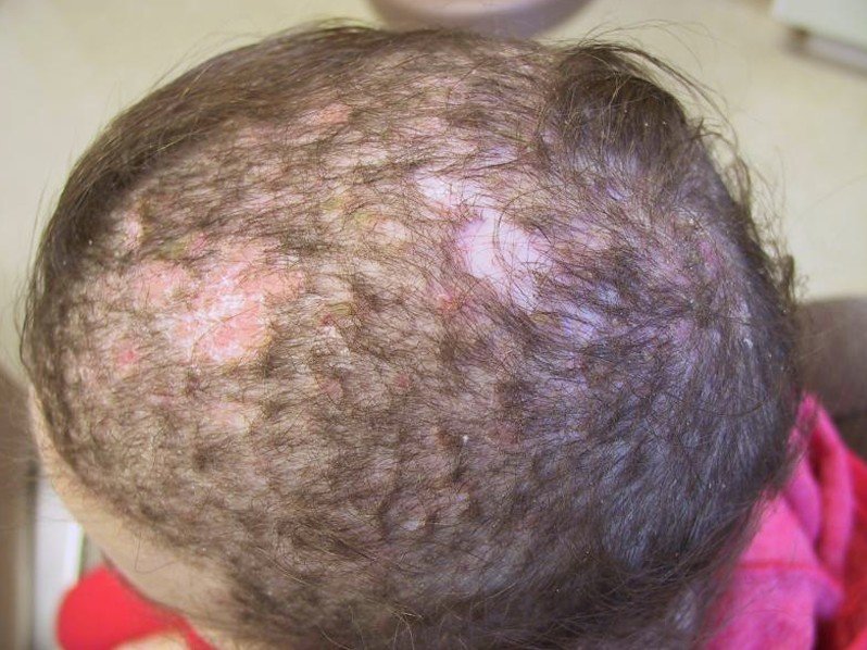
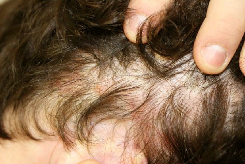
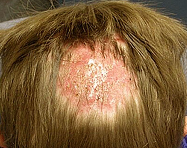
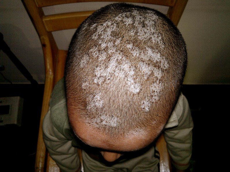
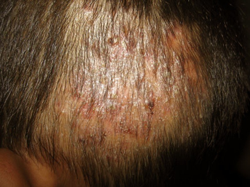
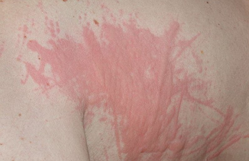
Lump on Back of Neck Pictures

Photos, Images and Pictures of Lump on Back of Neck…

lump on back of head

lump on back of head 2

No responses yet

Xiphoid Process

Feb 24 2017 Published by under General Health

What is Xiphoid Process?

Xiphoid process is a small projection of the lower part of the breastbone or known as the sternum and is made up of cartilage.

Xiphoid process is also known as metasternum, xiphisternum and xiphoid cartilage. It is located exactly at the center of the chest below the nipple line in a downward projection at the lowest portion of the rib that connects the sternum.

The xiphoid process has a sharp tip making it look like a sword. The primary function of which is in the attachment of a muscle that is significant for the breathing process which is called the diaphragm. Xiphoid process also serves as an attachment for rectus abdominus muscle and the transversus thoracis muscle.

Xiphoid process is generally an unsupported structure and unprotected by the ribcage that it is potential for an easy break off. The break in the xiphoid process is particularly potential during the cardiopulmonary resuscitation that extra care is necessary when performing CPR. The xiphoid process on the other hand is a safe landmark in determining the location of the apex of the heart when performing the cardiopulmonary resuscitation.

Xiphoid Process Structure

xiphoid processThe xiphoid process is the apparent protrusion in infant as it is basically a lump situated below the sternum notch during the early stage of life or during infancy. It is a soft and flexible cartilage during infancy and later fused to the sternum when it ossifies as the child is growing or developing. By the time an individual reaches the age of 15 to 29 years, the xiphoid process hardens and becomes bony.

The sternum is a flat and an elongated bone located in front of the chest. The xiphoid process is among the three parts that make up the sternum connected together by a cartilaginous joint known as synchondroses. The xiphoid process is triangular in shape with a sharp tip that resembles a sword.

Xiphoid process is deemed to be located at the 9th thoracic vertebra and at the T6 dermatome. The morphological variations of the xiphoid process tends to be inherited where in some people it can be bifurcated or split into two while it can be perforated to others. The variances in morphology of the xiphoid process on the other hand are not detrimental to the overall health status of an individual.

Xiphoid Process Pain

Xiphoid process generally does not cause any discomfort especially during the early stage of life when the process is just a protrusion of a soft lump. Xiphoid process however can have pain that can cause undue pain. Pain of the xiphoid process can be caused by several factors and early treatment is necessary to prevent serious problems and further discomfort.

Symptoms

The Xiphoid process syndrome is a pain or discomfort in the xiphoid process. The pain and discomfort is usually felt at the lower region of the sternum. Xiphoid pain is not a common condition and the symptoms are usually intermittent that the syndrome is usually not distinguished right away and only after a scrupulous medical examination can xiphoid process pain can be confirmed.

The symptoms of xiphoid process pain are often intermittent that it is rather difficult to determine the condition at first and is sometimes mistaken for other medical conditions. The pain in the xiphoid process can make an affected individual experience the following common symptoms:

  • Pain, tenderness and discomfort in the chest and chest area
  • The shoulders can also have pain, tenderness and discomfort
  • The back can also have pain, tenderness and discomfort
  • Pain, tenderness and discomfort can also be felt in the epigastric area
  • Nausea and vomiting may also be experienced
  • Abdominal pain

Causes

Xiphoid process pain can be due to several factors although at times the pain is difficult to pinpoint directly to the xiphoid process. The xiphoid process is generally an unsupported bone and unprotected by the rib cage. This location of the xiphoid process makes it highly potential for damages or break. In general, however, xiphoid pain is often caused by undisruptive activities such as lifting of weights and other heavy objects and can also be triggered by bending.

The causes of xiphoid pain can be caused by the following:

  • Lifting of heavy weights and objects
  • Repetitive and strenuous bending
  • Eating of large meal
  • Medications
  • Certain medical conditions or diseases

Cardiopulmonary resuscitation is an emergency procedure that is generally carried out to revive an individual who has suddenly stopped from breathing. Cardiopulmonary resuscitation basically aims to save lives. The xiphoid process however is generally at risk for break or fracture during the process of cardiopulmonary resuscitation where compression technique is applied to facilitate restoration of breathing. The break or fracture of the xiphoid process due to its unprotected and unsupported location is highly potential. A hard blow in the chest can also cause fracture of the xiphoid process which in turn can cause pain.

Treatment

Xiphoid process pain is often a harmless condition that usually does not cause fatality. The danger of fracture in the xiphoid process however is the involvement of the other internal organs which can get punctured by the fragments of the bone of the xiphoid process. Treatment is generally necessary and pain in the xiphoid process should not be ignored.

The treatment for xiphoid process pain depends on the cause of the pain. It is especially important to consult a doctor to identify the cause of pain that is tantamount to the proper treatment method.

Lump on the Sternum

The sternum is essential for the attachment of the ribs and which runs vertically along the midline of the upper portion of the body. The structure of the sternum basically starts at the sternal notch or the base of the throat while it ends at the bony and cartilaginous protrusion known as the xiphoid process.

The lump on the sternum is often not due to any serious medical condition such as growth of tumor. For most of palpable lumps on the sternum, it is basically due to the xiphoid process. It does not mean however that palpable lump on sternum should be ignored. It is still necessary to consult a doctor especially when one suspects otherwise.

No responses yet

Tinea Capitis

Feb 23 2017 Published by under Diseases and Conditions

What is Tinea Capitis?

Tinea capitis is a fungal infection involving the superficial layer of the scalp and the hair shafts. It is also termed as ringworm of the scalp, scalp ringworm and ringworm of the hair.

Tinea capitis is a mild dermatophytosis affects the scalp and the hair shafts and is clinically presented as single or multiple patches of skin. This skin condition is commonly known as ringworm of the hair although the worm has nothing to do with the condition. Tinea capitis may occur as a scaly and non-inflammatory dermatosis resembling the appearance of a seborrheic dermatitis. It may also occur as an inflammatory disease clinically presented with scaly erythema associated with hair loss.

Tinea capitis is known to affect people from all parts of the world without racial predilection. The infection is found to be prevalent in pre-school children affecting both boys and girls although it is found to have a slight prevalence among boys.

The fungi that can cause the infection vary according to geographical region. There are at least eight species of the fungi that can cause tinea capitis and all are generally termed as dermatophytes. Tinea capitis is not a serious or life-threatening condition but it tends to become chronic and linger until adult life. The severity of the infection, however, depends on the site of infection. Tinea capitis on the other hand is a contagious skin condition which can be spread from either a direct or indirect contact with an infected person. Direct contact with an infected animal is also another way of spreading the disease.

Tinea Capitis Symptoms

Tinea capitis is generally marked by the formation of itchy and scaly patches on the skin surface of the scalp and on the hair shafts. The section of the hair that is affected will eventually break off and will leave the scalp scaly and reddish. The affected area will also create a bald spot which will gradually increase in size if no treatment is applied.

Tinea capitis is clinically presented in several ways.

  • Black dots – describes the tiny capitis as scaly where the hair has broken away from the surface of the scalp.
  • Dry scaling – is tinea capitis similar to the appearance of dandruff with hair loss as if eaten by an insect
  • Kerion – describes tinea capitis as severely inflamed and resembles an abscess
  • Favus – is characterized by matted hair with yellowish crusts

Tinea capitis can also appear as sections of bald spots with smooth texture while there are cases of tinea capitis which remains asymptomatic and is presented only with mild scaling.

The common signs and symptoms of tinea capitis include the following:

  • Growth of single or multiple scaly patches on the surface area of the scalp or in the areas where the hair has broken off
  • The affected area is somewhat reddish or grayish in color and is scaly
  • The skin patches may have black dots in the area where the hair has broken off
  • The hair is characteristically brittle that it can be easily pulled out
  • The scalp may be tender or may have pain
  • Swollen lymph glands may occur at the back or on the sides of the neck
  • Tinea capitis may also result to an allergic or id reaction

Tinea Capitis Causes

Tinea capitis is the result of infection from any of the several variants of dermatophytes. These dermatophytes are fungi that invade the outer layer skin of the scalp.

Tinea capitis is the consequence of an infection from either a Trichophyton or a Microsporum. Both are genus of fungi that can cause tinea corpus and other fungal infections of the skin. The source of fungi that can cause tinea capitis can be anthropophilic, zoophilic or geophilic.

Microsporum canis is one of the most common fungi dermatophyte that can cause tinea capitis. The source of this fungus is zoophilic or which that is normally found in animals. Microsporum canis is often found in kittens.

Trichophyton tonsurans is the anthropophilic source of fungi and is among the most common cause of tinea capitis. This dermatophyte is spread from one person to another as the fungi generally favor humans over animals.

Tinea capitis can also be caused by fungi that thrive in the soil or the source is rather geophilic. This, however, seldom happens.

Common zoophilic fungi that cause tinea capitis are the following:

  • Microsporum nanum
  • Trichophyton verrucosum
  • Microsporum distortum
  • Trichophyton mentagrophytes var. equinum

Anthropophilic fungi causing tinea capitis is the following:

  • Trichophyton rubrum
  • Trichophyton violaceum
  • Trichophyton megnini
  • Microsporum ferrugineum

Tinea capitis is also classified according to the manner the fungus has infected the hair shaft.

Endothrix infection

Endothrix infection is characterized by the infiltration or filling in of the hair shaft with the fungal spores and branches. This type of infection is commonly caused by Trichophyton tonsurans, Trichophyton soudanense and Trichophyton violaceum.

Ectothrix infection

Ectothrix infection is described with infection covering only the outside of the hair shaft. This type of infection is commonly caused by Microsporum canis, Microsporum ferrugineum.

Tinea Capitis Pictures

Collection of Photos, Images and Pictures of Tinea Capitis…

tinea capitis pictures

tinea capitis pictures 2

tinea capitis pictures 3

tinea capitis pictures 4

tinea capitis pictures 5

tinea capitis pictures 6

Tinea Capitis Treatment

Tinea capitis is generally treated with an antifungal medication given orally. Griseofulvin is the drug of choice for the treatment of tinea capitis. It acts by stopping the fungal cell from dividing and is especially effective when the infection is caused by Microsporum canis. It is generally recommended for children as the medicine is well tolerated. Griseofulvin has been in use since 1958 although this drug does not kill fungi right away and treatment is necessary to be taken for several weeks to several months.

Terbinafine, Lamisil is the new drug of choice for treating children with tinea capitis. This drug was approved in the year 2007 and is prescribed for children ages 4 years and above. This drug is also to be taken for several weeks.

Is Tinea Capitis contagious?

Tinea capitis is a highly contagious disease which can be spread through the following:

  • Direct skin-to-skin contact with an infected person
  • The fungi causing tinea capitis can be acquired through contact with a contaminated surface such as sharing of hair brushes and comb
  • The fungi of tinea capitis also thrive in animals that the infection can be spread through petting or coming into close contact with an infected animal

No responses yet

Crusted Scabies

Feb 22 2017 Published by under Diseases and Conditions

What is Crusted Scabies?

Crusted scabies is a severe and contagious variant of scabies. The disease is characterized by the uncontrollable proliferation of the mite that causes the scabies. This variant of scabies is distinctive from the regular scabies wherein the human skin is infected with millions of mites as these mites literally reproduced in the skin.

Crusted scabies is also referred to as “Norwegian scabies” which was so called due to the outbreak that occurred in Norway during the year 1800. The disease was first recognized by the two physicians named Dr. Danielssen and Dr. Boeck who both recognized the disease among lepers. The name Norwegian scabies has nothing to do with the disease except that the outbreak was first recognized in the country. It is now an outdated name and the disease is now referred to as Crusted Scabies.

Crusted scabies is a highly contagious disease and is marked by crusted lesions. It usually affects people who are immunocompromised and those who have weakened immune system due to an existing condition.

The infestation of the mite that causes the crusted scabies often takes a portion of the body where it will proliferate dramatically. The web space between the fingers and the toes, the areas under the arms and groin are the usual place for infestation of the mite where it will continuously reproduce.

The regular scabies usually has a dozen or more of scabies that will invade the body at the same time whereas in crusted scabies, millions of the same mites that caused the regular scabies will take infest the body. The contagious property of crusted scabies is not the only issue but also the secondary infections. The affected person is more prone to secondary infections when their skin is left vulnerable due to incessant scratching and break from the skin as the result of crusting.

Crusted Scabies Symptoms

The classical scabies is hallmarked by the presence of itching but in crusted scabies which is a variant of classic scabies, no itching is usually present or it may be minimal in case there is itchiness. Crusted scabies has no acute eruption but is rather gradual in onset and the progression is rather insidious. The onset of symptoms usually takes about 3 to 6 weeks from the time of infestation.

It is rare for crusted scabies to cover the entire body. The onset is usually limited to the scalp, face, finger, sole of the foot and genitalia. The appearance of the scabies may be horny plaques or may be erythematous. The plaque is often parakeratotic crust with varying thickness of about 3mm to 15mm. Crusted scabies has a porous appearance when the crusts are removed. When the crusts are also removed, it will reveal the undersurface which rather has a smooth texture, moist and reddish in color.

Crusted scabies appears in multiple layers of skin that are rather thick and crusty. It is generally an unsightly appearance that the self-esteem of the patient is usually compromised due to significant embarrassment brought by the unappealing appearance of the crust.

Itchiness is often not present in crusted scabies or may be minimal despite the fact that millions of mites have infested the skin. This is what makes crusted scabies different from the classic scabies which is generally noted with itchiness. The formation of the crust may also result to deformities of the affected area of the body such as the fingers and the toes. The skin on the face may also flake off in crusted scabies while significant hair loss may also occur. Crusted scabies may also be in the form of psoriasis dermatitis or may assume a wart-like appearance. The finger nails are usually discolored and thickened while the nails may have subungual hyperkeratosis.

Crusted scabies respond to treatment gradually unlike the classic scabies. The disease may or may not respond well to treatment that it is sometimes treated in combination with other methods of treatment.

Crusted Scabies Pictures

Photos, Images and Pictures collection of Crusted Scabies…

crusted scabies pictures

crusted scabies pictures 2

crusted scabies pictures 3

crusted scabies pictures 4

crusted scabies pictures 5

crusted scabies pictures 6

Crusted Scabies Causes

People at risk for crusted scabies are the elderly, people who have poor immune system and neurologic diseases and those with mental incompetence. HIV or AIDS patients are also prone to acquire crusted scabies.

Crusted scabies is an extremely contagious variant of scabies. The disease can be transmitted via direct or non-direct contact with the affected individual. Close body contact such as hugging and sexual contact with an infected person is a direct transmission of the disease. Sharing of personal belongings is also included in the direct method of transmitting the harmful pathogen that causes the crusted scabies.

Crusted scabies is generally implicated on the infestation of a certain mite which is the same mite that causes the classic scabies. Sarcoptes scabiei var hominis is an obligate parasite that thrives in the stratum corneum. The main perpetrator of crusted scabies is the female mite that burrows in the surface of the skin. The mite has a rounded body with a length of approximately 400 microns. It has 4 legs with the front composed of two pairs of antenna or suckers. The mite can survive for 30 days in the burrows and feeds on melted human tissues. 2 to 3 eggs are being laid by the female mite daily and which usually takes 3 to 4 days before the eggs can hatch. In crusted scabies, the number of mite increases drastically as a result of uncontrolled proliferation.

It is believed that the antigen of the mite that stimulates the immune response is contained in the saliva of the mite. When a person is immunocompromised, it cannot control the proliferation of the mite thus, the development of crusted scabies.

Crusted Scabies Treatment

The primary aim of treatment of crusted scabies is to isolate the patient to control or prevent the spread of the disease which might result in an outbreak. Necessary and adequate precautions should be done by people who are in contact with the patient including the medical staff who will attend to the patient.

Treatment of crusted scabies has been difficult, often due to the present status of the immune system of the patient.

Topical Medication

Various topical medications can be used to effectively treat the scabies. Topical scabicidals is the preferred treatment although removal of the crusted skin is necessary to allow the medication to seep in and to bring down the number of the mites.

Antibiotics

Broad spectrum antibiotics are indicated for crusted scabies with secondary infection especially those associated with septicemia. Another form of treatment for crusted scabies is directed towards the relief of symptoms such as dry skin and itchiness.

Ivermectin is the drug of choice for crusted scabies in the aim of eliminating the mite in the body. This drug is taken orally and acts highly against the mite although it is safe in human. Ivermectin is also used in combination with other scabicidal to effectively eliminate the mite while effectively treating the patient.

No responses yet

Dermographism

Feb 22 2017 Published by under Skin Conditions

What is Dermographism?

Dermographism is among the most common type of urticaria. This skin condition is characterized by wealing of the skin in a rather exaggerated manner. Dermographism is also referred to as dermatographic urticaria, dermatographism and skin writing. The term skin writing generally describes the characteristic of the disorder where a person affected can literally write on their skin as a result of the exaggerated response from a stimulus where a slight scratch can create a raised weal resembling a hive.

Dermographism is prevalent among young adults although it can occur at any age with the peak onset on the second to third decade of life. It can affect both the male and female gender and is found to be more common in female children. Prevalence in racial variance is unclear as most people with dermographism usually ignore the incidence.

Dermographism is considered to be the most common form of physical urticaria. Urticaria on the other hand is described as a distinct skin rash that often results from an allergic reaction. The rash or the hives produced in urticaria are very similar to the appearance of the rash in dermographism which is a raised bump that is itchy and pale red in color.

Dermographism is not a serious and life-threatening disease. It is not a contagious skin disease although it is a life-long disease. The disease however has a good prognosis, especially the simple dermographism which is asymptomatic. People with dermographism may remain healthy despite the onset of the hives except for those who are already sick prior to the onset of dermographism. Dermographism on the other hand lingers for several months to several years and may occur sporadically. It will, however, resolve on its own after it has lingered for several years.

Dermographism Symptoms

Dermographism tends to occur sporadically and an individual may suffer from this for some period of time. The disease however, is benign and usually resolves on its own after some time or after it has lingered for several months to several years.

The signs and symptoms of dermographism usually take a few minutes to occur after the skin has been scratched or rubbed. A friction from the clothing such as from the cuffs or collar of the clothes can initiate the onset of hives. There are cases however, that the onset will take a few days before it develops although this is rather rare.

The onset of dermographism may be asymptomatic or may be symptomatic. Simple dermographism is usually asymptomatic and wealing occurs without significant itchiness. Symptomatic dermographism is usually associated with itching which is often worse at night. The symptoms can be irritated further by heat such as from a hot shower or it can be aggravated with minimum pressure such as from light scratching. Emotional stress, exercise and other forms of stress are also considered to aggravate dermographism.

Dermographism can affect the entire skin surface of the body with the scalp and genitalia seldom involved in the process. The weal of dermographism is not the typical weal which is roundish but instead has a straight line. The lines of the hives in dermographism are elevated with pale-red to red in color. Inflammation and swelling are also present and itching may also be associated especially in symptomatic dermographism.

The response to stimuli of dermographism is exaggerated where a firm stroking or scratching of the skin will result to a prominent red line associated with swelling down the line including a red flare up in the adjacent skin. Itching may also be present in some cases while pain is often absent.

Dermographism also has different forms although these forms seldom occur and such are:

Red dermographism

It produces a more palpable wealing which is easily established with the stretching of the skin. This form of dermographism is easily induced by rubbing the skin. The weal formed is more prominent in the trunk. Red dermographism is thought to be associated with seborrheic dermatitis.

Delayed dermographism

Delayed dermographism takes about 3 to 8 hours from the time of dermographic response before the symptom shows up. It produces a deep red line with tender and burning weal. This form of dermographism is closely linked with pressure urticaria.

Cholinergic dermographism

Cholinergic dermographism produces a band of large erythema that is studded with small weal similar to cholinergic urticaria.

Follicular dermographism

Follicular dermographism elicits transitory, follicular and discrete papules of urticaria.

Familial dermographism

This is dermographism deemed inherited in an autosomal dominant pattern.

Dermographism Causes

The exact cause of dermographism remains unclear while several factors are being considered to contribute to the incidence. The exact mechanism of how dermographism is being provoked by the factors considered remains unknown as well.

Factors considered to trigger dermographism include the following:

Histamine

Histamine released by the mast cell in the surface of the skin is the most implicated in the onset of dermographism. The histamine is released in the absence of an antigen and as a result of the existence of a weak membrane that envelops the mast cells. The swelling of the skin affected by dermographism is believed to be due to the released histamine.

Allergic reaction

Allergic reaction to certain medication such as penicillin and other stimulants are considered to the onset of dermographism although the exact mechanism is not clearly understood.

Infection

Infection from certain microbes or harmful pathogens is believed to trigger the onset of dermographism. Helicobacter pylori, a Gram-negative microaerophilic bacterium and usually causes peptic ulcer and other digestive illnesses are also among the implicated to the onset of dermographism.

Genetic factor

The skin condition is believed to be an inherited disease which is passed on in an autosomal dominant pattern. A genetic factor is strongly suggested to the onset of dermographism although this factor still remains unclear.

Dermographism Pictures

Collection of Pictures, Pics and Photos of Dermographism…

dermographism pictures

dermographism pictures 2

dermographism pictures 3

dermographism pictures 4

dermographism pictures 5

dermographism pictures 6

Dermographism Treatment

Dermographism is a benign skin condition without a known cause. It is a skin condition usually come and goes and lingers for several months to several years. The disease usually resolves without treatment after several years.

Antihistamines such as diphenhydramine, cetirizine and fexofenadine are found to be beneficial in the relief of symptoms. Treatment is generally given in severe cases or if the disease proved to be bothersome to the patient.

No responses yet

Soapy Taste in Mouth

Feb 20 2017 Published by under Oral Health

It is an odd experience to feel a soapy taste in mouth, right? Yet some people pass through this ugly experience without knowing what to do or how to salvage their situation. This caption may be catching your attention either because you are suffering the same thing or you know someone who does.

Whatever is the case, soapy taste in mouth is not a palatable story to tell and sufferers always wish they could wake up from their nightmares. Like a lot of other health challenges, this condition has a solution. And that is what we are going to show you in this post, and we believe that this post will hopefully solve your problem once and for all.

What Causes Soapy taste in Mouth?

soapy taste in mouth causes and treatment

Soapy taste in mouth is caused by several factors. Below are some of the reasons why you notice this funny taste.

Poor oral hygiene

Sometimes, neglecting our oral hygiene could create its own avalanche of problems. Poor oral hygiene leads to the tongue turning white with plague. The taste of plague can be very bad, including it tasting like you have eaten soap.

Medication

Some medications can produce untoward effects that are really nasty to the mouth. So, soapy taste in mouth could just be a side effect of some medications being used for treating certain diseases or handling other health challenges. One good example of drug that can cause soapy taste in mouth is Telavancin, which is used in the treatment of pneumonia. Some medications used in treating diabetes also cause soapy taste in the mouth.

Food contamination

Nothing can give you this taste more than when your food or drink is contaminated by soap. If you wash your dishes and fail to rinse them properly with enough water, you could have soapy taste in the mouth. This is one of the reasons why you have soapy taste after drinking coffee. In addition, if the soap directly contaminates your drinks or juice, you could also feel a soapy taste. Sometimes, if the quantity is much, you could have the bad taste after vomiting. Your body could equally react to it and this leads to diarrhea. So, watch what you eat or drink.

Sodium fluoride poisoning

According to the Centers for Disease Control (CDC), a soapy taste in mouth could be the first sign that there’s sodium fluoride poisoning. Because this crystalline salt also has sodium component, soapy taste in the mouth could be accompanied by a salty taste.

Other symptoms that can be present when there is excessive consumption of sodium fluoride include, vomiting, collapse, diarrhea, pallor, numbness of the mouth, dilated pupils and shock.

Sodium fluoride poisoning generally comes from being treated with so much fluoride by the dentist, drinking tap water that has too much fluoridation, contamination of foods such as flour, cake mixes, dry milk and baking powder.

Pregnancy

This symptom can be a sign of early pregnancy. A lot of women experience it in their first trimester of pregnancy and the reason for this is not fully understood. In some cases, the women are able to find a simple solution to the problem themselves by eating foods with sour taste such as lemon juice. Taking a lot of water can also help clear off the taste. Pregnancy can also be accompanied by symptoms like dizziness and sometimes heartburn.

Smoking

Soapy taste in mouth can also be as a result of cigarette smoking. One can feel this soapy taste after smoking due to the high level of contaminants in cigarette.

How to Get Rid of Soapy taste in mouth

Reduce tap water

If the symptoms of sodium fluoride poisoning are mild, you could stop them by reducing the amount of tap water you consume daily or you could completely stop taking tap water. That will put an end to the symptoms. You can check for the level of fluoridation in your tap water and have your water company reduce the level if it is too high.

Reduce dental fluoride treatment

If the problem is as a result of dental office fluoride treatment, the level should be adjusted downward. In addition, you can use toothpaste that has no sodium fluoride.

Rinse your dishes properly

Sometimes, soapy taste in the mouth comes from improperly rinsed dishes containing traces of soap. It is, therefore, advisable to wash and rinse your dishes thoroughly with clean water to get rid of those traces of soap. This will put an end to the soapy taste.

Prevent food contamination

Since sodium fluoride poisoning can come from the insecticides used in the home, you need to stop using insecticides in controlling insects at home. Instead, you can fit your doors and windows with nets. Besides, you can keep your surroundings clean in order to prevent the insects from breeding around your home.

See the doctor

It is possible that the tips mentioned above may not solve your problems completely, especially if the level of poisoning is high. If that is the case, you need to see your doctor immediately for further investigation and treatment.

Soapy taste in mouth after eating

If the food you eat contains sodium fluoride, you are likely to experience soapy taste in mouth after eating as a result of ingesting the chemical. Like we mentioned earlier in the course of this write-up, contamination of foods could come from poorly rinsed utensils and by the use of insecticides which contain sodium fluoride.

Conclusion

Soapy taste in mouth is caused by several factors. Before you think of the right treatment for the condition, you first need to identify the actual cause. We have outlined the various causes of this funny experience and we advise that you try out the different solutions we provided.

However, if the solutions here fail to address your condition or if the symptoms are getting worse, there is need for you to see the doctor for the right tests and treatments to be carried out. A stitch in time, they say, saves nine. Never wait until the situation gets completely out of hands.

No responses yet

Erythema Marginatum

Feb 19 2017 Published by under Diseases and Conditions

What is Erythema Marginatum?

Erythema marginatum is a rare skin condition characterized by the onset of distinctive rash which is being associated with rheumatic fever. Erythema marginatum is marked by the presence of pink rings in the torso or upper portion of the body and the inside surface of the extremities and usually spares the face and the outward portion of the extremities. Erythema marginatum is also marked by a distinct rash that appears and disappears over the course of several months.

Erythema marginatum is more common among children than in adults, but may affect people from all age levels. It affects both the male and female gender, although there is a slight prevalence among the male gender. The color of the rash is usually pinkish to reddish, which is more obvious among fair skinned individuals, but rather difficult to spot among dark skinned individuals. The rash also neither itches nor creates a burning sensation. A few numbers of cases of rheumatic disease are being associated with the onset of erythema marginatum.

Erythema marginatum is more of a symptom signifying an underlying condition and it is not considered a disease or a disorder. It is a rare skin condition that seldom causes discomfort and is neither a life-threatening condition. It is believed to be triggered by several factors including an infection and allergic reaction.

Erythema Marginatum Symptoms

Erythema marginatum is marked by the development of macular rings that has a pale core or more like the appearance of a doughnut. The macula has irregular borders which are due to the gradual development of the macula. The rash or the lesion itself is rather elevated while the center of the ring is rather flat and small and is usually clear. The color of the ring is pinkish to reddish while the core of the ring is pale or clear.

The rash of erythema marginatum generally occurs in the torso and the inner surface of the extremities. It usually spares the face and the outside portion of the extremities. The size of the rings varies ranging from 0.5 cm to 8 cm in diameter. Overtime, these rings will increase in size and eventually spread and which will later assume an irregular shape or may have a displaced margin. The rash initially develops in the lower portion of the trunk then gradually disappears as it moves upward towards the upper portion of the trunk where it will become more visible.

Erythema marginatum causes no discomfort and often does not cause itchiness and burning sensation. The rash is however getting worse when aggravated by heat, such as in hot showers or extreme heat in temperature and in hot climates. The aggravated rash assumes a reddish discoloration which usually lightens up as soon as room temperature is achieved.

Erythema marginatum is also divided into two types which are based on the characteristics of the rash and the location of their onset.

Erythema marginatum rheumaticum

Erythema marginatum rheumaticum occurs in the superficial layer and is often asymptomatic. This type of erythema marginatum is being associated with rheumatic fever. The rash is usually seen all over the skin of the trunk and the inner surface of the limbs. The exact cause of erythema marginatum rheumaticum is unknown and is also deemed a type of gyrate erythema.

Erythema marginatum perstans

Erythema marginatum perstans is presented with lesion distributed on the entire surface of the skin of the arms and the legs. It can also extend to the other areas of the body with the palms of the hands and soles of the feet spared from the rash distribution. The deep layer of the skin may also get affected by the rash. The incidence of erythema marginatum perstans is being linked to an existing underlying condition which triggered the eruption of the rashes.

Erythema Marginatum Causes

The onset of erythema marginatum is implicated on the inflammation of the blood vessels as triggered by the lymphocytes histiocytes. The mechanism in the alteration of the epidermal layer of the skin resulting to erythema marginatum is not clearly understood. The exact cause of erythema marginatum therefore remains vague.

Rheumatic fever

This is the most associated disease to the onset of erythema marginatum and basically involving Group A streptococcus that causes the complication of rheumatic fever.

Although erythema marginatum has no identified cause, several factors are being considered that triggered the onset and such factors include the following:

Rheumatic fever is the most associated with the incidence of erythema marginatum. The disease is a bacterial infection particularly of Group A streptococcus. The onset of erythema marginatum is deemed a complication of rheumatic fever and is also considered as among the clinical presentation of the disease. Most cases of erythema marginatum are found to be associated with rheumatic fever.

Allergies

Allergy is another consideration in erythema marginatum where the allergic reaction from various stimuli is believed to trigger the inflammation of the blood vessels that later causes the rashes in erythema marginatum.

Lyme disease

This is a disease caused by an infection with Borrelia burgdorferi. The harmful pathogen is transmitted to humans through bites from infected ticks. Erythema marginatum is present as part of the symptoms of Lyme disease.

Infections

Infection from various causative agents can result to eruptions of rashes which may include erythema marginatum. Candida albicans and Escherichia coli are among the infectious agents that can trigger erythema marginatum.

Adverse reaction

Adverse reaction from certain medications can trigger the eruption of rashes including erythema marginatum.

Malignancy

Malignancy such as breast cancer, leukemia and other life-threatening diseases are believed to activate erythema marginatum where in some cases it is part of the symptoms.

Erythema Marginatum Pictures

Collection of photos, images and pictures of Erythema Marginatum…

erythema marginatum pictures

erythema marginatum pictures 2

erythema marginatum pictures 3

erythema marginatum pictures 4

erythema marginatum pictures 6

Erythema Marginatum Treatment

Erythema marginatum is deemed a symptom rather than a disease or disorder. The incidence is believed as part of the symptoms of an underlying condition. The treatment therefore, is not directed towards erythema marginatum but rather in the underlying condition that influenced the eruption of rashes in erythema marginatum.

Erythema marginatum usually resolve without treatment although the rash may appear or disappear for several months to several years. Erythema marginatum involved in rheumatic fever is basically addressed with antibiotic treatment which is intended for rheumatic fever and not directly to erythema marginatum.

No responses yet

Stasis Ulcer

Feb 19 2017 Published by under Diseases and Conditions

What is Stasis Ulcer?

Stasis ulcer is a skin condition characterized by the ulceration of the skin that is deemed to result from venous insufficiency due to a dysfunction in the venous valve.

Stasis ulcer is also known as venous ulcer or varicose ulcer. Stasis ulcer is the most common ulcer in the lower extremities. It is a chronic skin condition that is more common in women and older adults and is also common among obese individuals. Stasis ulcer is open and shallow ulcer that usually takes 4 weeks to 6 weeks before it can heal.

Stasis ulcer occurs when there is the presence of valve incompetency in the veins of the lower leg. The valve incompetency causes the blood to leak into the superficial veins during contraction of the muscle of the calf and thus preventing the blood from flowing upward.

Older people, women and obese individuals are at high risk for developing a stasis ulcer. The disease is known to occur all over the world regardless of racial group. It usually takes a long time for stasis ulcer to heal and is rather costly while potential for recurrence is highly possible. Proper care and prevention of recurrence is necessary to avoid further complications such as gangrene which can cause irreversible tissue damage.

Stasis Ulcer Symptoms

Stasis ulcer often occurs in bony prominence such as in the medial distal leg and in the ankle. The onset can be painful although some patient experiences no pain. The ulcer in the skin is unsightly and discomfort is common that it can affect the quality of life. The pain is particularly felt when assuming a standing position or when standing for a prolonged period of time. Elevating the affected is beneficial in relieving the pain and in reducing the swelling.

Stasis ulcer is characterized by large and shallow skin ulcer in the medial distal leg or in the ankle. The incidence may also be associated with signs and symptoms of venous hypertension such as the onset of venous flare and varicose veins. The wound is sunken and shaped asymmetrically and the borders are well defined. The skin surrounding the ulcer is firm and hard which makes the affected feel hard causing a heavy feeling in the leg.

The skin of the affected area has a reddish color and is scaly. An enlarged and swollen vein may also be seen. Pus or yellowish-whitish exudates may also happen signifying an onset of infection. The skin ulcer may also have a foul smelling discharge which can be disturbing.

Onset of high fever may also be experienced which signifies infection. The pain is also worsening and the ulcer can be seen with a greenish discharge that emits a foul smell.

The presence of signs and symptoms of stasis ulcer should prompt the affected individual to seek medical consultation. It is also advisable to seek a specialist when infection is present. Stasis ulcer does not resolve on its own that medical treatment is necessary to address the condition and to prevent further medical complications.

Causes

The exact cause of stasis ulcer is still unclear. The etiology is thought to be the result of increased pressure in the veins of the lower leg subsequently leading to blood leaking out of the skin. The blood pooling on the skin surface causes the skin to break, leading to ulceration. The increased pressure of blood in the lower leg is the consequence of an incompetent valve. When valve incompetency occurs, the deoxygenated blood is prevented from flowing back to the heart and is rather brought back to the valve that is damaged where it will later collect in the veins of the lower leg.

Stasis ulcer involves the vein and the valve. The vein is responsible for carrying deoxygenated blood to the heart then to the lungs. The valve on the other hand is responsible in preventing the blood from flowing backward. The blood flow along the veins is helped by the muscle contraction.

The lower leg is composed of a calf which is the pumping function of the leg to help the blood from flowing along the veins. Both the venous hypertension and valve incompetency are the primary mechanism in the formation of stasis ulcer.

Although there is no exact cause that can be pinpointed in stasis ulcer, several factors are considered that increases the risk for developing the disease.

The factors considered at the onset of stasis ulcer include the following:

Age

Age is considered to increase the chances for stasis ulcer. Older people are predisposed to the incidence which could be related to poor peripheral circulation as part of the normal aging process. The risk for stasis ulcer also increases as one advance in age as well. Age advancement generally makes it harder for an individual to be moving about.

Obesity

Obesity greatly increases the chances for stasis ulcer. This could be related to the increased risk for pressure buildup in the vein. The pressure buildup could be associated with the heavy weight or the weight bearing of an obese individual.

Varicose vein

Varicose vein can damage the valve that prevents the blood from flowing backward into the vein. This increases the risk for stasis ulcer when the blood collects in the vein later causing damages in the skin.

Cigarette smoking

Cigarette smoking can cause the blood vessels to constrict thereby interrupting the blood flow and in turn increases the pressure and damages the vein.

Treatment for Stasis Ulcer

The primary goal of treatment in stasis ulcer is to promote wound healing while reducing edema and preventing recurrence.

Various treatment methods are available for stasis ulcer and this includes the following:

  • Compression bandaging
  • Leg elevation
  • Debridement and cleaning of the ulcer
  • Wound dressing
  • Antibiotics
  • Aspirin
  • Topical steroids
  • Pentoxyfilline
  • Surgical procedures such as skin grafting

Stasis Ulcer Pictures

Collection of pics, images and pictures of Stasis Ulcer…

stasis ulcer

stasis ulcer 2

stasis ulcer 3

stasis ulcer 4

stasis ulcer 5

Stasis Ulcer Prevention

Stasis ulcer often recurs after it has resolved. To prevent recurrence, wearing of compression stocking is recommended. It is recommended to be worn during the daytime over the period of five years from the time the ulcer has healed.

A diet change is necessary for those who have heavy weight. Heavy weight individuals should also consider losing weight.

Cigarette smoking is considered a factor in the incidence of stasis ulcer. It is therefore recommended to stop from smoking especially in predisposed individuals.

Exercises prevent the onset and progression of stasis ulcer. People who are often immobile are at greater risk for stasis ulcer. Exercising and mobilizing will help the blood from flowing in the legs properly.

No responses yet

Leukocytoclastic Vasculitis

Feb 18 2017 Published by under Diseases and Conditions

What is Leukocytoclastic Vasculitis?

Leukocytoclastic vasculitis is a variant of vasculitis characterized by the inflammation of the tiny blood vessels of the body. The disease often leads to damages in the blood vessels resulting in bleeding and damages of nearby tissues.

Leukocytoclastic vasculitis is also referred as hypersensitivity vasculitis or hypersensitivity angiitis. The disease is presented by small spots of discolorations known as purpura. The spot is caused by the bleeding beneath the skin which in turn causes the reddish or purplish discoloration of the skin.

Vasculitis refers to a group of disease characterized by inflammation of the blood vessels leading to damages in the blood vessels. It is an inflammatory disorder that involves both the veins and the arteries. There are various forms of vasculitis with symptoms that vary significantly and with different duration and severity. The severity of vasculitis ranges from mild to life-threatening and may leave a life-long or permanent damage to the body.

Leukocytoclastic vasculitis is deemed an uncommon disorder. It can affect many people without racial predilection. Men and women are equally affected although some studies revealed a slight prevalence of leukocytoclastic vasculitis among men. The disease can occur at any age with children below the age of 10 years often affected with Henoch-Schonlein purpura.

Leukocytoclastic vasculitis has a good prognosis except when the GI tract, kidney, heart, lungs and the central nervous system are affected. Leukocytoclastic vasculitis not only involves the skin but can also involve the internal organs and the joints. The involvement of the internal organs and the joints are generally a concern which necessitates an immediate medical intervention as this can be life-threatening.

Leukocytoclastic Vasculitis Symptoms

Leukocytoclastic vasculitis is presented by the development of rash in the lower limbs, trunk and buttocks. The inflammation of the tiny blood vessels occurs beneath the skin although there are cases where the internal organs and the joints are also involved in the process of the disease.

A palpable purpura is the most common complaints of patients. The purpura is a reddish or purplish discoloration in the skin that resulted from bleeding under the skin. Purpura is a small spot that is elevated from the skin. The spots may merge together to form a larger spot, although in some cases it may remain small. The purpura may tend to obstruct the flow of blood in the skin leading to necrosis or death of tissue.

The size of the purpura is around 1mm to 3mm in diameter. The lesions can grow bigger when they merge together. In some cases, the purpura may form bullae which can later develop into ulceration. Any areas of the skin can get affected although the common area of development is the lower legs and the areas with trauma and tight fitting clothing. The purpura may not also be palpable although this seldom happens.

In rare cases, urticarial lesions may occur along with leukocytoclastic vasculitis. Urticarial lesion may persist longer than a day and may leave pigmentation. Most patients with the onset of urticarial lesion complain of burning rather than itching.

Leukocytoclastic vasculitis may be presented with pain, itchiness and burning sensation but there are cases where leukocytoclastic vasculitis remains asymptomatic. Some patients with leukocytoclastic vasculitis may experience systemic symptoms such as fever, stomach pains or stomach upset and joint pains. Blisters may develop or there may be the presence of ulceration or open sores. Necrosis is potential especially when the purpura has obstructed the blood flow in the skin leading to deprivation of blood supply in the tissue.

Leukocytoclastic purpura associated with an underlying condition and the signs and symptoms may be reliant or not on the disease process. The signs and symptoms may include the following:

  • Onset of fever
  • Flu-like symptoms
  • Chronic cough
  • Abdominal pain or stomach upset
  • Presence of blood in the stool
  • Diarrhea
  • Weakness
  • Myalgia or arthralgia

Leukocytoclastic Vasculitis Causes

The majority of cases of leukocytoclastic vasculitis are idiopathic or the cause is unknown. There are identifiable causes however that are being implicated to the onset of leukocytoclastic vasculitis. Leukocytoclastic vasculitis is also being thought to be an error in the immune response where the very blood vessels of the body are mistaken as a foreign object and should therefore be warded off from the body.

Several drugs are considered to be the cause of leukocytoclastic vasculitis and often as an allergic reaction to certain drugs.

Antibiotics

Antibiotics are generally prescribed to inhibit the bacterial growth in the body. There are antibiotics however that can cause leukocytoclastic vasculitis often as an adverse reaction towards the drug.

Other drugs that can cause leukocytoclastic vasculitis include the following:

  • Diuretics
  • Nonsteroidal anti-inflammatory drugs
  • Orlistat
  • Letrozole

Several forms of infections can also cause the onset of leukocytoclastic vasculitis.

  • Leukocytoclastic vasculitis may occur along with bacterial endocarditis
  • Upper respiratory infection is mostly implicated in leukocytoclastic vasculitis especially an infection from beta-hemolytic streptococci
  • Viral hepatitis is also among the identifiable cause of leukocytoclastic vasculitis
  • Hepatitis C is also commonly implicated to the onset leukocytoclastic vasculitis especially with the presence of cryoglobulinemia

There are some diseases that are being associated with leukocytoclastic vasculitis such as:

Crohn’s disease

Crohn’s disease is an inflammatory bowel disease associated with the onset of leukocytoclastic vasculitis. The inflammation involves the lining of the digestive tract leading to symptoms of abdominal pain and severe diarrhea.

Ulcerative colitis

Ulcerative colitis is also an inflammatory bowel disease that causes abdominal pain and diarrhea. It has also been implicated in the onset of leukocytoclastic vasculitis.

Food additives and other substances can also cause the incidence of leukocytoclastic vasculitis.

Leukocytoclastic Vasculitis Treatment

It is important to establish the diagnosis first prior to the treatment of leukocytoclastic vasculitis. The patient has to be carefully evaluated and the cause of the disease should be identified first to determine the proper treatment necessary.

The treatment of leukocytoclastic vasculitis depends on the overall health status of the patient and the underlying cause.

The general measures for treating leukocytoclastic vasculitis include the following:

  • Elevation of the affected leg or legs or wearing of compression stockings are beneficial
  • Immediate discontinuation of the drug that causes leukocytoclastic vasculitis is required although a visit to the doctor is necessary to discuss the condition prior to discontinuation of the medicine

The pharmacological treatment of leukocytoclastic vasculitis includes the following:

  • Colchicines or dapsone
  • Antihistamine
  • Non-steroidal anti-inflammatory drugs
  • Prednisolone
  • Broad spectrum steroid
  • Immunosuppressive drugs

Leukocytoclastic Vasculitis Pictures

Collection of photos, images and pictures of Leukocytoclastic Vasculitis…

leukocytoclastic vasculitis

leukocytoclastic vasculitis 2

leukocytoclastic vasculitis 3

leukocytoclastic vasculitis 4

leukocytoclastic vasculitis 5

leukocytoclastic vasculitis 6

No responses yet

Lichen Striatus

Feb 18 2017 Published by under Diseases and Conditions

What is Lichen Striatus?

Lichen striatus is rare and benign skin condition that commonly affects young children. The cause of this skin condition is unknown and is characterized by the onset of pink papules in linear band. It is a self-limited skin disorder that primarily involves the arms and the legs but can also affect any part of the body.

Lichen striatus is a benign skin condition but can leave a temporary hypopigmentation or hyperpigmentation after it has resolved. The rash of lichen striatus often affects a single arm or leg only but may also exist in the neck and torso. It is a self-limiting disease that resolves on its own and is usually asymptomatic although there are some cases where mild or severe itchiness can be experienced.

Lichen striatus is prevalent among young children specifically between the ages of 5 years to 15 years and rarely affects infants and adults but the disease however, can affect people at any age. It can also affect people worldwide without racial predilection. Lichen striatus is found to be prevalent among girls approximately 3 folds compared to incidence in boys although there are some reports revealing an equal distribution of the disease in both boys and girls.

Lichen striatus occurring in adults is more aptly termed as Blaschkitis or acquired Blaschkoid dermatitis. This adult form of lichen striatus looks similar to eczema and is acquired. It is an inflammatory condition of the skin that is not present at birth.

Lichen striatus is not a life-threatening skin condition but can be disturbing due to the onset of visible rashes along the lines of Blaschko. The onset can be unappealing and it can be itchy for some although the disease is generally asymptomatic. It usually resolves without a need for treatment except in cases where the rashes are itchy and causing discomfort to the patient.

Lichen Striatus Symptoms

The hallmark of lichen striatus is the development of pink rashes in a form of a linear band where it involves the lines of Blaschko. The rashes develop as a single linear band in either arm or leg but can also develop in the neck and torso.

The lines of Blaschko are invisible lines on the skin that correspond to the pattern of developmental growth during the process of epidermal cell migration. It is different from other morphological lines of the skin as the lines of Blaschko do not correspond to the lymphatic, vascular and nervous structures of the body. It is rather a distinct line that is only made visible by several acquired or inherited diseases.

The upper spine of the body has a U-shape pattern of lines of Blaschko while the abdomen has S-shaped pattern. An inverted U-shape pattern can be found on the breast and the upper arms while the lower arms and the legs have a perpendicular pattern of lines of Blaschko.

Lichen striatus is unilateral or affects only one side of the body and solitary down the lines of Blaschko in the extremities. It can affect either of the arms and either of the legs. The development of small papules is rapid and is often asymptomatic. Pruritus is the main complaint of patients when lichen striatus becomes symptomatic. The pruritus can be mild or it can be severe in some cases. The eruption of papules usually lingers for several days to several weeks at the most.

The papules follow a linear band and may emerge in a continuous or intermittent pattern. The linear band on the other hand is composed of small lichenoid papules approximately 1 mm to 3mm in size. The colors of the papules ranges from tan, pink to skin colored while the texture may be scaly and smooth.

The linear band is about 1 cm to 2 cm wide while the length is about a few centimeters or may extend up to the full length of the extremity. In rare cases, the linear band may appear bilaterally or may be on multiple lines. In light skinned individuals, the linear band may appear as hyperpigmentation while in dark skinned individuals the linear band appears as hypopigmentation.

The nails are seldom involved in lichen striatus but is usually the only area involved and without the lesions. The involvement of the nails may occur before or after the development of linear bands in extremities or may occur along the linear band rash. The onset of lichen striatus is often limited to one nail only. Changes in nail may be seen with ridging, splitting, nail loss and pitting. The nail bed is seen with hyperkeratosis while the nail plate may have thinning or thickening or may be seen with an over curvature.

Lichen Striatus Causes

The exact cause of lichen striatus has not been established. Several predisposing factors on the other hand are being considered. Both the genetic factors and environmental stimuli are considered in the incidence of lichen striatus.

A family history of asthma and atopic dermatitis are deemed as predisposing factors in lichen striatus. This has been based on the reported cases of lichen striatus where three fourth of patients have a family history of asthma or atopic dermatitis.

The environmental stimuli such as trauma and infection are being linked to the onset of lichen striatus. The involvement of viral infection in lichen striatus is based on the elevated levels of interleukin 1-beta as revealed in the biopsy of lichen striatus specimen. The concept however has not been proven.

Pregnancy is also believed to be involved in the process of lichen striatus. It is thought to trigger the autoimmune response leading to the eruption of papules in lichen striatus.

Lichen Striatus Pictures

Pictures collection of Lichen Striatus…

lichen striatus

lichen striatus 2

lichen striatus 3

lichen striatus 4

lichen striatus 5

lichen striatus 6

Lichen Striatus Treatment

Lichen striatus is a benign skin condition that is generally harmless and non-life threatening. Treatment is not necessary and the lesion usually resolves on its own. It takes about several months before the lesions disappear. Hypopigmentation or hyperpigmentation on the other hand is common after the lesion has dissipated although only temporary.

If treatment is necessary, it is usually geared towards the relief of itchiness and dryness. Topical steroids and emollients are the usual medication for lichen striatus.

Topical steroids may help in clearing the lesion although this may take about several weeks. Emollient on the other hand is beneficial in relieving the dryness of the skin and reducing the itchiness.

No responses yet

Next »

© 2011-2019 MDDK.com - Medical Tips and Advice. All Rights Reserved. Privacy Policy
The health information provided on this web site is for educational purposes only and is not to be used as a substitute for medical advice, diagnosis or treatment.