Diseases General Health Skin Conditions

Archive for January, 2017

Hypercholesterolemia

Jan 07 2017 Published by under Diseases and Conditions

What is Hypercholesterolemia?

This medical condition involves the high cholesterol levels found in your blood. It is not an illness by itself but actually it is an imbalance of your metabolism. It can also be a factor that contributes to many other diseases, especially arteriosclerosis, which is the hardening and thickening of your arteries. Cholesterol is a natural component of all the cells in your body and is a waxy, soft, fat-like material and is something your body does need. In fact, your body makes all the cholesterol that you need so when you get extra cholesterol from the food you eat it can cause problems. Normal levels of cholesterol will range between one hundred forty to two hundred mg/dL. When it gets about two hundred forty mg/dL this is an indication of hypercholesterolemia. This medical condition can affect anyone of any age, race, or gender.

Hypercholesterolemia Symptoms

In the early stages of hypercholesterolemia there are no symptoms so it is important that you have your blood tested on a regular basis to find out your cholesterol level.

When the cholesterol level becomes high you may have these symptoms:

  • The tendons could become thick because of all the accumulating cholesterol. This is called xanthoma.
  • Having a whitish discoloration in the peripheral part of your cornea called arcus serilis.
  • Having a yellowish discoloration around your eyelids called xanthelasma palpabrum.
  • Due to pancreatitis you may have acute pain in your abdomen. This can happen when your pancreas has deposits of triglycerides.
  • Your liver and spleen can become enlarged. This may be able to be felt by your physician when they do an examination.
  • Because of the cholesterol building up in the walls of your blood vessels you could experience pain in your chest or even a stroke or heart attack.
  • Because of the narrowed or blocked vessels in your leg you may have pain in your calves when walking.

Hypercholesterolemia Causes

There are many different reasons as to while a person has such a dangerously high cholesterol level but the main culprit are have to due with your diet.

Some of these factors include:

  • Saturated fats – eating a diet that is high in saturated fats can lead to the levels of cholesterol in your blood to become very high.
  • Tran’s fatty acids – these are produced by the hydrogenation of unsaturated fatty acids which you will find in a lot of the processed foods, foods that are baked commercially, etc. This is a very important risk factor.
  • Carbohydrates – it is just coming to light that a diet that is high in carbohydrates can also lead to a high level of cholesterol and triglyceride levels. This is especially true of refined carbohydrates..

There is a rare form of hypercholesterolemia that is hereditary and can run in families. If you have this form of hypercholesterolemia you do not have the ability to properly metabolize cholesterol. Your liver may be making too much cholesterol. Others factors can include the lack of exercising, an increase in your body weight, not being very active physically, and more.

There are also some risk factors you may have that could lead to high cholesterol levels include:

  • Having a family history of heart disease.
  • Having high blood pressure.
  • Diabetes
  • Smoking

Hypercholesterolemia Treatment

The best treatment for hypercholesterolemia is by eating a well balanced diet and exercise.

Some healthy diets include:

  • Cutting back on all the Trans fat and saturated fats in your diet. No more than ten percent should come from your calories on a daily basis. If possible you should try to avoid trans fats completely.
  • You should start to eat more whole grains like oatmeal, pasta, and whole wheat bread.
  • Make sure that you are eating the recommended servings of vegetables and foods which is at least five servings of each one each day.
  • Because you body makes all the cholesterol it needs you want to make sure that you do not take in more in the form of food.
  • Eat fatty fish at least two times a week.

You also want to make sure that you are being careful about getting enough exercise and do not become a couch potato. If your cholesterol level is still too high after following the healthy diet your physician may put you on a prescription medication. When starting out and finding that you have high cholesterol it is the time to start making changes to your diet so you do not have to go on medication.
No responses yet

Diabetic Retinopathy

Jan 07 2017 Published by under Diseases and Conditions

What is Diabetic Retinopathy?

Diabetic Retinopathy is a chronic and progressive disease of the retinal microvasculature as a result of complication from Diabetes. It is a common complication and manifestation of prolonged diabetes or for those patients suffering from Diabetes for more than 10 years.

Diabetic retinopathy is characterized by damage in the blood vessel of the retina or the light sensitive tissue found at the rear of the eye. It is regarded as the leading cause of blindness among people 27 to 74 years of age and is commonly associated with diabetes type 1 and type 2. Patients suffering from long-term diabetes are especially at high risk for diabetic retinopathy.

In type 1 diabetes the onset of diabetic retina is mild and is usually recognized in the 7th year on the average from the onset of diabetes. Type 2 diabetes is the more common type of diabetes where the onset is rather gradual in which the retinal changes may have taken place right before the diagnosis of diabetes.

Diabetes is a metabolic disease described with high levels of blood glucose as a consequence of the inability of the pancreas to produce sufficient amount of insulin in the body or as a result of dysfunction of the pancreas. This metabolic disease is divided into two main types known as diabetes mellitus type 1 and diabetes type 2. Type 1 of diabetes mellitus is also known as juvenile onset diabetes and formerly known as insulin dependent diabetes. It is an autoimmune disease where the pancreas is mistakenly being attacked by the autoimmunity itself leading the pancreas to the inability to produce insulin. Type 2 diabetes on the other hand is known as adult onset diabetes mellitus or the non-insulin dependent diabetes. This type of diabetes is characterized by an overproduction of insulin by the pancreas and is also characterized by the insensitivity of the muscle cells and fat of the body. People over the age of 30 years are at risk for developing diabetes type 2 and the risk increases as the age is also increasing. Diabetes type 2 is commonly associated with obesity where it is found that people who are overweight are at greater risk for developing diabetes.

Diabetic Retinopathy Symptoms

The onset of diabetic retinopathy usually goes unnoticed especially in the early stage of the retinopathy. It is rare for the early stage of diabetic retinopathy to have any symptoms although the progression of the condition may lead to the onset of symptoms. The symptoms on the other hand depend on the stage in which the retinopathy has progressed and with the onset of the symptoms in a gradual manner.

Minimal changes in eyesight may be noticed although most patients still have the normal eyesight despite the onset of retinopathy. Diabetic retinopathy generally affects both eyes and the signs and symptoms are dependent on the stage in which the retinopathy has progressed. Common signs and symptoms on the other if retinopathy has progressed and manifested include the following:

  • Gradual reduction of central vision which is usually associated with the formation of cataract.
  • Floaters in vision are usually the result of hemorrhages.
  • Although painless, severe hemorrhages may lead to loss of vision.

Other symptoms of diabetic retinopathy characterized by changes in vision include the following:

  • Double vision
  • Blurry vision
  • Vision of spots
  • Loss of night vision
  • Acute pain in the eye may also be experienced which is usually due to the acute attack of glaucoma that is hastened by rubeosis iridis.
  • Color perception may also have difficulty as a manifestation of diabetic retinopathy.
  • Fluctuation of vision may also occur.

Permanent blindness and retinal detachment usually occur as a complication of diabetic retinopathy during the later stage of the condition.

Causes of Diabetic Retinopathy

Diabetic retinopathy occurs when there is a high level of glucose or sugar in the blood. The tiny blood vessels that supply the retina is thereby damaged by the high levels of blood sugar or may completely obstruct the tiny blood vessels. The damages and obstructions in the tiny blood vessels later result to the retina being cut off with the blood supply.

Insufficient blood supply or in the absence of blood supply in the tiny blood vessels that nourish the retina, changes in vision or a vision loss occur as a response of the retina to the insufficiency or absence of the blood supply. New blood vessels are being formed as an attempt of the eye to improve blood circulation. The attempt however resulted in abnormal formation of tiny blood vessels which are rather fragile and can leak and cause obstruction in the retina or at the back of the eye.

The obstruction of the retina is the result of the swelling that occurred during the course of fluid leakage into the macula. The swelling of the macula directly affects the color perception and the ability to see minute details resulting to changes in vision particularly a blurry vision.

The lenses of the eyes can also get affected by the high levels of blood sugar. Long period of high blood glucose can later cause the lenses to swell thereby resulting to changes in vision or a blurry vision. Formation of scar is also another concern which can later pull on the retina causing a retinal detachment.

People with long-term diabetes are at high risk for developing diabetic retinopathy. A blood sugar that is not managed properly or improperly controlled can put an affected individual at risk for diabetic retinopathy as well. Pregnant women who later developed gestational diabetes are also at risk for diabetic retinopathy as they increase in age. Other medical conditions such as elevated blood pressure and increased blood cholesterol can also put one at risk for developing diabetic retinopathy overtime.

Stages

Diabetic retinopathy is classified into two types which described the stage of retinopathy among patients. The two stages of diabetic retinopathy are as follows:

Non-proliferative diabetic retinopathy

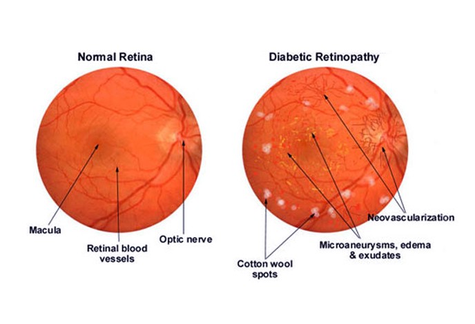
This is the early stage of retinopathy. It is characterized by mild and non-existence of symptoms. This stage of retinopathy has no significant formation of new tiny blood vessels that are rather abnormal and formed improperly. Microaneurysm or the protrusion of the tiny bulges in the weakened walls of the blood vessels of the retina may occur during the early stage of retinopathy. The protrusion can cause fluid and blood leakage into the retina and may have the tiny blood vessels obstructed as the condition progresses. The obstruction and the swelling can affect the retina or the lenses of the eyes that leads to the changes in vision or the blurry vision.

Proliferative diabetic retinopathy

This is the advance stage and the most severe form of diabetic retinopathy. This stage is marked by the formation of new tiny blood vessels that are abnormal and improperly formed. The retina during this stage is already deprived with oxygen supply which triggered the eye to form the tiny blood vessels in an attempt to sufficiently supply the eye with oxygen.

Diagnosis

Dilating the eyes is the initial step in diagnosing diabetic retinopathy among patient preceded by medical history taking. During the exam the eyes of the patients are dilated and an eye doctor takes note of the presence or the absence of the cataract that could possibly be causing the changes in vision. Formation of new and abnormal blood vessels are being evaluated including the swelling and deposits of fats and blood in the retina. Other abnormalities formed such as scar formation, retinal detachment and bleeding are also evaluated during the examination.

Additional tests may be done to confirm diabetic retinopathy and such tests include the following:

  • Measurement of visual acuity
  • Measurement of pressure
  • Optical coherence tomography
  • Fluorescein angiography

Diabetic Retinopathy Treatment

The treatment for diabetic retinopathy depends on the stage of the retinopathy including its extent. Non-proliferative diabetic retinopathy or the early stage of retinopathy usually require no treatment but close monitoring is required to evaluate for progression of the condition and if treatment will then be warranted.

Proliferative diabetic retinopathy is the most severe type of retinopathy that requires surgery. Surgical procedures however depend on the problems in the retina that occurred.

Laser photocoagulation is a procedure utilized to impede the fluid leakage or blood leakage into the retina. The leaks are being sealed with the laser beam which burns to seal the tiny blood vessels causing the leakage.

Panretinal photocoagulation is another method used to treat proliferative diabetic retinopathy that has caused widespread growth of blood vessels into the retina. Saving the central vision is the goal of this procedure and some vision on the peripheral on the other hand may be lost after the procedure.

Vitrectomy is a procedure utilized to remove the blood vessels that formed in the vitreous and other areas of the retina. Scar tissues and tiny blood vessels that formed in the retina are being removed with the use of delicate instruments while salt solution is being placed to maintain the shape of the eye.

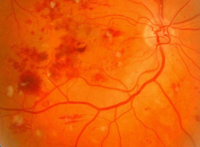
Diabetic Retinopathy Pictures

diabetic retinopathy

diabetic retinopathy 2

No responses yet

Scaphoid Fracture

Jan 07 2017 Published by under Fractures

What is a Scaphoid Fracture?

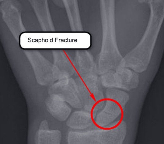
On the thumb side of your wrist there is a small bone. A scaphoid fracture is a break in that small bone. In your wrist there are eight carpal bones and of these eight bones, the scaphoid is the one that you would most likely break if you broke any of these bones. The bone is boat-shaped and in your wrist joint this is a very important part. Although in young males between the ages of twenty and thirty years of age this is a common fracture it can happen to people of all ages and this includes children.

Scaphoid Fracture Symptoms

The main symptoms of a scaphoid fracture are swelling and pain at the base of your thumb. It may not be obvious that you have a scaphoid bone fracture unless you have a wrist that is deformed. There are other times that physicians mistake this type of fracture for a sprain because they pain is not too severe. If within a day of an injury to your hand and wrist you still have pain in that wrist that just will not go away you should see your physician to rule out a scaphoid fracture.

Other symptoms you may have in addition to pain can include:

  • You may also have tenderness just below your thumb when you press on your wrist.
  • People who are thin may see a bulging of their joint capsule because of the blood that is filling the wrist joint from the scaphoid fracture.
  • Rapid swelling at the back of your wrist.
  • Pain if you compress your thumb inwards toward your wrist.
  • You may have bruises around your wrist.

If you have a nonunion scaphoid fracture, which is where there is no healing, the symptoms are more subtle. Some of these symptoms may include:

  • Having pain when you use your wrist such as trying to grip something.
  • The pain may be minimal.

Sometimes people will have these nonunion scaphoid fractures for many years and just did not realize it was a fracture because they thought it was a sprain. Overtime you can get degenerative arthritis in the wrist joint due to a nonunion fracture.

Causes of Scaphoid Fracture

The main cause of a scaphoid fracture is when you start to fall and you put your hand out in front of you to keep you from actually falling all the way down and you actually fall on your outstretched hand with most of your weight landing on the palm of your hand. They can also occur if your wrist is hit very hard or your wrist gets twisted severely. Many times these types of fractures happen when a person is person is playing sports such as basketball, football, or soccer. It could also be caused by doing other activities like skateboarding, riding your bike or motorcycle, rollerblading, etc. You could also have a scaphoid fracture if you have a punching incident or some type of vehicular accident.

If you have a nonunion scaphoid fracture it can happen because two pieces of bone have not healed together or the lower half of your fractured bone loses the blood supply and dies. When the second cause is the reason for a nonunion scaphoid fracture the condition is called avascular necrosis. The scaphoid bone is at risk for this condition because there is only one small artery that enters the bone closest to your thumb. If you tear the artery when you have a fracture you are going to lose your blood supply. There are no specific diseases or risks that increase your chances of having this type of fracture.

Scaphoid Fracture Treatment

There are two ways in which you can treat scaphoid fractures.

Non-surgical treatment

  • Fracture occurs near your thumb – most of these fractures will heal on their own within a few weeks after having this accident with the right protection. Most likely your physician will put your hand and arm in a cast or splint but it is usually below your elbow and the cast may or may not include your thumb being in a cast.
  • Fracture that happens near your forearm – with this fracture the physician will put a cast on that may go above your elbow and may include casting your thumb.

Surgical Treatment

You may have to have surgery to implant a metal device such as wires and screws if you have a scaphoid fracture near your forearms or in the middle of your bone. The metal implants will hold the fractured scaphoid in place until it is completely healed. How large the incision will be and whether the incision is on the back or the front of your wrist will depend on what part of the bone is broken. If the bone is broke in more than a couple of pieces the physician may do a bone graph. This is where they take a new bone and place it around the one that is broken to help stimulate the healing of the fractured bone. Once healed the bone will be a solid bone. They usually get this bone graft from the forearm in the same arm where the fracture occurred.

Right after you have fractured your scaphoid it may be too swollen to have a cast put on so the physician may just use a splint until the swelling subsides. You may also have to wear a splint if the physician is not sure if you actually have a scaphoid fracture they may just have you wear a splint. To help reduce swelling you should use ice packs.

Healing time

With a fracture near your thumb the time it will take for it to heal will vary with each individual. With this type of fracture the physician will periodically take images such as x-rays to check the healing process. If the fracture is near your forearm or in the middle of the bone the healing process is more difficult.

When you have a cast on your scaphoid fracture and you did or did not have surgery the actual healing time does vary but you can wear the splint or cast for as long as six months.

Scaphoid Fracture Pictures

scaphoid fracture

No responses yet

Nephrolithiasis

Jan 06 2017 Published by under Kidneys and Urinary System

What is Nephrolithiasis?

Nephrolithiasis is characterized by the formation of crystalline material in the kidney and the urinary tract. Nephrolithiasis is also known as kidney stones and is formed as a result of a decrease in the volume of urine or an increase in the substances in urine that can form stones in the kidney or in the urinary tract. Nephrolithiasis is also called renal calculi and the stone formations often results in blood in the urine. Severe pain in the abdomen, flank or groin is also included in the manifestation of renal calculi.

Nephrolithiasis mainly affects the general population without racial predilection. It occurs more commonly in male gender that in the female gender. The incidence can occur between the ages of 20 to 49 years with peak onset in the age of 35 to 45 years although the disease can also affect people from all age groups including children. Nephrolithiasis on the other hand is far more common in Asians and to those living in hot and dry areas. The prognosis is generally good as most cases of kidney stone spontaneously pass through the urine. Morbidity of kidney stones are those associated with urinary tract obstruction and upper urinary tract infection.

Nephrolithiasis Symptoms

The symptoms of Nephrolithiasis usually occur when the stones move within the kidney and passes through the ureter. Kidney stones generally originate from the kidney and extend distally and lodge in narrow areas adjacent to the kidney. When symptoms occur this may include the following:

Pain is the most common and initial symptom of kidney stones. The onset of pain is characterized as rapid and excruciating. The pain may be felt in the lower back and radiates to the sides, groin and the abdomen. The pain is also characterized as colicky pain which cannot be relieved with a change in the body position. The location and quality of pain are relative to the position on of the stone within the urinary tract while the severity is dependent on the degree of obstruction brought by the formation of kidney stones.

Nausea and vomiting usually accompany the rapid and excruciating pain.

Painful urination is also experienced and is accompanied by a blood in the urine.

Frequent urge to urinate is also noted and the urine while the characteristic of the urine can be noted with foul smells and cloudy appearance.

Fever and chills may occur if infection is present during the process of kidney stones.

The pain in kidney stones goes through different phases. The first phase is onset of pain that normally attacks early in the morning or during at night time that can disturb the sleep of an affected individual. The constant phase of pain is the second phase where the pain has reached the maximum intensity where it will linger until treatment or medical intervention has been applied. Abatement or relief phase is the third and last phase of pain characterized by spontaneous relief anytime following the initial onset. The relief of pain usually lasts for an hour and a half to 3 hours after the patient has awakened following administration of analgesic.

Nephrolithiasis Causes

Nephrolithiasis has no single cause and the stone formation results when the urine has more stone forming substances than fluid composition that the urine can dilute. Several risk factors on the other hand are considered to result in the formation of kidney stones.

There are different types of kidney stones that can form within the kidney and this includes the following:

Calcium stones

Calcium stones are the most common type of kidney stone that usually develop as a result of high intake or consumption of certain substances such as salt. Calcium stones are usually in the form of oxalate which is a substance that is naturally occurring in the food.

Struvite stones

Struvite stones commonly form in women suffering from urinary tract infection. The struvite stones grow rapidly and become large and manifested with few symptoms.

Uric acid stones

Uric acid stones potentially occur to individuals suffering from gout or went through chemotherapy. This is also potential for individuals who take in an insufficient amount of fluid and loses large amounts of fluid. This is also potential for individuals who include high protein in their diet.

Cystine stones

Cystine stones can develop in people with cystinuria or an inherited disorder marked by an elevation in the formation of stones in the bladder, kidney and the ureter.

Risk factors predisposing an individual to kidney stones include the following:

Dehydration

Dehydration from decreased fluid intake or from strenuous activities without sufficient fluid replacement can put one at risk for kidney stone formation. People living in hot and dry climates are also at high risk for developing Nephrolithiasis due to the risk of dehydration.

Diet

Diet can also affect or influenced the incidence of Nephrolithiasis. Diet that includes high protein, sugar and sodium potentially increases the risk for kidney stone formation.

Digestive disorders

Digestive disorders including surgery is a potential risk for Nephrolithiasis due to changes in the digestive process that can affect the absorption of calcium and water and thus increasing the levels of stone forming substances in the urine.

Gout

This is potential for uric acid stone formation as the condition is generally the consequence of an increase in the amount of uric acid in the blood and urine.

Family history

This increases the risk for kidney stones including personal history of previously suffered from kidney stones.

How is Nephrolithiasis diagnosed?

Suspicion of kidney stones or Nephrolithiasis is diagnosed with the onset of the pattern of symptoms. Diagnostic test procedures are the following:

Urinalysis

Urinalysis or Urine test requires a 24-hour urine collection for evaluation of stone forming minerals and a decreased in the levels of substances that inhibits the formation of stones.

Blood test

This is done to evaluate for calcium or uric acid content in the blood which can reveal an elevation in the levels of these substances.

Imaging tests

Imaging tests are done to confirm the formation of kidney stones. A helical CT scan is the imaging test of choice that is usually done without contrast materials. This imaging test can identify stones and obstruction within the urinary tract.

Nephrolithiasis Treatment

Most kidney stones usually resolve within 48 hours with sufficient amount of fluid intake to help wash away the stone through the urine. Small stones that have minimal symptoms can be treated with the following:

  • Increase fluid intake
  • Pain relieving medications
  • Alpha blocker medication facilitates the passing of stone rapidly as the medication act by relaxing the muscles of the ureter

A large stone formation that leads to manifestation of symptoms can be treated with the following:

  • Extracorporeal shock wave lithotripsy is a procedure that utilizes sound waves to break the stones.
  • Percutaneous nephrolithotomy is a surgical removal procedure recommended for large stone formation.

No responses yet

Myalgia

Jan 06 2017 Published by under Bone, Joints and Muscle

What is Myalgia?

Myalgia is commonly known as muscle pain or muscle ache. It is an extremely common condition that almost all people get to experience. Myalgia is not a disease or a disorder but rather a symptom signifying an existing underlying condition. The pain in the muscle can range from mild to excruciating and can linger for a few days or even longer. It can affect almost any part of the body including the neck, hands, legs and the back.

The muscles are soft tissues containing protein fibers that create contraction to change both the length and shape of the cell. The muscle is responsible for creating force and motion to allow human movement. It is also composed of three types of tissue known as the skeletal muscle, cardiac muscle and smooth muscle that act either as voluntary or involuntary. Both the cardiac and the smooth muscle as considered involuntary while the skeletal muscle is considered voluntary. The movement of the muscle or the action it generates is based on the location of the insertion and the location where it originated.

Myalgia is basically non-serious and non-life threatening but can be discomforting. It usually affects a small figure of muscles and usually one at a time. The muscle pain however can be felt anywhere as every part of the body composed of muscle tissues.

Myalgia Types

Almost every part of the body composed of muscle that the pain can occur in any part. Myalgia composed of several types which define the part of the body that is being affected and the number of muscles that are involved in the process of pain including the cause of pain.

Epidemic myalgia

This is the type of myalgia that is also known as Bornholm disease. It is muscle pain caused by a viral infection and affects the upper abdomen and the lower chest. The pain is characterized as spasmodic and develops suddenly. The pain is also worsening with every movement and deep breathing causing shortness of breath to the affected individual.

Fibromyalgia

It is defined as myalgia that is widespread that usually involves the joints, muscles and the ligaments. The pain can be perceived on both sides of the body and is described as dull and constant ache. This form of myalgia is often aggravated by pressure applied on the affected site while this form is commonly associated with insomnia, osteoarthritis, lupus, irritable bowel movements and headaches.

Trapezius myalgia

Trapezius myalgia is myalgia that involves the muscle of the neck and is triggered by a problem with the trapezius muscle. The pain is described as long-lasting and is particularly reactive to stressful situations and repetitive work tasks. This form of myalgia is more common in women who are working for longer hours in front of the computer.

Polymyalgia rheumatica

Polymyalgia rheumatica is a type of myalgia that involves several muscles. It is an anti-inflammatory disorder that usually affects older people and rarely occurs in individual below the age of 50 years. The pain is often associated with stiffness and inflammation and occurs in the muscle of the neck, shoulders, upper arms, hips and the buttocks.

Myalgia Symptoms

Myalgia has numerous symptoms while the manifestation of symptoms also depends on the area that is being affected.

  • Pain that is often severe is the most prominent among the symptoms of myalgia. The onset is usually sudden and lingers for a few days to several weeks depending on the intensity and the cause of the pain. The pain is also associated with stiffness and spasm.
  • Acute case of myalgia can severely incapacitate an affected individual.
  • Chronic case of myalgia is marked with pain and stiffness after periods of inactivity and pain usually eases after reasonable exercise.
  • Myalgia affecting the cervical manifests with heaviness of the head accompanied with occipital headache.
  • Cervical myalgia can also manifest with impairment of vision and problems with swallowing and numbness of the face may also be experienced.
  • Difficulty in breathing in and breathing out may also be experienced with chest involvement in myalgia.
  • Irregular heart beat is also experienced when myalgia involves the heart and is also potential for cardiac arrest.
  • Tenderness of the affected area may also be observed.

Other signs and symptoms of myalgia may include the following:

  • Nausea and vomiting
  • Onset of fever
  • Anxiety and depression
  • Stiffness
  • Vertigo
  • Numbness and tingling sensation in the arms or other parts of the body.

Myalgia Causes

The most common cause of myalgia is tension or stress that results from an injury or excessive use of the affected muscle. Myalgia that developed from this cause is usually localized and can involve a single or multiple muscles. Physical damage to muscle due to compulsive exercises can also cause myalgia. Severe or strenuous pulling activity of the ligaments can also result to pain in the muscles.

Myalgia can also develop as a consequence of some diseases and disorders which may be in existence in the individual suffering from myalgia. Such diseases and disorders that can cause myalgia include the following:

  • Chronic fatigue syndrome is a disorder marked by extreme fatigue and includes muscle pain as one of the 8 official symptoms of the disorder.
  • Chronic exertional compartment syndrome is a syndrome that causes nerve and muscle pain induced by an exertion of exercise.
  • Eosinophilia-myalgia syndrome is a rare inflammatory and autoimmune disease that causes pain and inflammation of the skin, nerves, lungs, heart, blood vessels and muscles.

Diagnosis

Myalgia is a symptom rather than a condition that direct diagnosis is not recommended. The development of muscle pain is often associated with other conditions or disorders and muscle pain is usually an inflammatory response.

The diagnosis of myalgia is directed towards the existing underlying condition that triggered the onset and this may include the following diagnostic method:

Medical history taking is the initial step in identifying the trigger of myalgia. The process includes the complete health history that includes the previous and current illnesses, injuries and including the medications being taken.

Physical examination is also included in the diagnostic procedure. The process is beneficial in identifying the possible cause of myalgia. Evidence of stiffness or weakness may be observed through the gait and posture of the affected individual.

Blood test is helpful in detecting inflammation and ruling out the underlying conditions that is causing the muscle pain.

Treatment

The aim of treatment of myalgia is through alleviation of pain to provide relief and comfort to the patient. The treatment is also dependent on the underlying condition that triggers the onset of myalgia. It is therefore necessary to initially identify the cause of myalgia prior to administration of treatment.

Myalgia can be alleviated with natural treatments and can be treated at home.

  • Enough rest is necessary to help reduce the inflammation that is causing the muscle pain.
  • Warm or cold application can reduce the inflammation and relax the muscle thus alleviate the muscle pain.
  • Gentle massage can relax the muscle and reduce the pain and it can also help in reducing the muscle inflammation.
  • Over-the-counter pain relievers such as ibuprofen.

No responses yet

Agranulocytosis

Jan 05 2017 Published by under Blood and Heart Diseases

What is Agranulocytosis?

Agranulocytosis is a condition marked by the inability of the bone marrow to produce enough or sufficient white blood cells leading to its decrease in count or the condition known as leukopenia. It is an acute and a rare condition that most commonly affects the neutrophil content of the white blood cells in which the decrease in count can lead to neutropenia.

Neutrophils are among the granulocytes that make up the white blood cells. It is classified according to its staining characteristic marked by neutral subtle granules. Neutrophils outnumbered all types of granulocytes found in the white blood cells and the absolute neutrophil counts are in relation to the production of the white blood cells. Neutrophils are the most abundant type of white blood cells and are generally an integral part of the immune system which are the first to respond during and inflammatory process and during an infection from harmful microorganisms.

Agranulocytosis is a severe deficiency in the production of white blood cells that the onset can greatly affect the immune system of an individual. People with this deficiency are potentially at risk for infection as a consequence of suppressed immune system. The minor infection can become serious in individuals suffering from agranulocytosis while harmless microbes can potentially become strong enough to cause harm and attack the own body of the immune system.

The decrease in the production of white blood cells is the defining characteristic of agranulocytosis. The decrease in the white blood cell of an individual puts one at greater risk for infection as the function of which is to defend the body from harmful effects of microbes or other microorganisms. It is an essential component of the immune system that act by identifying harmful microbes, destroys them and remove the pathogens including the damaged cells and other harmful foreign materials in the body.

Symptoms of Agranulocytosis

The onset of agranulocytosis can be asymptomatic but if clinically present the symptoms may include the following:

  • Sudden onset of high fever and chills.
  • Incidence of sore throat.
  • Weakness of the limbs may also be experienced.

People with agranulocytosis and suffering from an infection in any part or organ of the body will have a rapid progression of infection. Agranulocytosis can also hasten the progression of a life-threatening medical condition known as sepsis.

The main affected in the process of agranulocytosis is the production of a type of white blood cells called neutrophils. The alteration in the white blood cell count affecting the neutrophils can lead to neutropenia where the number of neutrphils in the blood stream is greatly diminished thereby resulting to symptoms such as:

  • Gum disease that includes gum bleeding, mouth ulcers, halitosis and an increase in the production of saliva.
  • Bacterial pneumonia
  • Urinary tract infection.
  • Gastrointestinal tract infection.
  • Fungal infections

The infection in neutropenia may be localized to a certain area of the body but it can also spread via the bloodstream which in turn can spread to other areas and organs of the body.

Agranulocytosis Causes

The cause of agranulocytosis is classified into two forms namely:

Congenital agranulocytosis which is a form of agranulocytosis that is present at birth and is implicated on genetic imperfection.

Acquired agranulocytosis is a form of agranulocytosis as a consequence of medication and other form of treatment which can affect the bone marrow and its production of the blood cells. Acquired agranulocytosis may be due to the following treatments:

  • Anticonvulsant drugs that are used for treating or managing epileptic seizure and this drug may include barbiturates, aldehyde, aromatic allylic alcohol, benzodiazepines and other anticonvulsant drugs.
  • Antithyroid drugs such as methimazole, carbimazole and propylthiouracil.
  • Antibiotics such as cotrimoxazole and penicillin.
  • Nonsteroidal anti-inflammatory drugs such as metimazole and naproxen.
  • Mirtazapine is an anti-depressant drug considered in agranulocytosis due to its side effect of bone marrow suppression.
  • Antipsychotic drugs such as clozapine.

Other causes of acquired agranulocytosis include the following:

  • Cocaine abuse
  • Chemotherapy treatment for cancer.
  • Diseases involving the bone marrow such as aplastic anemia.
  • Cancers or tumor growth involving the bone marrow such as leukemia.
  • Chemical toxins and radiation exposure.
  • Vitamin B-12 and folate deficiency.
  • Diseases involving the autoimmune such as systemic lupus erythematosus.
  • Spleen enlargement
  • Autoimmune diseases

Agranulocytosis Treatment

Agranulocytosis often responds well to treatment. The prognosis is positive for those diagnosed and treated promptly while it is potentially life-threatening if left untreated. Mortality in agranulocytosis is associated with infection due to the rapid progression and multiple organ involvement.

Close monitoring of serial blood counts is necessary in agranulocytosis that is asymptomatic. Withdrawal from medications and other offending agent affecting the bone marrow and the white blood cell production is also necessary but upon the advice of a doctor.

Antibiotic and antifungal treatment is usually given for agranulocytosis that resulted from an infection from bacteria and fungus and other form of infection.

Colony-stimulating factor encourages more production of neutrophils in the bone marrow and is beneficial for agranulocytosis patient undergoing chemotherapy treatment.

Leukocyte transfusion may also be beneficial for agranulocytosis patients to replace the deficit in the granulocytes especially the neutrophils

No responses yet

Polydipsia – Psychogenic, Causes, Diagnosis, Treatment

Jan 04 2017 Published by under General Health

What is Polydipsia?

Polydipsia is not a disease or disorder but a symptom of many a condition characterized by an excessive need to take in fluid or the excessive thirst. The excess in need to quench a thirst therefore leads an affected individual to take in lots of water or any fluid which can potentially set forth an imbalance in the fluid and electrolyte of the body.

Polydipsia is often associated with polyuria which is defined as the excessive amount of urine released against the amount of fluid intake or relative to the amount of fluid intake.

Thirst is the physiologic need to take in an amount of water or any form of fluid. It is a corrective mechanism of the body to correct and control physiologically the fluid balance in the body. The excess in thirst can be a symptom of various medical conditions and recognizing the physiology of thirst will help in appreciating the significance of thirst as a symptom.

  • Thirst will drive an individual to take in fluid as a regulatory component in which balance of fluid will keep the proper secretion of the antidiuretic hormone that is secreted by the hypothalamus when there is an increase in plasma osmolality.
  • Drinking water or other fluid as a response to thirst thus helps in the maintenance of blood osmolality.
  • The amount of thirst and the amount of fluid needed to quench the thirst is relative to the blood osmolality while the brain is being sent a signal to inhibit fluid overload.
  • A decrease in the blood pressure or blood volume also stimulate or trigger thirsts.
  • Vasopressin and human chorionic gonadotrophin are both thought to play a role in stimulating thirst during the process of pregnancy.
  • Age advancement dulled thirst stimulus thus a decrease in primary thirst which on the other hand is recompensed by secondary drinking.

Psychogenic Polydipsia

Psychogenic polydipsia is a condition characterized by the excessive urge to drink or take in fluid in an excessive amount even with the absence of stimulus. This form of polydipsia is associated with mental disorder such as Schizophrenia, depression and bipolar disorder. The etiology of the disorder among psychiatric patients has not been clearly understood but is thought to have resulted from neurochemical imbalances.

An extended or prolonged period of taking in excessive amounts of water or any fluid is potential to dilute or affect the concentration of sodium in the blood thereby resulting in low levels of sodium in the blood. Psychogenic polydipsia is also potentially life-threatening the extent of sodium dilution has reached the extent of causing seizure and cardiac arrest. The condition of the extreme amount of water intake may lead to hyponatremia and progress to water intoxication which can manifest with lethargy, confusion, psychosis, seizure and potentially death.

Patient with psychogenic polydipsia can be observed with utmost desire to seek water sources to quench their excessive thirst. Weight gain is also often apparent as a result of the inability of the kidney to compensate for the fluid overload. The manifestation of psychogenic polydipsia may be observed with undue thirst and the need of the patient to quench the thirst, nausea and vomiting, confusion and seizure. Polyuria or the condition of incessant need to urinate is also observable and is often associated with both the primary and psychogenic polydipsia.

Polydipsia Causes

Polydipsia can be influenced or caused by a lot of factors including numerous diseases and disorders that can trigger incessant thirst and need to quench that thirst.

Diabetes

Diabetes is the most implicated in the incidence of polydipsia. Both diabetes mellitus and diabetes insipidus can trigger polydipsia often as a side effect of diabetes medication. Polydipsia is also among the symptoms of diabetes and is also the effect of failure to take diabetes medication including the inadequacy in the dosage of the medication.

pH level

Deviation of pH level in normal plasma can also lead to polydipsia. The deviation in the levels is often the result of imbalance in the normal acid and base level of the body. This will lead an affected individual to a level of dehydration that will trigger excess need to quench the thirst.

Diarrhea

Diarrhea is a condition characterized by increased looseness in the stools and increased bowel movement. This condition is potential for dehydration as a result of increased secretion of fluid into the intestine accompanied by a reduction in the absorption of fluid from the intestine thereby disrupting the fluid and electrolyte balance.

Severe burns

Severe burns can also lead to loss of fluid in the body which can cause an imbalance in the fluid and electrolyte of the body. The imbalance of fluid in the bloodstream can result to increase in the osmolality of the plasma thus the physiological driving force of thirst to maintain balance osmolality.

Pregnancy

Pregnancy is also another factor considered to trigger excessive thirst and need to quench that thirst. The incessant thirst is triggered by the low level of osmolality which is thought to be affected by both the vasopressin and the human chorionic gonadotrophin.

Polydipsia Diagnosis

Polydipsia is not a disease but often a symptom of an underlying medical condition. It is mostly associated with both diabetes mellitus and insipidus. Polydipsia is also often associated with polyuria that directly diagnosing the condition is rather difficult. Investigating or direct diagnosis of diabetes mellitus and diabetes insipidus can both be helpful in diagnosing polydipsia. Differential diagnosis proved helpful in diagnosing polydipsia to isolate it from different disorders related to the onset.

The blood serum test is also useful in diagnosing polydipsia through evaluation of osmolality of the extracellular fluids of the body. The revelation of decrease in the concentration of serum in the red blood cells, blood urea nitrogen and sodium signifies excessive water intake therefore polydipsia.

Polydipsia Treatment

There are numerous treatments for polydipsia as the treatment is dependent on the cause or the underlying condition associated with the onset of polydipsia.

Desmopressin is the drug of choice for polydipsia associated with diabetes and nocturnal polydipsia. It is a synthetically produced drug that acts by replacing the vasopressin and aids in controlling both polydipsia and polyuria by reducing the production of urine.

Treatment of psychogenic polydipsia is different from non-psychogenic polydipsia. Psychogenic polydipsia involves dietary controls and monitoring of daily weight fluctuations including evaluation and management of damage to the urinary system based on the extent and damage to functions.

Early diagnosis and prompt treatment of polydipsia is essential in preventing further complications and irreversible damages which can be potentially life-threatening.

No responses yet

Ecchymosis – Symptoms, Causes, Treatment, Pictures

Jan 04 2017 Published by under Skin Conditions

What is Ecchymosis?

Eccyhmosis is characterized by a reddish or bluish discoloration of the skin that is often non-raised and spontaneously caused by a medical condition. The onset of reddish or bluish discoloration of the skin is due to the escape of blood from ruptured blood vessels into the capillaries. It is a subcutaneous purpura with a diameter of 1cm or more and is different from a bruise.

Ecchymosis is a degree of hematoma that is larger than 10mm or more than 1cm in diameter. It is an extravasation of blood in the thin layer of the skin that resulted from a rupture within the blood vessel causing the blood to leak into the layer of the skin. It is sometimes mistaken or erroneously identified as a bruise but the two are entirely different in terms of the etiology. The bruise is a blood leaking into the thin layer of the skin as a result of trauma while ecchymosis occurred spontaneously as a consequence of an underlying medical condition.

Purpura is a purplish colored patch of the skin as a result of blood leaking into the capillaries and the large degree or size of which is known as ecchymosis while the small size is known as petichiae. Purpura involves the platelets thus with ecchymosis. The condition of purpura and ecchymosis may neither have a normal or low platelet count. The platelets are the smallest of the blood types and are a disc shaped cell of clear fragments and does not have a nucleus. Platelets are not true cells but are fragments of blood cells that are generally light and are usually pushed on the walls of the blood vessels where it serve its function.

Ecchymosis Symptoms

Ecchymosis and hematoma are both the consequence of blood leaking into the thin layer of the skin as a result of damage to the blood vessels. The difference however is in not only in the appearance but also in the location of its occurrence. Ecchymosis occurs in the thin layer of the skin and in the mucus membrane while hematoma can occur not only under the skin but also in the different organs of the body and in the mucous membrane as well. Ecchymosis also has a flat character while hematoma has a three dimensional character.

Ecchymosis does not have numerous symptoms to exhibit although the symptoms that may occur if ever may be associated with the existing condition that influenced the development of ecchymosis. The general symptoms of ecchymosis on the other hand include the following:

  • Onset of skin discoloration that is reddish or purplish.
  • Purplish or reddish skin patch that has a diameter of an inch or more.
  • Perception of pain on the affected site although pain may not always be present in some patients.
  • Inflammation of the unaffected skin surrounding the site of ecchymosis depending on the degree of damage in the tissue.
  • Ecchymosis may also extend to surrounding areas from the original site depending on the location and the severity of ecchymosis.

Ecchymosis Causes

Ecchymosis is not a condition or a disorder and is generally harmless. It is however, a symptom of one or more serious underlying medical condition that needs medical attention and intervention. The onset of ecchymosis can be a mild inflammatory response of the body or can be a more complex medical condition.

Ecchymosis is generally the result of blunt trauma that caused the blood vessels to rupture and seep into the thin layer of the skin or in the mucus membrane in the absence of an incision or any break in the skin to allow the blood to go out of the skin surface. The accumulation of blood within the skin layers causes the reddish or purplish discoloration of the skin.

Ecchymosis is not just an inflammatory response of the body but is also the result of some underlying medical condition that causes rupture of the capillaries. Such medical conditions resulting to ecchymosis includes the following:

Leukemia

Leukemia is a malignancy of the blood cells including the tissue that produces blood. Easy bruising and bleeding are among the symptoms of leukemia including that of ecchymosis. The increase in the levels of white blood cells influences the development of ecchymosis.

Acute renal failure

This is the rapid loss of the function of the kidney. The inability of the kidney to perform its function of expelling waste from the body leads to the progression of waste product in the blood. Ecchymosis is among the potential result of waste accumulation within the blood as a consequence of abnormalities that resulted from the degeneration of the kidney.

Multiple myeloma

Multiple myeloma is the cancer of plasma cells in the body. This plasma cell is a type of white blood cells that are normally found in the bone marrow. The abnormal growth of plasma cell in the bone marrow as the character of the disease progresses to an aberration in the blood thus causing ecchymosis.

Liver cirrhosis

Liver cirrhosis is characterized by an abnormal function and structure of the liver as a consequence of the complication of various liver diseases. Ecchymosis is among the result of any alteration in the blood composition as a consequence of impairment of in the normal function of the liver.

Myelofibrosis is the formation of fibrosis in the bone marrow tissue. The development of fibrosis results to a disruption in the normal production of blood cells of the body.

Treatment

Ecchymosis usually resolves without treatment and can be managed at home. The frequent onset of ecchymosis associated with severe pain on the other hand requires medical attention. Other treatment or ecchymosis depends on the existing underlying condition that influenced the onset.

Treatment of ecchymosis includes the following:

  • Rest promotes tissue healing that enough rest is recommended to hasten the tissue repair and healing of ecchymosis.
  • Ice application facilitates vasoconstriction of the ruptured blood vessels which in turn prevents ecchymosis from extending to the nearby unaffected site.
  • Elevation of the affected site can help in inhibiting inflammation as the elevation facilitates a proper venous return while improving the circulation of the affected site.
  • Pain relievers such as ibuprofen and other form of analgesics can help reduce the pain associated with the onset of ecchymosis.

A light massage and stretching exercises can help improve tissue repair as long as the activities do not exacerbate the condition of ecchymosis causing further damage to the tissue and the blood vessels.

Ecchymosis Pictures

No responses yet

« Prev

© 2011-2019 MDDK.com - Medical Tips and Advice. All Rights Reserved. Privacy Policy
The health information provided on this web site is for educational purposes only and is not to be used as a substitute for medical advice, diagnosis or treatment.