Diseases General Health Skin Conditions

Archive for August, 2016

Fibrous Dysplasia

Aug 24 2016 Published by under Bone, Joints and Muscle

What is Fibrous dysplasia?

This is a medical condition that disturbs the process of bone regeneration. It causes normal bone to be replaced with fibrous tissue that is abnormal. It is characterized by a heavy build-up of scar tissue, also known as fibrous tissue, within your bones. This is a very rare medical condition and is not a form of cancer.

Forms of Fibrous dysplasia

There are two forms of this medical condition which are:

  • Monostotic fibrous dysplasia – with this type only one bone is affected and accounts for approximately seventy percent of all cases of fibrous dysplasia
  • Polyostotic fibrous dysplasia – with this type it affects several bones and is often associated with McCune-Albright syndrome, which is a genetic disorder that affects not only the bones but also many endocrine (hormone producing) tissues and your skin. In addition to fibrous dysplasia this syndrome may also lead to early puberty and skin lesions. Having McCune-Albright syndrome is the most severe form of polyostotic fibrous dysplasia and is seen in approximately three percent of all cases of polyostotic fibrous dysplasia.

Pathophysiology

Although this medical condition starts during the development of the fetus is it not detected until later. It could be detected in childhood or adolescence or even as late as adulthood. As the affected bone continues to grow, the weaker fibrous tissue enlarges and causes the whole bone to become brittle, painful, even deformed, and weak. Fibrous dysplasia can affect any bone in your body. They can also be several bones that are affected at the same time. When several bones are affected it has been observed that they are all located on one side of your body. Fibrous dysplasia does not spread from one affected bone to another.

Some of the bones that are normally affected include:

  • Skull
  • Face
  • Humerus, which is the bone in your upper arm
  • Tibia, which is the your shinbone
  • Femur, which is your thigh bone
  • Pelvis
  • Ribs

It can also affect your spinal vertebra but this does not happen that often. Although it can affect any bone it is more commonly found in the long bone in your legs or arms or in your skull.

Symptoms

The symptoms of fibrous dysplasia may be experienced differently by different people but there are some common symptoms that everyone may experience. When people who have monostotic fibrous dysplasia they often do not develop symptoms.

  • Due to the expansion of the fibrous tissue there is pain in the affected bone
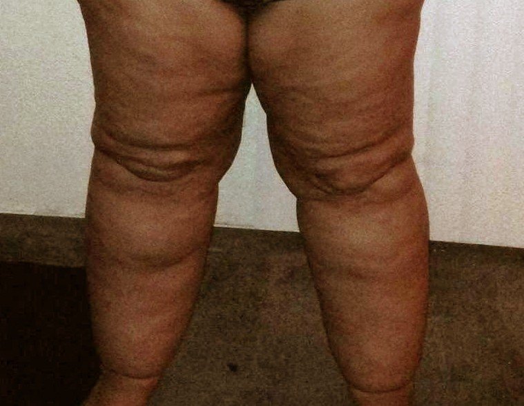
  • Bone deformity
  • Fractures of the bone affected
  • An abnormal lateral curvature of your spinal column called scoliosis. This can result in someone seeming to lean towards one side
  • A tottering walk
  • Nerve entrapment

When a person has these symptoms, especial the fractures, bone pain, and bone deformities, begins in early childhood, usually by the age of ten. The tottering walk is often experienced by people who have polyostotic fibrous dysplasia when their pelvic, tibia, or thigh bones are affected.

Causes

Because the medical condition develops during the fetal stage its occurrence has been associated with a mutation of a gene that affects the cells that make your bones. It is not known exactly what causes this mutation but it is known that it is not a hereditary disease but happens spontaneously. The gene that mutates is referred to as the GNAS1.

Even after you stop growing bones continuously renew themselves. In this renewal process there are particular types of bone cells called osteoclasts that reabsorb bone minerals or break down the bone. There are also other types of bone cells that build it up again called osteoblasts. When fibrous dysplasia affects a bone it causes a disruption in this process and results in old bone breaking down at a faster rate than the rate which it was built. To be able to cope with this your normal bone tissues gets replaced by weak, soft fibrous tissue.

Diagnosis

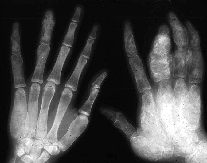
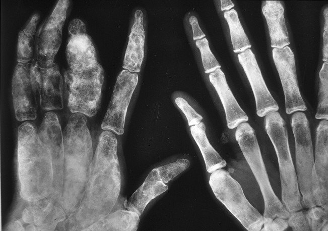
The preliminary diagnosis is usually made on the basis of the symptoms you might be experiencing such as recurrent bone fractures and bone pain. To confirm a diagnosis of fibrous dysplasia the physician will order imaging tests like MRI scans and x-rays. These imaging tests can help pinpoint the location of the bones that are affected along with any bone deformities that could have developed. Your physician may also order a bone scan, which uses radioactive tracers injected into your bloodstream. The parts of your bones that are damaged will take up more of these tracers and show up brighter on the scan so they can see which bones and how much is affected.

Treatment

At this point there is no known cure for fibrous dysplasia so the treatment mainly involves relieving any symptoms that you are experiencing. If there are no symptoms then the medical condition is just observed with periodic x-rays taken. If the x-rays show that the medical condition is not progressing then there is no further treatment needed. Sometimes your physician will have you use a brace in order to prevent the affected bone from fracturing but the brace may not be able to prevent the bone from becoming deformed. If the x-ray shows that the medical condition is progressing it is usually then treated with medication and surgery.

Medication

The medications that are used to treat progressing fibrous dysplasia are called bisphosphonates, which help to slow down the breakdown of the bone, increase the density of the bone in your spine and hip, and maintain bone mass. This will help to lower the chances of the bone fracturing. Not much is known about how this medication affects children and adolescents with fibrous dysplasia but it is thought that it may help to relieve pain.

The medication is normally used in adults to help increase the density of your bones and to treat osteoporosis. Sometimes the medication can even help improve the formation of bone and help to reduce the pain. Two of the types of the bisphosphonates medications used are Alendronate and Pamidronate. This medication is taken orally but it also available in intravenous injections. This is for those that cannot tolerate the oral type because of gastrointestinal irritation. Your physician may also have you take over-the-counter anti-inflammatory medication and analgesics or give you a prescription for them to help with the pain. Your physician may also have you do pain management therapy.

Surgery

When doing surgery it can include the excision of the affected area of the bone and may be followed by bone grafting. This is where a healthy bone from another area of your body is transplanted into the area affected by this medical condition. Unfortunately over a period of time this grafted bone is absorbed and replaced with fibrous tissue. The surgeon can also stabilize the affected bone with the help of a rod that is placed inside the bone along with plates and screws. This procedure can be useful in fixing a fracture or deformity. It can also help prevent the bone from breaking. Surgery may also be done to relieve pressure on a nerve, especially if it is a facial or skull bone that is affected.

Complications

Although fibrous dysplasia does not usually cause any complications besides bone fractures or deformities in severe cases of fibrous dysplasia you may see complications that can include:

  • Hearing and vision loss – if the nerves to your ears and eyes are surrounded by an affected bone(s) and the facial bones become severely deformed it could lead to loss of hearing and vision but it is a complication that is rare.
  • Arthritis – if your pelvic and leg bones are deformed you could have arthritis form in the joints of these bones.
  • Cancer – although this rarely happens it is possible that an affected area of the bone could become cancerous but is a complication that usually affects only people who have had prior radiation therapy.

Pictures

fibrous dysplasia

fibrous dysplasia pictures

No responses yet

Gastric Volvulus

Aug 22 2016 Published by under Digestive System

What is Gastric Volvulus?

This medical condition happens when all or part of your stomach twists around by more than 180 degrees and causes a blockage of your gut. It is sometimes possible for your stomach to rotate far enough so the blood supply could become cut off and lead to gangrene. Approximately twenty percent of the cases of gastric volvulus happen in children and more than half happen before they are one year old but are more common in adults. You will find this medical condition more commonly in the elderly as their stomach’s supporting ligaments are becoming more lax as they age.

Types

  • Acute gastric volvulus – with this type it happens suddenly and is considered an emergency needed surgical treatment. When a physician tries to pass a tube down into the stomach they will find it impossible to do so.
  • Organoaxial gastric volvulus – this type involves your stomach revolving along its length and more frequently associated with the blood supply being cut off to your stomach. Fifty-nine percent of all cases of gastric volvulus are this type.
  • Mesenteroaxial gastric volvulus – this type occurs when your stomach flips upside down with the back of your stomach ending up facing the front of your body. This happens occasionally and is less likely to completely obstruct the blood supply to the stomach or block the gut. It can give rise to what is called a chronic condition, which means that over a long period of time the symptoms come and go. Approximately twenty-nine percent of the cases of gastric volvulus are mesenteroaxial type.
  • Combined type – this is a rare form in which the stomach twists organoaxially and mesentericoaxially.

Gastric Volvulus Symptoms

The symptoms that a person displays with gastric volvulus depend on which type they are experiencing.

  • Acute gastric volvulus – severe pain just below your breastbone, retching without being able to properly vomit. You may also have breathlessness and swelling of the upper abdomen with the lower abdomen remaining flat and feeling soft. There are some cases where the pain can travel from the chest along your arms and up to your neck.
  • Mesenteroaxial gastric volvulus – when this type gives rise to a chronic condition you may have pain below your breastbone that you feel now and then along with a feeling fullness soon after you start to eat. You may also have problems with swallowing and breathlessness.

Causes

Approximately one third of all gastric volvulus cases are associated with a hiatus hernia, which is a protrusion of the upper part of your stomach into the area between your neck and stomach called the thorax. It comes into your thorax through a weakness or tear in your diaphragm. Gastric volvulus is also more commonly found in people who have birth defects, also known as congenital abnormalities, of their diaphragm.

It can also be caused by:

  • Having no spleen
  • Gastric, or stomach, ligaments that are too long
  • Weak muscles, referred to as motor neurone diseases
  • Tumor of the stomach
  • Any other defects that can affect your stomach

If you have a defect of your diaphragm you would most likely suffer from organoaxial volvulus.

Diagnosis

It is usually diagnosed by having x-rays taken, which will show the gastric volvulus as an air-filled sac behind the heart shadow on a chest x-ray. If they do an x-ray of your stomach it may show that a part of your stomach as being massively distended. Your physician may have a barium meal done, which is a special type of x-ray. You will be given a toothpaste-like gel that you swallow to provide contract. After you have swallowed it the x-ray will be taken. It will show the absence or narrowing of the barium substance in the part of your stomach where the obstruction is located. You may also have a CT-scan done to show the entire anatomy of your stomach, which makes precise diagnosis possible along with showing what the obstruction is. A CT-scan may also give the physician a clue as to what caused the gastric volvulus to begin with.

Treatment

The treatment needed depends on which type of gastric volvulus you are experiencing.

Acute gastric volvulus – the treatment for this type is surgery

Surgery and Repair

When you have surgery for acute gastric volvulus it is done to repair the problem by untwisting your stomach and fixing it into place so this does not happen again. If there is any presence of gangrene during the surgery that tissue is also removed. Before your stomach can be fixed to the diaphragm and anterior abdominal wall it will have to be tested for viability, which means they need to make sure that it has not turned gangrene and will still function the way the stomach should function. The surgery to repair gastric volvulus is called anterior gastropexy and is considered an emergency surgery. After surgery you will be in the hospital for a couple of days before you are discharged to make sure that the surgery fixed the problems and there are no complications from the surgery.

In some cases of gastric volvulus the surgeon may use an endoscope, which is a thin long instrument that is similar to a telescope, to do keyhole surgery. With this type it is repaired by rotating your stomach back to the original position but the drawback with keyhole surgery is that there is a risk of perforating your stomach wall. This type of surgery is useful for those who are not fit enough to have open surgery, as what is done for acute gastric volvulus. Keyhole surgery may be done as a temporary measure and have the traditional surgery done later. If there have been any defects in the wall of your diaphragm that may have caused your case of gastric volvulus they will also have to be repaired.

Surgery Recovery time

If you have had open surgery to fix your case of gastric volvulus it can take two to six weeks before you are fully recovered. During this time you should not be putting this area under a lot of stress and stretches and taking it easy.

No responses yet

Ventrogluteal

Aug 21 2016 Published by under Pain Management

Definition

The ventrogluteal area is the area in which intramuscular injections are performed.  The ventrogluteal muscle area is often preferred for the administration of intramuscular injections, as it allows for the rapid absorption of the injected medication.

Ventrogluteal vs Dorsogluteal

The dorsogluteal approach was used for many years for the administration of the intramuscular injections but recent research has demonstrated an increased risk of injury. Today, it is known that the dorsogluteal approach can lead to a series of complications, especially in patients who receive intramuscular injections for prolonged periods of time. Among these complications, there are: abscess, cellulitis, necrosis at the site of the injection, formation of granuloma, muscle fibrosis, muscle contracture, hematoma, injury of the blood vessels or nerves in the area.

The dorsogluteal area is found in the superior and lateral part of the gluteal muscles, which is known in the medical field as the upper outer quadrant. The area can be more easily identified by dividing the buttock into four imaginary quadrants.

The constant administration of dorsogluteal injections presents an increased risk of damage to the sciatic nerve, as well as to the superior gluteal artery. The damage can cause complications such as the dropping of the foot or even the paralysis of the entire limb. Apart from that, given the fact that there is a lot of fat in the area, there is always the risk of the needle not reaching the muscle. The dorsogluteal injections are also known to be more painful, given that there are many pain receptors in the subcutaneous layer of the skin. There is also a reduced absorption rate of the administered medication and the risk of the drug building up under the skin, causing the effects of an overdose.

The ventrogluteal site is preferred for the administration of intramuscular injections, as there is a lot less fat in the area. Apart from that, the muscular mass is more generous (gluteus medius and gluteus minimus muscles), so the needle can certainly reach the muscle. Another advantage of the ventrogluteal approach is that there are no major blood vessels or nerves in the area, presenting thus a reduced risk of injury. The ventrogluteal site can be found at a half point between the head of the femur and the hip. It is important to remember that up 3 ml of fluid can be administered through the ventrogluteal approach.

Ventrogluteal IM injection site

ventrogluteal injection site
In order to correctly identify the ventrogluteal IM injection site, there are several steps that have to be followed. First of all, you need to place the heel of the hand on the greater trochanter of the patient’s limb. You will need to palpate the anterior superior iliac spine using the index finger. Keep in mind that the left hand has to be used in case you are planning on administering the injection on the right buttock, while you will be using the right hand in case of the left buttock.

The next step will require that the middle finger slides across, thus making a peace sign and pointing in the direction of the iliac crest. The ventrogluteal IM injection site will be found exactly in the middle of the peace sign. Apply alcohol to the site of the injection and insert the needle at an angle of 90 degrees. Inject the medication without hurry – each ml should take 10 seconds and then remove the needle from the skin, applying pressure to the injection site for about 10 seconds (to stop the bleeding).

Ventrogluteal injection technique

The most common technique for the ventrogluteal injection is represented by the Z-track. The main advantages of this technique are related to the reduced pain experienced by the patient and the correct distribution of the medication (prevent the irritation of the tissues caused by the leaking of the medication into the subcutaneous tissue).

Before injecting the medication, you have to aspirate with the plunger of the syringe and check for blood return. If no blood results from the aspiration process, this means that you have inserted the needle correctly, meaning into a muscle and not into a blood vessel.

In order to administer a correct ventrogluteal injection through the Z-track technique, you will have to start by applying gentle traction on the skin. Pull the skin from the injection site – about two or three cm – using your non-dominant hand. Inject the needle at a 90 degrees angle, in a slow manner. Administer the medication as required and then withdraw the needle rapidly. The last step will be to release the skin. You can cover the injection site with a dry gauge and apply pressure to stop the bleeding. It is strictly forbidden to massage the site of the injection, as this can lead to unnecessary complications.

The clinical guidelines for nurses indicate that the ventrogluteal site should be used whenever it is possible, in preference to the other ones. The medication is recommended with a needle that has a sufficient length in order to reach the said muscles groups and without affecting the surrounding structures. The patient should maintain a position that allows for the gluteal muscles to be relaxed – a contracted musculature increases the risk for injuries. Also, the Z-track technique should be used in all patients.

When administering an injection in the ventrogluteal site, it is recommended that one assesses the site to see whether there is enough muscular tissue or not. Apart from that, it is forbidden to administer intramuscular injections in the areas where there are lesions, healing injuries or bony protuberances. The frequent change of the ventrogluteal site (from one side to the other) is recommended as well, so as to prevent the damaging of the tissues, especially if the patient has to undergo a long-term treatment.

So, you see, it is really important that you always use the ventrogluteal area when administering intramuscular injections. The patient experiences a lot less pain and the risk of complications is genuinely reduced, which is a clear advantage of this method. Plus, the Z-track technique is easy to follow, no matter your experience in the field.

No responses yet

Tattoo Numbing Cream

Aug 20 2016 Published by under Pain Management

If you have decided to get a tattoo but you are not certain you can handle the pain that comes with having your skin punctured, perhaps you should consider using a tattoo numbing cream. This is especially recommended for those who have a low threshold to pain or if you have decided to have your tattoo made in an area that is quite sensitive. It is for the best that you talk to the tattoo artist about the possibility of using a tattoo numbing cream – you might find out that there are tattoo artists who do not want to use such products, as they believe that one should endure the small amount of pain that comes with having a tattoo made. If you are suffering from various conditions of the skin, you should also talk to your doctor about the potential risks of using the tattoo numbing cream.

What is the tattoo numbing cream?

The tattoo numbing cream is a product designed to reduce the sensitivity of the skin to pain. Containing active ingredients such as lidocaine, benzocaine, prilocaine and tetracaine, the tattoo numbing cream actually has similar effects with the ones of local anesthetics. The cream is applied to the skin before the making of the tattoo, numbing the nerve terminations in that specific area.

Does the tattoo numbing cream work?

The answer is yes. As it was already mentioned, the tattoo numbing cream contains a number of active ingredients that increase the threshold to pain. One of the most important roles of the tattoo numbing cream is to deaden the nerve endings. The active ingredient in the cream that serves to this purpose is lidocaine. It is important to understand that the cream will only penetrate the superficial layers of the skin and, thus, the nerve endings are not going to be 100% deadened. This means that you probably still feel some pain but it will be a duller sensation. The effect of lidocaine is generally increased by adding other ingredients into the mixtures, such as the ones presented below.

The tattoo numbing cream also works by blocking the nerves, with the active ingredient responsible for such purposes being tetracaine. This means that the nerves will be able to feel the pain (not as intense thanks to the lidocaine) but the pain signal will not be further transmitted to the brain. Apart from the tetracaine, tattoo numbing creams might also contain benzocaine, which is also a proficient nerve blocker. The combination of nerve deadeners and nerve blockers seems to be responsible for the efficiency of the tattoo numbing cream.

Some of the tattoo numbing creams on the market also contain vasoconstrictors as active ingredients. Epinephrine is often included because of its properties – as it causes the constriction of the blood vessels, it also prevents the excess bleeding that may occur during a tattoo. The addition of epinephrine to the tattoo numbing creams also means less inflammation resulting from the tattoo.

Side effects

These are the most common side-effects that can occur with using the tattoo numbing cream:

  • Irritation of the skin
  • Redness
  • Itchiness (aggravated by scratching)
  • Inflammation
  • Burning or tingling sensation

These side-effects usually appear in people who have extremely sensitive skin or different disorders of the skin. If you know yourself to have a sensitive skin or you have been diagnosed with a skin disorder, it is for the best that you talk to the doctor about the possibility of using the tattoo numbing cream. The symptoms presented above are suggestive of an allergic reaction, developed after the application of the tattoo numbing cream on the skin. The itchiness can occur immediately after the application or at a later time, while the redness is commonly described as the wheal and flare. You can expect the rash that results from the application of the tattoo numbing cream to subside in approximately one day. Antihistamines might help to reduce the negative consequences of the recently developed allergic reaction.

In rare cases, one can suffer a severe allergic reaction to the tattoo numbing cream. If that happens, immediate medical intervention is necessary, before the symptoms exhibited become life-threatening. Among the symptoms that are suggestive of a highly severe allergic reaction, there are:

  • Difficult breathing or swallowing
  • Nausea
  • Vomiting
  • Rash present on the entire surface of the skin, with severe inflammation
  • Heart palpitations
  • Fainting or vascular collapse (shock).

Where to buy?

If you are interested in buying tattoo numbing cream, you should know that there are plenty of reliable online stores that sell such products. At the same time, you may discover that there are numerous tattoo shops that sell such products. No matter from where you decide to buy the tattoo numbing cream, it is important that you choose a shop or an online virtual store that is reliable and professional. Beware of counterfeit products, as these might cause irritations of the skin and other similar problems.

If you have found a certain tattoo numbing cream but you are not sure whether to buy it or not, you can always go online and read the opinion of others who have tried it. In this way, you can see how efficient the cream actually is and decide if it is the right product for you to purchase or not. Product reviews are also found on the websites that sell such products, do not hesitate to read those as well.

How long does tattoo numbing cream last?

In general, the tattoo numbing cream is applied with one or two hours before making the tattoo. The numbing effect of the cream varies from one product to the other but, on average, you can expect it to last between one hour and one hour and a half.

In conclusion, the tattoo numbing cream can be an efficient product to use when having a tattoo made, especially if you have an extremely sensitive skin or you want a tattoo in an area that is highly sensitive. Use it with caution if you know that you are allergic to skin care products in general.

No responses yet

Hobo Spider Bite

Aug 17 2016 Published by under Diseases and Conditions

The hobo spider is a member of the funnel web spiders, of the Eratigena agrestis family. The spider often lives around human habitats, hence the risk of being bitten unknowingly. It is known that the spider has its origins in Europe, having reached America in the 1920s, being brought on by the cargo shipments and vessels traveling across the ocean. They have slowly expanded throughout America, being often confused with other types of spiders, such as the brown recluse spider.

What does a hobo spider bite look like?

Picture 1
Studies have shown that half of the hobo spider bites are actually dry, which means that no venom has been injected through the bite. Given the fact that no venom is injected, no symptoms are going to occur. Often times, one does not even realize he/she was bitten by a spider. However, if it happens that the hobo spider injects venom through the bite, the respective area will become red and inflamed. In the first twenty-four hours, the bite will resemble the one of a mosquito. After that period has passed, it is possible that a blister will form in the center of the bite. The blister will break open partially or totally – liquid will ooze from the blister, while the respective area will remain ulcerated. It may takes approximately three weeks for the ulceration to heal, leaving a scar instead.

Symptoms

These are the most common symptoms caused by the hobo spider bite (in case of venom injected through the bite):

  • Redness at the site of the bite
    • Small area initially
    • Slowly starts to expand, covering an area between 2 and 6 inches
  • Blisters appear in the center of the bite
  • The rupturing of the blisters leads to liquid oozing out of the wound and subsequent ulceration
  • The healing of the ulceration leaves a scab on the skin
  • In rare cases, the necrosis of the tissues can occur due to the spider bite (high risk in those who have pre-existing medical problems)
  • Systemic symptoms can also be caused by the bite of the hobo spider, with some of the most common including:
    • Headaches
    • Nausea
    • Vomiting
  • Symptoms of severe allergic reactions to the venom include:
    • Dry mouth
    • Lethargy
    • Blurry vision
    • Joint aches
    • State of general weakness
    • Hallucinations.

Stages

As it was already mentioned, in the first stage of the bite, redness will appear at the bite site, extending in the next couple of hours. For the second stage of the bite, a blister will form in the center of the bite (within 24 hours – until then, the hobo spider bite can be easily mistaken for the one of a mosquito). In the third stage of the bite, the blister will break open and liquid will ooze from it, leaving an ulceration instead (within 24-36 hours). The fourth stage is represented by the healing period, which can last as far as three weeks, leaving a scab at the site of the bite.

Identification

Hobo spiders are brown in color and they measure approximately between 12 and 18 mm in length. Their legs are brown as well, with no rings on them and the hairs present on them are short. Instead, they have characteristic markings on the abdomen. One can distinguish the male from the female hobo spider by the fact that the males present two large palps. These are often confused as fangs or sacs of venom but, in reality, these are the genital organs of the hobo spider. The females present these palps as well but they are smaller in size than in males and they do not present the characteristic, swollen appearance. Also, the females can be distinguished from the males, due to their larger abdomen size.

Treatment

If the hobo spider bite is dry (no venom injected), no treatment is required. In general, it is recommended that the site of the bite is cleaned with soap and warm water, as soon as you notice it. This will reduce the risk of secondary infections, as bacteria love nothing more than to enter the body through the site of the bite. If you notice that there is a lot of redness and inflammation in the area, it is for the best to address a doctor and seek out specialized assistance. Antivenin might be administrated, so as to counteract the effects of the venom inside the body. The doctor might also decide to administer a tetanus shot or antibiotics, so as to reduce the risk of infection.

Can you die from a hobo spider bite?

Even though the hobo spider bite can lead to some serious complications, including aplastic anemia and necrosis of the tissues, it is not fatal. The risk for complications increases if the person suffers from pre-existing medical conditions or in those who are suffering a severe allergic reaction to the bite. However, with prompt medical intervention, the risk for complications can be reduced down to a minimum.

How long does it take for a hobo spider bite to heal?

If a person is healthy and does not suffer from other pre-existing medical problems, he/she can expect the healing of the hobo spider bite to occur in approximately three weeks. A scab will remain at the area of the bite, as the ulceration heals. In those who suffer from the complications of the bite, the healing period might be prolonged. The longest healing period is encountered in those who have suffered from complications, such as the necrosis of the tissues. This can take for several months to heal, with a slow recovery process.

In conclusion, if you notice that you have been bitten and you are not certain if the bite comes from the hobo spider, you should seek immediate physical attention. If you are in a restricted area, such as in the woods, try at least to wash the bite site with clean water and apply a bandage or cover, so as to protect yourself against the risk of infection.

Pictures

Collection of pictures of Hobo spider bite…

No responses yet

Mean Arterial Pressure

Aug 15 2016 Published by under Blood and Heart Diseases

Definition

The Mean Arterial Pressure refers to the average pressure of the blood circulating through a person’s arteries, during the cardiac cycle. The value of the mean arterial pressure is normally derived from the systolic blood pressure and diastolic blood pressure of the patient.

The mean arterial pressure is often used for the indication of the blood flow, being considered a more faithful and accurate measurement than the systolic blood pressure. This is because the mean arterial pressure takes into account that 2/3 of the cardiac cycle is represented by the diastole. The mean arterial pressure can be calculated using the formulas presented below, in all patients for which the systolic and diastolic blood pressure values can be obtained.

Normal Value of Mean Arterial Pressure

The normal value of the mean arterial pressure is between 70 and 110 mmHg.

Formula

The mathematical formula for the calculation of the mean arterial pressure is:

Mean arterial pressure = (cardiac output x systemic vascular resistance) + central venous pressure

Note: given the small values of the central venous pressure, this can be neglected in calculating the mean arterial pressure. However, both changes in the cardiac output and the systemic vascular resistance can affect the values of the mean arterial pressure.

In real life medical practice, the mean arterial pressure is not determined by knowing the values of the cardiac output and the ones of the systemic vascular resistance. Instead, the arterial pressure is measured, either through direct or indirect measurements. The most faithful measurement of the mean arterial pressure is obtained with the help of specialized devices, these presenting a reduced risk for errors. It is important to remember that the true value of the mean arterial pressure can be measured using invasive techniques; this is why, if there is no need for the value of the systemic vascular resistance to be identified, in practice the values of the systolic and diastolic blood pressure are going to be measured.

Given the fact that the mean arterial pressure is calculated with the help of the cardiac output and the systemic vascular resistance, it should be mentioned that the variations of the latter can influence the relationship between the mean arterial pressure and cardiac output negatively, making the measurement unreliable. For example, if a patient is in cardiogenic shock, he/she might have a poor cardiac output but a high systemic vascular resistance – this will result in an acceptable value for the mean arterial pressure, even though the patient’s cardiac output is obviously too reduced in order to provide the necessary perfusion with oxygen to the vital organs and tissues.

Calculation

In patients who have normal heart rates, the mean arterial pressure can be calculated with the help of the systolic and diastolic pressures. It is recommended that the mean arterial pressure is calculated when the clinical scenario of the patient indicates the need for such measurements, rather than using the values of the systolic blood pressure. The mean arterial pressure should also be calculated in patients who are suffering from different acute medical problems, with a high concern being presented for the adequate perfusion of the internal organs with oxygen (risk of hypoxia or ischemia).

Mean arterial pressure = diastolic pressure + 1/3 (systolic pressure – diastolic pressure)

Other variants for calculation include:

Mean arterial pressure = 2/3 (diastolic pressure) + 1/3 (systolic pressure)

Mean arterial pressure = (2 x diastolic pressure) + systolic pressure / 3

Mean arterial pressure = diastolic pressure + 1/3 (pulse pressure)

The calculation of the mean arterial pressure for patients who have high heart rates is done through the arithmetic media, given the fact that there will be a change in the shape of the arterial pressure pulse.

Mean arterial pressure = (diastolic pressure + systolic pressure) / 2

Clinical Significance

The clinical significance of the mean arterial pressure lies in the fact that it represents the perfusion pressure of the different internal organs. As it was already mentioned the normal values of the mean arterial pressure vary between 70 and 110 mmHg but, in general, it is believed that even a pressure of 60 mmHg could provide the internal organs with the necessary supply of blood (adequate tissue perfusion – oxygenation).

The measuring of the mean arterial pressure offers vital information in patients who are suffering from generalized infections (sepsis) or those who have gone through trauma. It can also be useful for assessing the vital state of those who have suffered a stroke, a hemorrhage at the level of the brain or hypertension due to a medical emergency.

The decrease of the mean arterial pressure under the value of 60 mmHg, especially when it occurs for a longer period of time, signifies that the vital organs do not receive the adequate supply of blood and thus no oxygen. In the medical literature, this condition is known as hypoxia or ischemia, leading to damage and negative consequences within the affected organs.

Regardless of the condition the patient is suffering from, it can represent an objective of the treatment (short-term or long-term) to bring the values of the mean arterial pressure within normal limits. The values of the mean arterial pressure guide the treatment that the doctor is going to administer to the patient – for example, if a patient is suffering from sepsis (generalized infection), the doctor will recommend vasopressors based on the values of the mean arterial pressure.

In conclusion, the mean arterial pressure can deliver useful information about the state of perfusion of the vital organs, signifying the need for immediate medical intervention. The mean arterial pressure can be calculated through mathematical formulas but it is important to remember that it can be negatively influenced by the changes in the cardiac output or the systemic vascular resistance. At the same time, the true value of the mean arterial pressure can be calculated only through invasive methods, so the measuring of the systolic and diastolic blood pressure is often preferred in the general clinical setting.

No responses yet

Lipedema

Aug 10 2016 Published by under Diseases and Conditions

What is Lipedema?

Lipedema is a chronic disorder, in which the excess adipose tissue is deposited on different parts of the body, such as legs or the arms. The condition can be transmitted from one generation to the other (inherited condition), affecting the female population almost exclusively. The excess adipose tissue can affect women of different ages and weights (from underweight to those who suffer from severe obesity – morbid obesity). The adipose tissue is deposited on the body in a specific pattern, affecting both sides of the body and being symmetrical. In general, it starts at the waist and it continues down to the ankles.

This type of excess adipose tissue is very difficult to be lost just through physical exercise and dieting. Lipedema has three main stages, depending on the quantity of excess adipose tissue but it is known that in none of the three stages the feet are affected. It is estimated that 11% of the general population suffers from lipedema, with more than 370 million women being diagnosed with this condition.

Lipedema Symptoms

These are the most common symptoms of the lipedema:

  • Excess adipose tissue in the areas where fat is commonly stored (these are known as lipidemic areas)
  • Reduced adipose tissue in areas where there is commonly no fat (these are known as non-lipidemic areas)
  • Inner thigh pads – frequently one of the first signs of lipedema (when recognized, allows for the diagnosis of the condition since the pre-puberty period)
  • Fat starts to build up around the knees – the appearance is of the excess adipose tissue draping over the knees
  • The excess adipose tissue on the inferior limbs give a column-like appearance, with the greatest build-up being around the knees
  • The fat is also distributed around the waistline, affecting primarily areas such as the hips, stomach or buttocks (disproportionate appearance)
  • The majority of the excess adipose tissue is found in the lower part of the body
  • The excess adipose tissue that continues to expand can have a negative impact on the lymphatic vessels, leading to the appearance of a condition that is known as lipo-lymphedema
  • Lymphedema increases the risk for infection and fibrosis
  • Lymphedema can also restrict the mobility of the patient.

Causes

At the moment, the exact cause that leads to the appearance of lipedema is not known. It is believed that the condition can be transmitted through the genetic inheritance, based on the idea that more women in a family can suffer from the same condition (especially if they are first or second degree relatives). Researchers have also found that a connection between lipedema and the hormonal imbalances (estrogen and progesterone) – this is based on the fact that the condition occurs for the large part in women and especially in those who have gone through puberty already.

In the majority of the cases, the condition is triggered by puberty but there are other factors that can trigger or worsen the condition, such as: pregnancy, the period before entering menopause (peri-menopause) and surgical interventions (especially those in the gynecological sphere – surgery of the uterus, fallopian tubes and ovaries). Some studies have demonstrated that any surgical intervention that has been made with general anesthesia can trigger the appearance of lipedema but further research is required to confirm such associations.

It is also believed that situations of extreme stress can trigger the appearance of lipedema, such as the death of a loved one or even getting a divorce. This occurs as the stress has a negative impact on the adrenal glands, leading to an increase in the cortisol levels – as the condition is just at the beginning, most people mistake it for the common weight gain and try to solve it through dieting. Even though it is very hard to diagnose lipedema at the first stages, a successful diagnosis at that moment can help patients manage their condition in a more efficient manner. This leads to a reduced expansion of the excess adipose tissue and thus to reduced consequences on the overall health.

Treatment

These are the most common courses of treatment taken for patients who suffer from lipedema and lymphedema:

  • Manual lymphatic drainage
  • Application of special bandages – these are usually recommended and applied by a drainage specialist
  • Custom-fitted compression garments – stockings/biker shorts
    • Prevent the recurrence of lymphedema
    • Reduce the pain caused by the excess adipose tissue (compressing blood vessels, nerves and lymph vessels)
  • Physical therapy
    • Frequent and gentle exercises to improve the blood circulation in the legs
  • Newer therapies include:
    • Diet + low impact exercise + herbal protocol
      • Benefit – inflammation reduction
    • Change of eating habits and food choices
    • Tumor necrosis factor (TNF) antagonists or blockers
    • Complex decongestive therapy
    • Pneumatic compression
  • Surgical intervention
    • Lymphatic-sparing gentle water-jet-assisted liposuction
      • Performed with tumescent local anesthesia
      • Integrity of the lymphatic system is preserved
      • The excess adipose tissue can be removed in an efficient manner.

Natural Cure

These are the most common natural cures that can be used for lipedema:

  • Seaweed (brown and green)
    • Reduce the excess lymph accumulated in the legs
  • Horse chestnut
    • Recommended choice – capsules
    • Pain relief (in the legs) and reduction of inflammation
  • Bio-rutin
    • Improving of blood circulation
    • Allows the small vessels of blood (capillaries) to remain open
    • Reduces the risk of bruising or bleeding, due to the capillary fragility
  • Cayenne pepper
    • Active ingredient – capsaicin
    • Recommended choice – tincture
    • Improves the blood circulation, reducing the risk of bleeding due to fragile capillaries
  • Colloidal silver
    • Reduced risk of infection
    • Recommended – daily intake
  • Bioflavonoids
    • Strengthen the walls of the capillaries
    • Antioxidant effect.

Diet

Even though the diet alone is not sufficient to improve the symptoms of lipedema, this does not mean that one should not consider a change of the eating habits. A healthy and balanced diet is essential in patients with lipedema, especially if they want to avoid complications. The diet, in combination with the physical exercise, the treatment and the natural cures can improve the overall quality of life. One should learn to include fresh fruits and vegetables on the diet, eliminating processed foods, those that are in rich in carbohydrates (sugar) and gluten. Unhealthy oils, fast food and spicy food should not be included in the diet. Sugary drinks and sweets are not recommended at all. Drinking plenty of water, as well as unsweetened herbal tea, is recommended at all times.

Lipedema Pictures

Take a look at some of the pictures of Lipedema…
lipedema pictures

lipedema pictures 2

No responses yet

IV Infiltration

Aug 08 2016 Published by under Treatments

Definition

The IV infiltration can be defined as a complication of the intravenous therapy, with the administered medication infiltrating into the surrounding tissues. The leakage of the IV-administered fluids is clearly unintended, the condition being often presented as extravasation. The IV infiltration most commonly occurs when the IV catheter is dislodged from its normal place, leading to the fluids infiltrating in the nearby tissues.

The infiltration of non-vesicant drugs into the subcutaneous tissues can be the result of numerous actions, such as:

  • Puncturing the vein wall during the insertion of the IV catheter
  • Moving the arm in which the catheter was inserted, which ultimately leads to the erosion of the vein wall
  • The restriction of the blood flow in the area near the IV site (due to thrombosis or other factors)
  • An inflammatory process – this can lead to the widening of the gap between the cells of the vein wall, leading in the end to leakage.

Signs and Symptoms

These are the most common signs and symptoms of the IV infiltration:

  • Inflammation at or near the site of insertion
  • The skin is taut and swollen
  • Intense pain is experienced by the patient
  • The skin around the IV site is blanche and cool
  • The dressing applied over the IV site is either damp or wet
  • The infusion has reduced its speed or it has stopped altogether
  • If the solution container is lowered, there is no backflow of blood into the IV tubing

Grading

In the clinical practice, the IV infiltration can be graded into five different categories:

Zero

  • No symptoms

First grade

  • Skin appearance – blanche skin
  • Edema – under 1 inch (in any direction)
  • The skin is cool upon touch
  • The patient can be with or without pain

Second grade

  • Skin appearance – blanche skin
  • Edema – between 1 and 6 inches (in any direction)
  • The skin is cool upon touch
  • The patient can be with or without pain

Third grade

  • Skin appearance – blanche and translucent skin
  • Edema – over 6 inches (gross – in any direction)
  • The skin is cool upon touch
  • The pain can range from mild to moderate
  • Numbness can be present

Fourth grade

  • Skin appearance – blanche and translucent skin
  • The skin can feel tight and leakage is present
  • The skin can be discolored or bruising is present
  • Inflammation is present at or the near the IV site
  • Edema – over 6 inches (gross – in any direction)
    • Deep pitting tissue edema can also be present in certain patients
  • The blood circulation in the area can be impaired
  • The pain can range from moderate to severe
  • Infiltration of: vesicant, irritant or blood product.

Treatment

The most common treatment measures in case of complications such as IV infiltration consist of:

Application of compresses (absorb the excess fluid, calm the pain, reduce the inflammation)

  • Cold compress
    • Recent infiltration
    • Hypertonic solution
    • Solution with increased pH
  • Warm compress
    • Infiltration occurring over time, reduced quantities of solutions that are not caustic
    • Isotonic solutions with normal pH

It is not recommended to elevate the affected limb of the patient, as this can lead to intense pain. On the other hand, one of the first measures that should be taken is represented by the discontinuance of the IV line.

Prevention

In order to prevent complications such as the IV infiltration, it is important that one follows the standard policy for the insertion of an intravenous catheter. If you are at the start of your nursing career, it is for the best that you practice the venipuncture skills on patients who do not suffer from chronic medical conditions and are well-hydrated. As you gain more experience, you can move on to the patients who have veins that are accessed a little bit more difficult. Keep in mind that there are certain medical conditions that change the integrity of the vein wall, such as diabetes or chronic hypertension. On the other hand, there are patients who receive intravenous therapy on a long-term basis – these are best handled by a nurse with experience.

The risk of IV infiltration can be seriously reduced by avoiding the insertion of the IV catheter in areas where there is a lot of movement, such as the hand, wrist or even the antecubital fossa. The most recommended point of access for the prevention of the IV infiltration is the forearm, where there are a lot of veins and the bones protect the catheter from the potential trauma generated by movement.

Management

If you suspect that the IV infiltration has occurred, the first and most important thing that you should do is discontinue the IV line. You might be reluctant to remove the IV catheter, especially if you have had difficulties in inserting it in the first place, due to the poor vein structure. However, you must not think about yourself but about the patient and how much discomfort and pain the IV infiltration generates. With patience and attention, you can place a new catheter, despite the poor vein structure, guaranteeing the comfort of the patient and the elimination of the systems caused by the IV infiltration.

As it was mentioned in the treatment section, you can apply either cold or warm compresses to the IV site. The warm compresses are recommended for non-vesicant drugs, increasing the blood flow in the area and the amount of interstitial tissue that comes in contact with the fluid. On the other hand, if the patient was administered fluids that are hypertonic or hyperosmolar, it is for the best to go with the cold compresses. These will reduce the contact with the interstitial tissues, reducing the damage done in the area. In case of fluids that are hypotonic or isotonic, you may choose the application of warm or cold compresses, according to the comfort of the patient.

If the fluids have infiltrated into the surrounding tissues, the administration on an antidote might be necessary. One of the most recommended antidotes to be administered is represented by hyaluronidase, which has fluid reabsorption as one of its main properties. In case of a gross IV infiltration, surgical intervention might be necessary to avoid the appearance of life-threatening symptoms. The surgical intervention generally consists of decompression in the area, along with the additional fasciotomy.

No responses yet

Double Jointed (Hypermobility)

Aug 06 2016 Published by under Bone, Joints and Muscle

What does double jointed mean?

When someone is double jointed, it means that person has joints that stretch beyond the regular range of motion. There are a lot of people out there who can bend their thumb a lot farther than it is normal, not to mention many people who are able to perform all sorts of strange movements due to their hypermobility. It is important to remember that these hypermobility can be encountered in one or more joints, being related to the excess range of motion and not to an anatomical difference in the respective joints.

Causes

These are the most common causes believed to be responsible for hypermobility:

  • A misalignment in the respective joints
  • The bones that take part in the affected joints have epiphyses that are characterized by an abnormal shape
  • Connective tissue disease
    • Ehlers-Danlos syndrome
    • Loeys-Dietz syndrome
    • Marfan syndrome
  • Impaired proprioception at the level of the joints
  • Stickler syndrome
  • Rheumatoid arthritis
  • Osteogenesis imperfecta
  • Lupus
  • Poliomyelitis
  • Down syndrome
  • Morquio syndrome
  • Cleidocranial dysostosis
  • Myotonia congenita.

Joint hypermobility syndrome

The joint hypermobility syndrome is a medical condition in which the persons who are double jointed, present a wide range of other symptoms, including myalgia or arthralgia. This condition is often encountered in children and, in adults, it is believed to be more common in the female population.

Symptoms

These are the most common symptoms those diagnosed with joint hypermobility syndrome experience:

  • Instability at the level of the affected joints
    • Increased risk for sprains
    • Can lead to the inflammation of the tendons (tendinitis) or the bursa (bursitis) in the area
  • Pain in the respective joints (arthralgia)
    • Most common – pain at the level of the knee joint
    • Back pain is also frequently encountered, with patients suffering from prolapsed intervertebral discs or spondylolisthesis
  • Osteoarthritis
    • Characteristic – early onset
    • Appears during the teenage years (puberty)
    • Another symptom of this condition – the movement of the affected joints leads to the production of certain noises (clicking sound)
    • In children – growing pains (these appear especially during the night)
  • Increased risk for subluxation/dislocation
    • Most commonly occur at the level of the shoulder (due to the multiple planes in which this joint is able to move)
  • Higher susceptibility to conditions such as whiplash
  • TMJ (temporomandibular joint syndrome)
  • Increased risk for nerve compression
    • At the level of the vertebral spine
    • Or at the wrist – carpal tunnel syndrome
  • Finger locking
  • Reduced response to the administration of analgesics or anesthetics.

Causes

These are the most common causes that lead to the appearance of the joint hypermobility syndrome:

  • Abnormally-shaped epiphyses of the bones
  • Protein deficiencies – elastin, collagen proteins
  • Hormonal imbalances
  • Pregnancy – relaxin (hormone) allows for the increased range of movement in the joints of the body (so that the baby can pass through the pelvis)
  • Muscle tone
  • Yoga
  • Intense physical exercise
  • Different athletic sports – this syndrome is often encountered in gymnasts
  • Impaired proprioception at the level of the respective joints – the body cannot detect the exact position in space, with the eyes closed
  • Disorders of the connective tissue
    • Ehlers-Danlos syndrome
    • Marfan syndrome.

These are the most common measures of treatment taken for the joint hypermobility syndrome:

  • Physical therapy
  • Occupational therapy
  • Podiatry
  • Anti-inflammatory medication
  • Cognitive behavioral therapy

Treatment

In general, you can consider the following measures of treatment for the double jointed condition:

  • Physical therapy
    • It can help to reduce the risk for recurrent injury
    • Water physical therapy is especially recommended, as the movements are easier performed in water
    • Different exercises are recommended with the purpose of strengthening the muscles (the stronger your muscles are, the better stability you will experience in the joints)
    • The physical therapist can help you stretch muscles that are tight or over-stretched
    • The exercises are also meant to teach the person to use the regular range of motion in the affected joints
  • Low impact physical exercise
    • Recommended choices – Pilates, Tai Chi
    • Reduced risk of injury (as opposed to the exercises with high-impact or the contact sports)
  • Moist hot packs
    • Purpose – pain relief at the level of the joints or muscles
  • Ice packs
    • Purpose – pain relief
  • Alternation between cold and warm applications
    • Recommended in some patients with the purpose of pain relief
  • Medication
    • Recommended choices
      • Analgesics
      • Anti-inflammatory medication
      • Tricyclic antidepressants
      • Steroid injections
      • Gabapentin
      • Tramadol (opioid pain reliever)
      • Benzodiazepines
    • Purpose – reduction of inflammation and pain relief
  • Other
    • Braces
      • Recommended in case of joints that are painful and injured (not indicated for prolonged periods of time, as they can reduce the muscle strength)
    • Prolotherapy injections
      • Strengthen tendons and ligaments (especially those affected by degenerative processes).

Living with Joint hypermobility

Living with joint hypermobility is possible but you will have to learn how to protect your joints from further damage. For example, instead of writing, you can consider typing (less pressure on the joints). If you have to type quite a lot, you can choose to use a professional software that recognizes your voice or you can purchase an ergonomic keyboard (reduce the risk of developing carpal tunnel syndrome).

You might also have to pay attention to the postures you adopt – for example, you should consider sitting instead of standing, so as to protect your joints. If you do have to stand, try to bend your knees from time to time. Discover the benefits of yoga – this ancient practice is going to provide a lot of advantages, for both your mind and your body. Try to spend as much time in the water as possible, as this is the best exercise for your joints. Avoid going over the normal range of movement, as this can do a lot of damage to the joints. Maintain an excellent posture, regardless of your activity and make sure to avoid sedentary activities – these are just as harmful, causing the joints to stiffen and the muscles to shorten. You need to take good care of your body and protect the joints that have been affected by the hypermobility.

No responses yet

Regional Enteritis

Aug 04 2016 Published by under Digestive System

Regional enteritis is also known as Crohn’s disease, being a medical condition in which the gastrointestinal tract suffers from chronic inflammation. The inflammation is commonly found at the level of the small and the large intestine but, in general, it can affect any part of the GI tract. The condition was named after the person who discovered it – Burrill Crohn – in 1932. Doctor Crohn was a gastroenterologist and he identified this condition in a series of patients who had chronic inflammation at the level of the small intestine (most common form of regional enteritis, even today).

Regional enteritis is more common in the countries of Europe and in North America, affecting approximately 3.2 in 1000 people. It is less commonly encountered in the regions of Asia or Africa. Each year, thousands of deaths are caused by the complications of this condition – it is believed that the life expectancy of those diagnosed with regional enteritis is slightly reduced. The onset of regional enteritis is often seen in the puberty period and there are no gender differences when it comes to the number of patients diagnosed with this condition. Despite extensive research, Crohn’s disease cannot be cured; however, the available treatments allows for the symptoms to be kept under control.

Regional Enteritis Symptoms

These are the most common symptoms that are encountered in patients diagnosed with regional enteritis:

  • Pain in the abdomen
  • Accelerated bowel movements (diarrhea)
  • Blood may be present in the stool (not in all patients)
    • Less common in the chronic inflammation of the colon
    • Symptom comes and goes
    • Bright or dark red in color
    • Severe cases – intense bleeding
  • Inflammation at the level of the ileum – the stools are characterized by a high volume and a water-like consistency
  • Inflammation at the level of the colon – the stools are characterized by a reduced volume and a high frequency; the consistency of the feces can range from watery to solid
  • Severe cases
    • More than 20 bowel movements per day
    • Also bowel movements at night
  • Flatulence and bloating accompany the other symptoms
  • Vomiting and nausea are also encountered in those who suffer from intestinal stenosis
  • Inflammation of the bile ducts
  • Perianal discomfort
    • Itchiness
    • Pain around the anus
    • Fistula or abscess in the anal area
    • Anal fissures
    • Perianal skin tags
    • Fecal incontinence
  • Aphthous ulcers at the level of the mouth
  • Rare cases – involvement of the esophagus or the stomach
    • Difficult swallowing (dysphagia)
    • Pain in the upper part of the abdomen
    • Vomiting and nausea
  • Systemic symptoms
    • Growth failure in children
    • Fever
      • Low-running fever is common, due to the chronic inflammation
      • High-running fever appears in case of complications (abscess, for example)
    • Weight loss – due to the reduced food intake
      • Patients often notice they feel better when they do not eat (reduced appetite)
    • In case of severe cases, with chronic inflammation of the small intestine
      • Malabsorption – carbs, lipids
      • The malabsorption contributes to the accelerated weight loss

Other symptoms

  • Uveitis (the interior portion of the eye is inflamed)
    • Blurry vision
    • Pain in the eye
    • Photophobia (increased sensitivity to light)
  • Episcleritis
    • The white part of the eye is inflamed
    • Can lead to loss of vision, if no treatment measures are taken
  • High susceptibility for stones at the level of the gall bladder
    • Often encountered in patients diagnosed with ileitis (chronic inflammation of the ileum)
  • Rheumatologic conditions (in association with regional enteritis)
    • Seronegative spondyloarthropathy
      • Inflammation of the joints (arthritis)
        • Affecting the joints that are responsible for the bearing of the weight (knees, hips, shoulders, wrists, elbows)
        • Affecting the small joints of the hands or feet
        • Arthritis of the spine – ankylosing spondylitis, sacroiliitis
      • Inflammation of the muscle insertions (enthesitis)
  • Modifications at the level of the skin
    • Erythema nodosum (nodules that are tender and red in color, appearing most commonly on the shins)
    • Pyoderma gangrenosum (ulcerating nodules that are extremely painful)
  • Increased risk for blood clots
    • Deep vein thrombosis
    • Pulmonary embolism
  • Autoimmune hemolytic anemia symptoms
  • Finger deformity
    • Clubbing (end of the fingers present an abnormal shape)
  • Increased risk for osteoporosis
    • Bones are thinner – increased risk for fractures
  • Iron deficiency due to the chronic blood loss
  • Neurological symptoms
    • Seizures
    • Cerebrovascular stroke
    • Myopathy
    • Peripheral neuropathy
    • Headache
  • Depression and anxiety
  • Bacterial overgrowth at the level of the intestines
  • Modifications at the level of the oral cavity
    • Cheilitis granulomatosa
    • Geographic tongue
    • Migratory stomatitis

Causes

These are the causes incriminated for the appearance of regional enteritis:

  • Environmental factors
    • Consumption of animal proteins and dairy products
    • Contraceptives usage
  • Genetic predisposition
    • Inherited risk for developing regional enteritis
    • Genetic mutation
  • Smoking
    • Increased risk for flare-ups
  • Impaired innate immunity
    • Due to the reduced immunity, the colon responses to the bacterial overgrowth through the chronic inflammation
    • Impaired ability to attack the bacteria that appears at the level of the colon
    • Modern hygiene – might have affected the strength of the immune system
  • Different microorganisms (especially in individuals with weakened immunity)
    • Mycobacterium paratuberculosis
    • E. coli
    • Blastocystis
    • Yersinia
    • Listeria
  • Other potential causes
    • Isotretinoin
    • Stress
    • Toothpaste ingredients.

Diagnosis

These are the most common methods used for the diagnosis of regional enteritis:

  • Colonoscopy – especially recommended in patients who are suspected of colitis (chronic inflammation of the colon)
  • Capsule endoscopy – indicated in patients who present symptoms of ileitis (inflammation of the ileum, part of the small intestine)
  • Imaging investigations – CT or MRI (better visualization of the structures affected by the chronic inflammation; can be used to determine whether there are other parts or organs of the body affected as well)
  • Endoscopic biopsy – can be useful for the identification of the chronic inflammation and the confirmation of the diagnosis.

Other tests

  • Gastroscopy
  • Barium X-ray
  • Barium enema
  • CBC (complete blood count)
  • Testing for antibodies (detection of previous infections).

Treatment

These are all the treatments and measures that can be taken in order to improve the symptomatology of regional enteritis:

  • Change of the diet
    • Increased fiber intake
    • Elimination of fatty or refined foods from the diet
    • No more dairy or wheat-based products in the diet
  • Increased hydration
  • Quitting smoking (reduced risk for flare-ups)
  • Reduction of portion size
  • Physical exercise to reduce the chronic fatigue
  • Getting enough sleep is also recommended
  • Keeping a food diary – recommended so as to identify the foods that might aggravate the existing inflammation
  • Medication
    • Antibiotics for acute infections
    • Anti-inflammatory medication – pain relief and reduction of inflammation (symptomatic treatment).
    • Corticosteroids
      • Cannot be administered for prolonged periods of time, as they can have negative consequences over a person’s health
      • Recommended choice – prednisone
      • Hydrocortisone – recommended only in severe cases of regional enteritis
    • Aminosalicylates
      • Also require the administration of immunosuppressive medication
      • Recommended choice – aminosalicylic acid
    • Immunomodulators
      • Azathioprine
      • Methotrexate
      • Infliximab
      • Adalimumab
      • Certolizumab
    • Iron supplements
      • Recommended in patients who present iron-deficiency anemia, due to the chronic blood loss
      • Parenteral iron is recommended to be administered, due to the better absorption and the reduced risk of GI effects
  • Blood transfusions
    • For anemic patients recommended as well (so as to compensate for the blood loss)
    • Often performed in those who also suffer from heart disease
  • Surgical intervention
    • In case of bowel obstruction (total or partial)
    • Other potential reasons for surgery – fistula, abscess
    • Can be recommended in case the symptoms are not alleviated by medication
    • Intestinal transplant surgery (high risk of infection or rejection of the transplant)
  • Other solutions
    • Acupuncture
    • Homeopathy

No responses yet

Next »

© 2011-2019 MDDK.com - Medical Tips and Advice. All Rights Reserved. Privacy Policy
The health information provided on this web site is for educational purposes only and is not to be used as a substitute for medical advice, diagnosis or treatment.