Diseases General Health Skin Conditions

Archive for March, 2017

Superficial Spreading Melanoma

Mar 17 2017 Published by under Skin Conditions

What is Superficial Spreading Melanoma?

According to the medical literature, this is one of the most encountered types of melanoma in the general population. Superficial spreading melanoma is a type of skin cancer, in which the pigment cells or melanocytes are transformed into malignant cells. It often affects more males than females and it is characterized by the fact that the malignant cells tend to stay within the initial tissue (epidermis). In many patients, the superficial spreading melanoma remains in the ‘in-situ’ phase for a long period of time, even decades.

In the initial phase, the superficial spreading melanoma grows on the surface of the skin in an horizontal manner. This is also known as the radial growth phase, when the skin presents lesions that are expanding (the area appears to be discolored). This type of skin cancer presents an increased risk to become invasive; the malignant skin cells can cross the border and breach into the other layers of the skin, including into the dermis (vertical growth phase). Such situations require aggressive treatments and the results are not always positive.

Symptoms

These are the most common symptoms of superficial spreading melanoma:

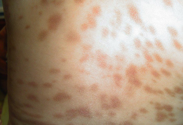
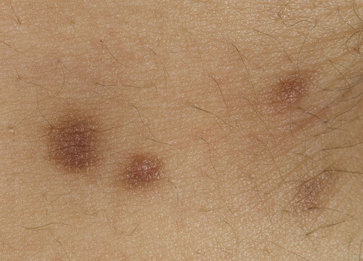
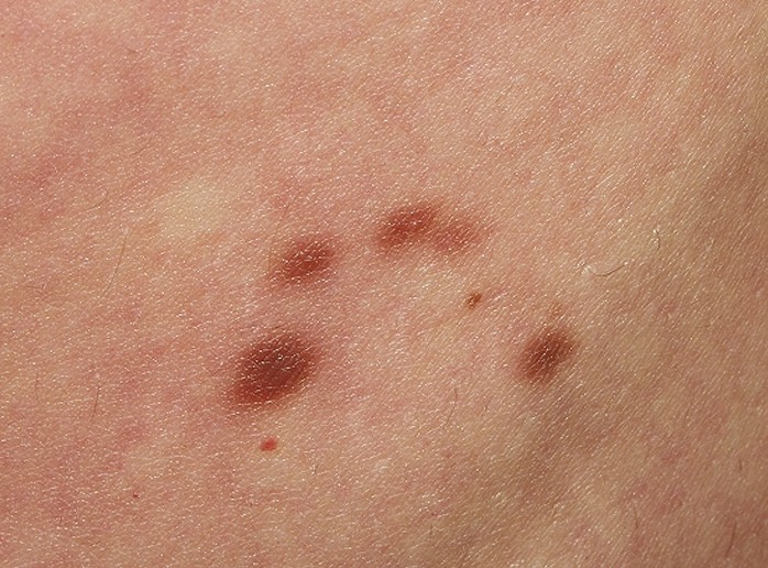
  • The skin presents patches where there is discoloration. These patches extend with the passing of time but the growth is slow (several decades can pass before the patient notices the lesions).
  • The patches of discolored skin are seen on the torso in males and on the inferior limbs in females.
  • Often confused for other skin formations, such as freckles, moles or lentigo.
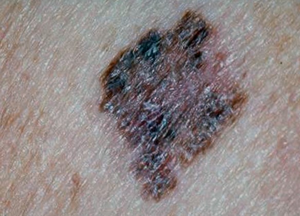
  • They are already large in size when they are diagnosed, having a diameter of one or two millimeters.
  • Their shape is irregular and the border is not always clear.
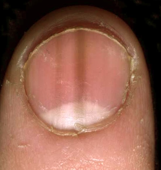
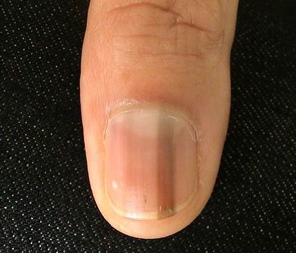
  • They can have various colors (because of the different pigmentation), ranging from different shades of brown (lighter, darker) to black and blue, not to mention lighter shades of gray, pink or red.
  • The discolored area might present streaks that have the color of the skin.
  • When these lesions appear, their surface is initially smooth. As the time passes and the lesions become more distinctive, the surface becomes thicker, irregular and it is characterized by increased dryness.
  • When the melanoma has breached the border and invaded other layers of the skin, the symptoms presented by the patient might be:
    • Partial thickening of the discolored patch of skin
    • More than one color present in the same area (often darker shades are noticed, including blue and black)
    • There may be bleeding from ulcerating lesions
    • The patient might feel a stinging sensation and also the need to scratch.

Causes of Superficial spreading melanoma

The main cause that leads to the appearance of superficial spreading melanoma is the development of pigment cells that are cancerous. These appear initially in the basal layer of the epidermis and they can breach this layer, entering the dermis. The interesting thing is that this type of cancer can appear on a healthy skin, without previous problems but in 25% of the cases it appears within an existing mole. This mole can be common but there are many situations in which the superficial spreading melanoma is linked to atypical, congenital or funny looking moles.

Specific gene mutations have also been incriminated as a direct cause and it seems that the damage caused by UV radiation (especially in case of sunburns and artificial tanning) is a cause of superficial spreading melanoma as well.

Besides the actual causes that lead to the appearance of this skin condition, there are certain risk factors that can add their contribution to the problem. These are:

  • Age – as the superficial spreading melanoma develops over several decades, it seems that old age is a risk factor (however, the lesion has existed for some time and only then it was noticed and diagnosed).
  • Existence of invasive melanoma or melanoma in-situ – patients who have already been diagnosed with such problems stand a high chance of receiving the same diagnosis all over again.
  • Existence of other types of skin cancer
  • Numerous moles of various colors and forms present on the skin. This becomes an even more obvious risk factor if there is constant and prolonged sun exposure
  • Family history of melanoma
  • Light skin and light colored eyes

Diagnosis

Superficial spreading melanoma can be diagnosed based on the clinical findings and the patient’s medical history. In the majority of cases, the dermatologist will first use a dermatoscope in order to take a better look at the lesion and then he will most likely recommend what is known as an excisional skin biopsy. The biopsy will be able to determine whether there are malignant cells or not. If a lesion is thick (over one millimeter thickness), then additional investigations will be recommended (complete blood count, biopsy of lymph nodes and CT or MRI).

Treatment

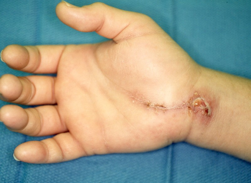
When it comes to superficial spreading melanoma, the first choice of treatment is surgical removal of the lesion. The surgeon will also take some safety margins, removing some healthy tissue surrounding the affected area, to be safe. However, as postoperative treatment the doctor might recommend radiotherapy, to make sure that the tumor is gone completely. In cases of metastatic melanomas, it is highly recommended that the lymph nodes are removed as well.

Survival rate

The prognosis for the superficial spreading melanoma is poor if the lymph nodes in the area are swollen or if there are metastases affecting the brain, the bone, the lungs or other parts of the skin. It is important to understand that the superficial spreading melanoma that stays within its border is not dangerous; it becomes serious when that border is breached and malignant cells enter other layers of the skin. The thickness of the melanoma upon surgical removal is also a good indicator for the survival rate. This is why it is highly important to go to regular follow-up controls and make sure that the lesion has not progressed or, in case of removal, that it did not appear again.
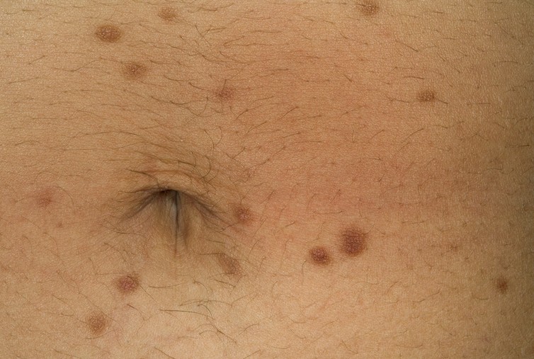
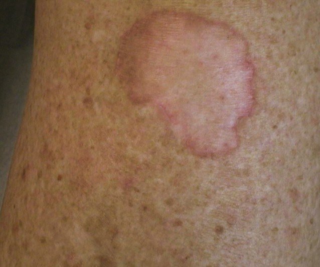
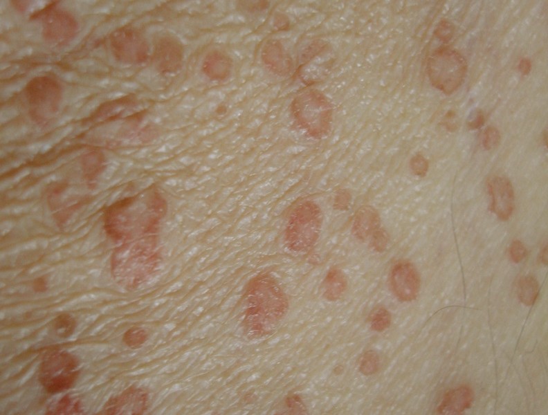
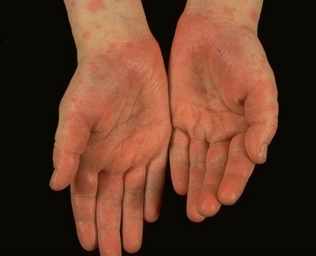
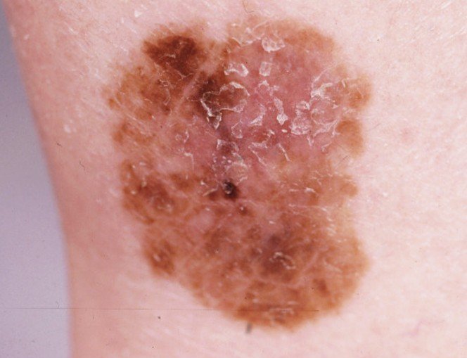
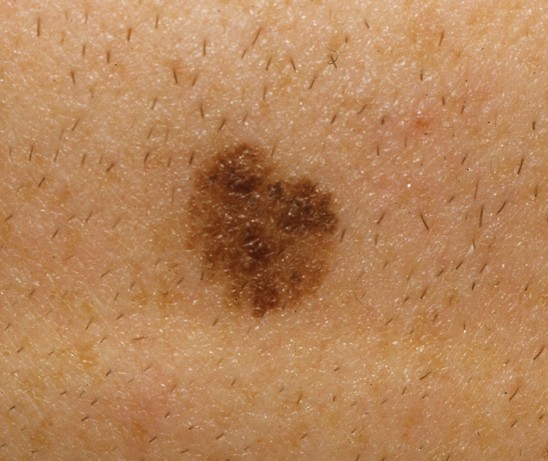
Pictures

Collection of photos, images and pictures of Superficial Spreading Melanoma…

superficial spreading melanoma

superficial spreading melanoma pictures

superficial spreading melanoma pictures 2

superficial spreading melanoma pictures 3

superficial spreading melanoma pictures 4

No responses yet

Sore Throat Without Fever

Mar 16 2017 Published by under Ear, Nose and Throat

Almost everyone has had a sore throat at one time or the other and I can tell for certain that it is not fun to have. You suddenly awake to see yourself caught up with swollen gland, difficulty with swallowing, pain and fever. However, on a few occasions, you might discover that you are sore in the throat but have no fever which usually accompanies a sore throat. Usually,a sore throat disappears after just a few days but when the condition persists for more than one week, it should become a cause for concern.

This article has been written to guide you as you seek to understand the factors responsible for sore throat without fever, the care and treatment options and when to see the doctor.

Causes of Sore Throat with No Fever

sore throat with no fever

The lymph nodes are located in different parts of your body. Their primary responsibility is to clean up your lymph fluids. They act as soldiers helping the body to prevent infections and some diseases like cancer. The lymph fluid contains lymph, white blood cells, water, fat and protein. As they filter the blood, collecting and destroying infections, they may in the process become infected as well.

Having sore throat is primarily as a result of infection of the pair of lymph nodes on the posterior part of your tongue. There are different reasons behind this swelling and sore occurring in the nodes. A sore throat usually heralds a cold and comes with fever as one of the presenting symptoms. However, we are looking at occasions where an individual has a sore throat but no fever. Some of the factors responsible for this condition are:

1. Common Cold

This is a common cause of sore throat with no fever. It is a health condition caused by a virus and can easily spread to other people. It causes inflammation of the membranes that line the nose and throat. Although it could be caused by a plethora of viruses but the most common causes are the rhinoviruses and coronaviruses. Other presenting symptoms of common cold include stuffy runny nose, scratchy itchy throat, sneezing, fatigue, mild hacking, cough, headache etc

2. Syphilis

Having sore throat with no fever may also be as a result of syphilis. Syphilis is a highly infectious STD contracted primarily through sexual activities. Engaging in oral sex and prolonged kissing with anyone who has syphilis is an avenue for contracting this contagious disease. Sore throat is one of the non-genital symptoms of syphilis.

3. Fifth Disease

Another possible cause of sore throat with no fever is the fifth disease. Fifth disease also known as erythema infectiosum and “slapped cheek’’ disease is a viral infection that causes rash. It is contracted through contact with fluid from the nose and throat as well as blood of an infected person. Although it is moderately infectious and common among children of school age, it can also be contracted by adults. Other symptoms are, nausea and vomiting, diarrhea, rash, itching and runny nose.

4. Leukemia

This is a medical condition that results in the abnormal growth of cells of the blood and bone marrow or simply put, it is the cancer of the blood. As a result of leukemia, some patients end up producing abnormal white blood cells which are not mature. These white blood cells are unable to fight off diseases and infections the way healthy cells should. The lymph nodes become susceptible to infections as a result also. This results in sore throat, anemia, easy bleeding and susceptibility to infections.

5. Allergies

Sometimes a mild sore throat can easily be treated with lozenges but on few occasions the sore throat refuses to go away lasting several days or even weeks after treatment. This could be as a result of exposure to some allergens. Allergic reactions to airborne particles such as pollens could cause irritation of the throat leading to sore throat. When an individual is exposed to an allergen, postnatal drip; a condition in which mucus of the sinus drains down the throat, occurs causing tickling and scratchy pain, sneezing, congestion, itchy eyes and coughing and also causing the throat to become sore in the process.

6. Bronchitis

The bronchial tubes are the main passages through which air is delivered to the lungs. Sometimes these tubes can become inflamed; a condition known as bronchitis, which leads to the buildup of mucus causing such symptoms as sore throat, coughing, shortness of breath etc. Bronchitis can either be acute or chronic depending on the duration. Acute bronchitis lasts a few days before clearing up while chronic bronchitis lasts longer. Chronic bronchitis is a type of chronic obstructive pulmonary disease (COPD) a respiratory disorder which occurs in cigarette smokers and former smokers and people who are constantly exposed to dust, fumes, ammonia, strong acid, chlorine, hydrogen sulfide, sulfur dioxide or bromine.

Treatment And Care Of Sore Throat With No Fever

The following tips can help to assuage the effect of the sore throat if they cannot totally eliminate the symptoms:

  • If sore throat is a result of allergic reaction, identifying the sources of the allergens is a vital way to alleviating the sore throat. Once identified, stay away from the allergens or irritants.
  • Drink water frequently to keep the throat moist at all times as dryness helps to aggravate the pains. Use warm fluid such as thin soup or warm tea to soothe and comfort the throat. Avoid caffeinated drinks.
  • Gargle with warm salt water to reduce the pain and fight the infection.
  • Use known over-the-counter painkillers daily. Antihistamines like Claritin and cetirizine are known to be very effective against pain. They also help prevent the body from mounting histamine responses to allergen attack in the body.

When To See The Doctor

Ideally, the sore throat should go away few days after home treatment, but if there is not much in terms of improvement and you experience any of the symptoms listed below or a combination then you need to see the doctor.

  • When the sore throat becomes severe and prolonged lasting up to two weeks
  • You have difficulty in breathing
  • You experience joint pain
  • You have difficulty in swallowing
  • You have earache
  • You start experiencing weight loss
  • When the sore throat becomes recurrent
  • When you have difficulty in opening your mouth
  • If you have an accompanying rash.

Conclusion

Whenever you have a sore throat, remember to pay attention to the signs and symptoms you have. Check if there is fever or not; this could make the difference. But should you have sore throat no fever, apply the remedies we listed in this post.

However, if these remedies fail to address your problem or if you notice the symptoms mentioned in the last part of this post, you should see a doctor immediately. You may need to undergo some tests in order to find out the root cause of the problem so that it can be properly addressed. Don’t wait until things get completely out of hands before you take action. Your life is too important to gamble with.

No responses yet

Bromhidrosis

Mar 16 2017 Published by under Diseases and Conditions

Bromhidrosis is also known as body odor and it is a common modification in individuals who have just gone through puberty. However, in some cases it can turn into a pathological and chronic condition, as the body odor becomes excessive and it interferes with the life of the person suffering from it. When an excessive and unpleasant odor is emanated from the skin, that person tends to refrain from certain activities and the quality of life is significantly impaired.

There are two main types of bromhidrosis. The first is the apocrine body odor, which is also the most common form. This appears when the sweat glands function excessively, leading to a characteristic body odor. To that excessive function is added an increase in the axillary bacterial flora, increasing the unpleasantness of the body odor (especially in the axillary region). The second is the eccrine body odor, which comes from the eccrine glands. These glands do not give out any odor in normal situations, but they can give a distinctive odor if a person consumes certain types of food (garlic for example), takes medication (penicillin) or drinks alcohol. Also, it seems that there is a genetic predisposition to bromhidrosis.

Bromhidrosis Symptoms

These are the most common symptoms of bromhidrosis:

  • Excessive, unpleasant body odor
  • Primary area from where the body odor comes: axillae
  • Other areas affected: soles of the feet, genital area
  • The odor can be described differently from one patient to the other. However, the most common words used for the description of BO were: pungent, acrid, musty or rancid.
  • Skin looks normal in the affected areas. Exception: association with other skin conditions (erythrasma, trichomycosis axillaris).
  • In case of eccrine bromhidrosis, the following modifications may occur on the skin in the affected areas:
  • Skin maceration (the keratin is consumed by bacteria)
  • Thick mat of moist keratin (physical examination will reveal it)
  • Modifications appear on the soles of the feet and in areas where the skin rubs together.

What Causes Bromhidrosis?

These are the most common causes that lead to the appearance of bromhidrosis:

  • Excessive secretion of apocrine or eccrine glands – the foul odor comes from the bacteria that actually feed on the keratin.
  • Inadequate bodily hygiene.
  • Dermatological conditions (two basic mechanisms can lead to bromhidrosis, meaning excessive sweating and bacteria overgrowth)
    • Excessive weight
    • Endocrine dysfunction – diabetes
    • Skin folds inflammation (intertrigo)
    • Trichomycosis axillaris (the hair shafts in the axilla are colonized superficially with bacteria)
    • Erythrasma (the areas where the skin rubs together become chronically infected)
    • Colonization of other parts of the body with different bacteria, including Sphingomonas paucimobilis.
  • Laser hair removal (potential adverse effect)
  • Metabolic disorders (in eccrine bromhidrosis):
    • Phenylketonuria
    • Fish odor syndrome
    • Sweaty feet syndrome
    • Odor of cat syndrome
  • In children – nasal foreign body.
  • Ingestion of certain foods (garlic)
  • Taking specific medication (penicillin)
  • Alcohol consumption
  • Toxic materials
  • Metabolite excretion in sweat (gout, scurvy, typhoid)

Treatment

These are the most common courses of treatment undertaken for bromhidrosis:

Change in hygiene habits:

  • Adequate washing of the axillary region
  • Removing sweaty clothing without delay. Also, try to refrain from wearing the same clothes for several days in a row.
  • Use of deodorant to reduce the foul odor and bacteria count
  • These hygiene changes are especially effective in the case of apocrine bromhidrosis

Reducing the bacteria count in the affected area by:

  • Regular shaving of the hair in the axilla
  • Using special methods for the removal of the hair in the axilla (such as electrolysis)

Topical antibiotics for cases of increased bacterial count (these inhibit the actual bacterial growth):

  • Clindamycin
  • Erythromycin

Other treatments include:

  • Antiseptic soap – this should be used on a regular basis in order to prevent the bacterial growth in the axillary region and not only.
  • Treatment of associated medical conditions (diabetes, intertrigo, trichomycosis axillaris, erythrasma etc.)
  • Antiperspirants with aluminum chloride – these are recommended in cases of bromhidrosis accompanied by hyperhidrosis
  • Iontophoresis for eccrine bromhidrosis (a weak electrical current passes through the skin)
  • Systemic anticholinergic agents (not the primary course of treatment because of the potential adverse effects)

Non-surgical treatments:

  • Laser (for the destruction of apocrine glands)
  • Botulinum toxin (recommended for eccrine bromhidrosis and in cases where the body odor is accompanied by excessive sweating)

Surgical treatments:

  • Surgical removal of apocrine glands (there are different surgical approaches and they may involve cutting into several layers of the skin). The surgical approach depends on the severity of the condition and the intensity of the body odor.
  • Carbon dioxide laser is used for the removal of apocrine gland residue
  • Superficial liposuction curettage
  • Ultrasound-assisted suction aspiration
  • Upper thoracic sympathectomy

Diet changes:

  • Eliminating certain foods from the diet or reducing the daily consumption (garlic, onion, spices)
  • Reducing the daily intake of alcohol.

No responses yet

Melanonychia

Mar 16 2017 Published by under Hair and Nails

What is Melanonychia?

Melanonychia is a medical condition in which the nail plate has a pigmentation band that is either black or brown in color. There are two main types of melanonychia, meaning longitudinal (this being often times a sign of subungual melanoma) and transverse. This medical condition appears within other medical conditions that are benign, such as the common mole.

As it was already mentioned, the band of pigment appearing on a nail has to be properly investigated, as it might be a sign of melanoma. It seems that individuals with darker skin and Japanese people present a higher risk, the percentage being reduced in the white population. As for age and gender, there is no particular category affected by melanonychia.

Melanonychia Symptoms

These are the most common symptoms of melanonychia:

  • Pigmentation band on the nail plate, of brown or black color
  • Single or multiple nail presence
  • The longitudinal type starts at the cuticle and ends at the free edge of the nail. Longitudinal melanonychia can be a sign of melanoma, being accompanied by the following:
    • Change in nail appearance
    • Alteration of nail color
    • Modification of nail pattern
    • Change in size of the pigmentation band
    • Pain – sudden onset
    • Ulceration or subungual blood
  • The transverse type runs across the nail
  • There are cases where the entire plate of the nail is affected
  • Periungual pigmentation can be present (this is also known as benign pseudo-Hutchinson sign)
  • Pigmentation bands are wider than 3 mm in 50% of the patients.

What Causes Melanonychia?

These are the most common causes of melanonychia:

  • Moles
  • Subungual melanoma
  • Melanocytic hyperplasia or activation, such as the melanotic macule of the nail plate
  • Dark skin – multiple bands
  • Pregnancy – multiple bands
  • Trauma
  • Bad shoes
  • Nail biting
  • Subungual foreign body
  • Radiation therapy
  • Systemic disease (Addison syndrome, Cushing syndrome, B12 deficiency)
  • Dermatological disease (psoriasis, lichen planus)
  • Chemotherapy
  • Drug related
  • Iatrogenic causes
  • Trauma
  • Post-inflammatory event

Melanonychia Treatment

The first and most important thing is to treat the underlying condition that has led to the appearance of melanonychia. These are the most common courses of treatment undertaken specifically for melanonychia:

  • Surgical excision of the total nail is recommended in case of melanoma
  • If the melanoma is aggressive and invasive, then the surgeon might decide to perform a complete amputation of the distal phalanx of the affected finger. This guarantees the best chance for the cancer not to spread to other parts of the body and affect vital organs
  • After the surgical excision, there will be other surgeries performed for the reconstruction of the area and the necessary skin grafting
  • In case there is a suspicion of melanoma, then the doctor will probably recommended a biopsy of the lymph nodes to be performed as well
  • If the melanonychia is caused by a benign condition and there are no other symptoms associated with the condition, then no treatment will be recommended by the specialist doctor
  • If the condition is caused by the taking of a certain drug, then the recommendation is stop taking that particular medication and find an alternative.

As you have seen for yourself, there are cases in which no treatment is required for melanonychia. However, if you have been diagnosed with longitudinal melanonychia, you need to keep a close eye on that nail. If you notice any change in it whatsoever, be sure to go to the doctor and request further investigations. The risk of melanoma is present in all patients with this type of melanonychia.

Melanonychia Pictures

Pictures collection of the medical condition Melanonychia…

melanonychia

melanonychia pictures

melanonychia pictures 2

melanonychia pictures 3

melanonychia pictures 4

melanonychia pictures 5

No responses yet

Carpal Tunnel Surgery

Mar 15 2017 Published by under Treatments

What is carpal tunnel surgery?

The carpal tunnel surgery is performed in patients diagnosed with carpal tunnel syndrome. This is actually a neuropathy in which the median nerve is compressed at the level of the wrist, causing a wide range of symptoms, including paresthesia, pain and numbness. The carpal tunnel surgery is performed in order to release the nerve from entrapment, eliminating the symptoms commonly associated with the condition. The patient will have to undergo physical therapy after the procedure, so as to promote faster healing in the area and have a normal range of mobility.

The carpal tunnel syndrome appears in people who suffer from a wide range of medical conditions and especially in those who work a lot of on computers. The procedure can be performed in an open manner or endoscopic and it guarantees promising results, as the pressure on the median nerve is released. The surgery is recommended in case of aggravated symptoms (constant numbness, weakness and atrophy of the muscles in the area) and also when other solutions have failed to provide the desired results (such as night splinting for example).

What to expect after carpal tunnel surgery?

carpal tunnel surgery

Carpal tunnel surgery

After the carpal tunnel surgery, your hand will be bandaged and you will have to undergo removal of the stitches after a period of two weeks. The symptoms commonly associated with the carpal tunnel syndrome will disappear gradually. However, if you undergo physical therapy, the exercises that the physical therapist will teach you are also going to help with the symptoms. The recommendation will be that you avoid heavy work involving the hands for at least three months after the carpal tunnel surgery. The endoscopic surgery guarantees a faster recovery, because there are no large cuts made to the hand.

This is a procedure that allows you to go home in the same day. Prescription pain medication will be recommended to deal with the pain, inflammation and other upsetting symptoms. Open carpal tunnel surgery will require that you wear a protective splint for a few days, in order to protect the recently operated area. Wrist braces might also be recommended for at least two weeks after the surgery. The doctor will make the exact recommendation when you can start to use the hand again but, once again, heavy activities should be avoided for a while. Also, it is important to understand that the expectations should be different if the surgery took place on your non-dominant or dominant hand.

Procedure

There are two main methods used for the carpal tunnel surgery:

Open carpal tunnel surgery

  • The transverse carpal ligament is cut and the pressure on the median nerve is released
  • The doctor will make an incision at the base of the palm, making the transverse carpal ligament visible. After the ligament is cut, the incision is closed with stitches. Scar tissue will form at the site of the cut and this is why physical therapy is so important. The hand will be bandaged and a splint or brace might be recommended.

Endoscopic carpal tunnel surgery

  • A small incision is going to be made at the level of the wrist and/or palm, depending on the approach technique
  • The endoscope will be introduced in order to visualize the transverse carpal ligament. This will be cut with tiny yet precise instruments. The recovery period for this type of procedure is better, as there are no large incisions made in the skin. The small incision is stitched as well.

Cost

The cost of the carpal tunnel surgery depends from one clinic to the other and from one geographic area to the other. Generally speaking, the cost is around $1.300 for the actual outpatient procedure but you will also have to pay somewhere around $900 for the physician services and $500 for the anesthesia. It is for the best to check your insurance and see which costs are covered for this type of procedure. Depending on your type of insurance, you might be covered for the whole thing.

Also, it is important to understand that the cost of open carpal tunnel surgery is less expensive than the one of the endoscopic carpal tunnel surgery, because of the differences in the technology being used. If there are complications during the surgery, then the overall cost might increase dramatically. As it was already mentioned, the costs for this kind of procedure are split into three categories: first, the services of the surgeon, second the ones of the clinic or hospital and third, the ones for anesthesia. Plus, you will have to take into consideration the costs for the physical therapy recommended after the actual surgery.

Complications

These are the most common complications of carpal tunnel surgery (low rate):

  • Infection – there are few chances of infection occurring but when it happens oral antibiotics might be recommended and a second surgery will be scheduled as well in order to clean the infected area.
  • Symptoms of the carpal tunnel syndrome persist (surgical failure to release the pressure on the median nerve) – this happens because the cut of the transverse carpal ligament was incomplete or because there was a lot of fibrous tissue in that area. Sometimes, the surgery fails because there was too much damage already done to the nerve or the nerve was compressed in other areas of the body (elbow for example). The median nerve also might suffer from different diseases and the patient will exhibit the same symptoms as if it was compressed.
  • Symptoms of the carpal tunnel syndrome disappear and recur after a short period of time – revision surgery might be recommended in such cases. Recurrent symptoms might also appear when there is too much scar tissue at the site of the surgery, plus there is always the possibility that the transverse carpal ligament applies pressure on the nerve again.
  • Damage to the nerve – if that happens, the patient will experience abnormal sensations or loss of sensation, not to mention the entire functionality of the hand will be affected.
  • Persistent numbness in the area
  • Loss of mobility because of the excessive scar tissue

Pictures

Collection of pictures of carpal tunnel surgery…

carpal tunnel surgery pictures

carpal tunnel surgery pictures 2

carpal tunnel surgery pictures 3

Recovery time

The recovery time depends on the type of surgery that was performed:

Open carpal tunnel surgery

  • Surgery performed on the non-dominant hand – the patient can return to his or her normal activities within one or two days. However, the recommendation is that the patient waits at least two weeks before resuming normal activities, so as to allow for the area to heal.
  • Surgery performed on the dominant hand – the patient is expected to make a full recovery in a period of at least six weeks until twelve weeks. Only then, he or she will be able to resume his or her normal work-related and household-related activities.

Endoscopic carpal tunnel surgery

  • Surgery performed on the non-dominant hand – the patient can return to his or her normal activities within one or two days. However, heavy activities performed with the hands should be avoided for a period of time.
  • Surgery performed on the dominant hand – the patient is expected to make a full recovery is a period of four weeks. Depending on the patient, the recovery time might last more but physical therapy is recommended for faster healing.

Video

Watch this Carpal Tunnel Surgery Video to learn more about this condition:

No responses yet

Yellow Nails

Mar 14 2017 Published by under Hair and Nails

Not many people are aware of this fact but the truth is that our nails can provide useful information on our body’s general health. Sometimes, the nail can present a certain discoloration and turn a yellowish shade. This can be a sign of infection or it can be a sign for various conditions of the skin. In some cases, the nail thickens and the growth might even be decreased. The affected nail might lack the protective cuticle and it might also detach itself from the nail bed.

Both men and women can suffer from yellow nails and it is always important to identify the underlying condition. This has to be treated first for the nails to return to their normal color. For many people, the concern is also of aesthetic importance, as having yellow nails can be a source of embarrassment. Whether the condition affects the fingernails, the toenails or both, the basic aspect is the same. You need to find what is causing it and then treat it.

Causes of Yellow Nails

These are the most common causes that lead to the appearance of yellow nails:

  • Respiratory disease (bronchitis)
  • Nail polish (the pigments used in the nail polish can stain the nails, especially when it comes to darker shades)
  • Fungal infection (with Trichophyton rubrum, for example)
  • Chronic liver disease
  • Excessive smoking
  • Poor lifestyle habits
  • Rheumatoid arthritis
  • Congenital heart disease
  • Yellow nail syndrome – this is a hereditary condition in which three major health modifications occur. The first is related to the nails, as these suffer from discoloration and dystrophy. The nail will grow more slowly and they will have a yellow color. The patient will also suffer from swelling of the hands (lymphedema) and chronic respiratory disease affecting the lungs.

How to treat Yellow Nails?

In order for the appearance of your nails to improve, you will have to follow a specific treatment for the medical conditions that were presented above. For the yellow discoloration presented in the yellow nail syndrome, the most efficient treatment turned out to be clarithromycin.

Apart from that, there are certain home remedies you can try in order to improve the appearance of the nails as well. These are:

Lemon juice

lemon juiceThis is recommended for whitening yellow nails, as the active substances contained in the lemon juice can act as bleaching agents. Squeeze some fresh lemon juice, put it into a bowl and soak your nails for a quarter of an hour. Then, use a soft brush in order to improve the color of the nails and scrub the yellow discoloration off. Rinse in warm water and apply a moisturizer made from natural ingredients. Repeat twice a day until the nails resume their natural color. The same process can be followed by using lemon essential oil, as an alternative to fresh lemon juice.

Hydrogen peroxide

hydrogen peroxide for yellow nailsThis is recommended because of its whitening properties but you need to be careful and use solutions that contain only 3% of this substance. The recommendation is that you mix the hydrogen peroxide with water and soak your nails for two minutes. Then, you will have to scrub them with a gentle toothbrush and rinse with warm water. You can also apply the substance directly to the nails, if the discoloration is severe and then follow the same process. Apply a moisturizer to prevent the skin from becoming dehydrated.

Baking soda

baking soda for yellow nails
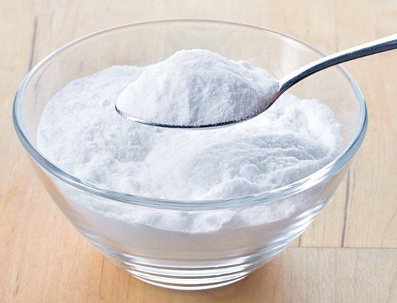
This is recommended because of its whitening and exfoliating properties. One indication is that you mix baking soda with hydrogen peroxide and then apply the mixture on the yellow nails. You keep it for three or four minutes, then rinse in warm water and apply moisturizer. Another indication is that you mix baking soda with olive oil and fresh lemon juice, apply the mixture and leave it on for five minutes. The first procedure should be repeated every six to eight weeks, while the second every two weeks.

Tea tree oil

tea tree oil for treatment of yellow nailsThis is recommended especially when the yellow discoloration is caused by a fungus infection. Tea tree oil is a natural fungicide and it has amazing antiseptic and antimicrobial properties. You can use it to fight against the fungi infection and also to restore the natural color of your nails. Using an eyedropper, apply the tea tree oil for a few minutes, then rinse in warm water and apply moisturizer. This procedure should be repeated two times a day. Or, if you want, you can mix the tea tree oil with olive oil and apply it to your nails.

Apple cider vinegar

apple cider vinegarThis is recommended because of its antifungal properties. Mix the apple cider vinegar with warm water and soak your nails for about twenty minutes. Dry your nails with a soft towel and repeat the procedure at least three times every day.


Pictures of Yellow nails

Collection of pictures of yellow nails, fingers and toenails…

yellow nails pictures

yellow nails pictures 2

yellow nails pictures 3

yellow nails pictures 4

No responses yet

Urticaria Pigmentosa

Mar 14 2017 Published by under Skin Conditions

Urticaria pigmentosa is a skin disorder in which the skin presents patches of dark color and the patient experiences intense itchiness. Rubbing or scratching the lesions can lead to the appearance of hives. This skin condition is also known as the generalized eruption of cutaneous mastocytosis and it most often affects children. Mastocytosis is a rare skin condition in which the number of mastocytes is excessive.

The mastocytes are the cells responsible for the elimination of histamines in case there is an allergen breaching into the body. Being diagnosed with any form of mastocytosis means that patient is more susceptible to serious allergic reactions, ranging from urticaria pigmentosa to severe anaphylactic shock.

Urticaria Pigmentosa Symptoms

These are the most common symptoms of urticaria pigmentosa, caused by the excessive amounts of mast cells present in the skin:

  • Red or brown spots or patches present on the skin
  • Patches are initially confused with moles or insect bites
  • Lesions persist and increase in number over the course of years
  • The areas that are most affected – chest, back and forehead
  • Darier’s sign – stroking the skin in a particular area will cause inflammation, redness and itchiness (histamine response)
  • Scratching can aggravate the condition and can also lead to the formation of blisters filled with liquids in children
  • Flushed face in certain cases

Symptoms can vary considerably from one person to the other:

  • Mild – redness, irritation
  • Moderate – accelerated intestinal transit, cardiac pulse changes, nausea, vomiting, headaches and fainting
  • Severe – anaphylactic shock, with difficult breathing and vascular collapse. This requires emergency medical treatment.

Possible Causes of Urticaria Pigmentosa

Urticaria pigmentosa appears in people who have too many mast cells in their skin. The main cause is determined by the mutation of an amino acid, this in turn leading to the division of more and more mast cells. Several mutations have been incriminated into the appearance of urticaria pigmentosa and it seems that each mutation leads to a different time of appearance for the skin condition. Familial cases of urticaria pigmentosa have also been identified in the medical literature. Patients who have received radiotherapy as a treatment for breast cancer have also developed certain forms of urticaria pigmentosa.  Two other conditions have been linked: HIV infections and disorders of the autism spectrum.

It is believed that there are many triggering factors that can also cause the condition to manifest itself. These are:

  • Stress
  • Emotion
  • Anxiety
  • Physical exercise
  • Excessive heat
  • Skin friction
  • Bacterial infections
  • Venom from different animals
  • Eye drops containing allergenic substances
  • Anti-inflammatory drugs – aspirin is one of the most controversial triggering factors, as a certain amount of the drug will prevent the cells from dividing while a bigger amount will cause a histamine response.
  • Alcohol
  • Treatments with morphine.

Treatment

The treatment for urticaria pigmentosa is aimed to provide relief from the symptoms experienced, both at the level of the skin and also at a systemic level. The recommended is that all substances that might trigger an allergic response and thus the release of histamine should be avoided. As for the conservative treatments, they are as it follows:

  • Antihistamines – these are recommended in order to reduce the itchiness, alleviate the flushing and also improve the symptoms experienced in the gastrointestinal tract.
  • Oral disodium cromoglycate – this is recommended for patients who experience both cutaneous and systemic symptoms. This medication can be used in order to ameliorate the itchiness and the flushing at the level of the skin but also to improve the systemic symptoms, such as the accelerated intestinal transit, the abdominal cramps and other similar problems.
  • Aspirin – this is recommended in a certain dose in order to maintain the mast cell degranulation. It is especially indicated in patients who have shown resistance to antihistamines but one must be careful as a high dose of aspirin could aggravate the symptoms of urticaria pigmentosa and even induce vascular collapse.
  • Topical corticosteroids are recommended for cutaneous symptoms restricted to smaller areas.
  • Intralesional injections with corticosteroids are recommended in more serious cases but the treatment cannot be performed for prolonged periods of time, as there is an increased risk of skin atrophy.
  • Systemic corticosteroids are recommended only in severe cases, where there are other medical problems present, including malnutrition, liver disease or grave skin disorders.
  • Oral psoralen in combination with UV-A therapy has shown promising results, improving the appearance of the skin affected by the lesions. However, prolonged treatments of this kind increase the risk for skin cancer.
  • Immune modulators
  • Patients who know that they present an increased risk for anaphylactic shock should wear medical alert braces and they should always have an epinephrine self-injector with them.

Urticaria Pigmentosa Pictures

Collection of photos and pictures of Urticaria Pigmentosa…

urticaria pigmentosa

urticaria pigmentosa pictures

urticaria pigmentosa pictures 2

urticaria pigmentosa pictures 3

No responses yet

Porokeratosis

Mar 13 2017 Published by under Skin Conditions

What is Porokeratosis?

Porokeratosis is a medical condition in which the process of keratinization is affected and disrupted. The patient presents patches of atrophy on the skin, these having distinctive clinical and histopathological features. The most distinctive feature is the border surrounding the patch, this being obviously hyperkeratotic. This border is also known as the cornoid lamella. Porokeratosis has more than just one clinical variant and you will find them presented below. Also, keep in mind that squamous cell carcinoma can develop within porokeratosis patches.

These are the most common forms of porokeratosis:

  • Classic porokeratosis of Mibelli
  • Disseminated superficial actinic porokeratosis (there is also a non-actinic variant identified)
  • Linear porokeratosis
  • Disseminated plantar and palmar porokeratosis
  • Punctate porokeratosis

Less common forms include:

  • Porokeratosis ptychotropica (verrucous variant, localized on the buttocks)
  • Porokeratoma (also known as porokeratotic acanthoma)
  • Porokeratotic adnexal ostial nevus (rare congenital disorder)
  • Pruritic papular porokeratosis

Porokeratosis Symptoms

The symptoms of porokeratosis depends on the actual form of the skin condition:

Classic porokeratosis of Mibelli

  • Small lesion located on extremities (more common) or in the genital area or mucous membrane (less common).
  • These lesions do not cause other symptoms in the majority of cases.
  • In some patients, mild itchiness may occur.
  • Lesions expand gradually but slowly over the course of years.
  • In immunosuppressed adults, lesions can appear and go through a rapid growth period.
  • Diameter of several cm (medical literature depicts cases of giant lesions, up to 20 cm in diameter).
  • Center of the lesion can suffer from modification of pigmentation (hyper or hypo).
  • In some cases, the center of the lesion presents scales. It can also be atrophic or characterized by the absence of hair.
  • Hyperkeratotic patches might also be present on the skin.

Disseminated superficial actinic porokeratosis

  • Multiple macules present on the arms and legs (distal extensor surface)
  • Presence of cornoid lamella
  • No lesions on the palms and soles
  • In some patients, facial lesions may occur
  • Hyperkeratotic patches might also be present in some cases
  • Lesions usually asymptomatic
  • Itchiness or burning sensation may accompany the lesions in certain cases
  • The non-actinic variant presents a generalized distribution of the lesions

Linear porokeratosis

  • Red or brown patches on the skin (unilateral)
  • Keratotic papules or plaques (linear distribution)
  • The distribution is along the embryonic lines of Blaschko
  • Less common – bilateral or generalized distribution
  • High risk of malignancy degeneration

Disseminated plantar and palmar porokeratosis

  • Areas affected – palms and soles
  • Generalized distribution or along the embryonic lines of Blaschko
  • Usually asymptomatic
  • May present itchiness or tenderness to palpation
  • Discomfort when walking in plantar lesions

Punctate porokeratosis

  • Multiple keratotic papules
  • Presence of cornoid lamella
  • Areas affected – palms and soles.

Causes of Porokeratosis

These are the most common causes and risk factors that lead to porokeratosis:

  • Genetic inheritance
  • Exposure to artificial ultraviolet light and prolonged exposure to the sun
  • Deficiency of the immune system
  • Renal transplants
  • Radiation therapy
  • Chemical and thermal burns
  • Dialysis
  • Photodynamic therapy
  • Cancer

Treatment

As there are so many different types of porokeratosis and they can all manifest differently in various patients, it should come as no surprise that the treatment of this condition is highly particularized. When prescribing a certain treatment, the doctor will take into consideration how spread the lesions are on the skin, where they are located and what are the risks of them becoming cancerous. However, apart from treatment, the patient will be recommended to avoid sun exposure, to use high SPF sunscreen when going out and to come to regular check ups so as any signs of cancer can be immediately identified.

These are the most common courses of treatment for porokeratosis:

  • Topical 5-fluorouracil – improves the symptoms and induces remission of all forms of porokeratosis.
  • Analogues of D3 vitamin – these are administered as topical treatments as well, being especially recommended for the disseminated superficial actinic porokeratosis. Two of the most recommended choices are calcipotriol and tacalcitol.
  • Anti-inflammatory creams, such as diclofenac, might also be used for the disseminated superficial actinic porokeratosis.
  • Modulators of the immune system – these are administered as topical treatments and the most recommended choice is imiquimod cream. This treatment is especially recommended for the classic form of porokeratosis (Mibelli).
  • Retinoids
    • Topical retinoids are recommended in the cases of porokeratosis where the cornoid lamella is present. They can reduce the level of excessive keratosis and reduce this distinctive border of the lesion. However, these are not recommended to be taken for prolonged periods of time, as they can have serious side-effects on one’s health.
    • Oral retinoids are recommended only in severe cases of porokeratosis, where there is an increased risk for malignancy degeneration.

Porokeratosis Pictures

Here are some of the pictures collection of the condition Porokeratosis…

porokeratosis

porokeratosis pictures

porokeratosis pictures 2

porokeratosis pictures 3

No responses yet

Waking Up Sweating

Mar 13 2017 Published by under General Health

When a person wakes up drenched in sweat, even if the room temperature is cool, then that person might be suffering from a medical condition that is known as sleep hyperhidrosis. This can occur in a localized manner (no underlying pathological condition) or generalized (often a symptom of other medical conditions). It affects persons of any age but most commonly appears in adults. Women are more affected by this condition than men, especially those who are going through menopause. Also, dark skinned individuals present a higher risk for sleep hyperhidrosis than those with a fair skin.

There are certain factors that increase the risk for a person to wake up sweating. These are, among others: obesity, drinking excessive quantities of caffeine-based drinks, alcohol and eating unhealthy foods. Also, it is important to understand that the excessive sweating during the night might represent a sign that there is an infection somewhere in the body. It is for the best to consult a doctor and identify the exact cause that leads to you waking up drenched in sweat. The doctor will recommend the necessary tests and investigations for the confirmation of the diagnosis.

Causes of Waking Up Sweating

These are the main causes that can lead to you waking up sweating:

  • Menopause – entering menopause, women experience an entire range of upsetting symptoms, including hot flashes, weight gain and excessive sweating, including during the night
  • Obstructive sleep apnea
  • Medication – anti-depressants, anti-inflammatory medication (aspirin), corticosteroids (prednisone), medication for sleep apnea, anti-psychotics, medication to lower high blood pressure, anti-histamines, hypoglycemic agents
  • Alcohol drinking
  • Addiction to drugs (heroin)
  • Low blood sugar (in diabetes)
  • Infection – tuberculosis, endocarditis, osteomyelitis, abscess (of lung, liver or spleen), HIV, cystitis, fungal infections , bacterial infections (brucellosis), viral infections (mononucleosis)
  • Cancer – lymphoma, leukemia, breast cancer
  • Hormonal dysfunction – overactive thyroid gland (Hashimoto’s disease), acromegaly
  • Digestive disorders – GERD, ulcerative colitis, inflammatory bowel disease
  • Idiopathic – the exact cause that leads to the appearance of sweating during the night is unknown
  • Environmental – not enough air in the room, too many blankets, synthetic bed sheets
  • Emotions, stress or anxiety
  • Burn out syndrome
  • Chest pain
  • Diabetic neuropathy
  • Pulmonary edema
  • Sarcoidosis – this is a medical condition in which nodules appear in various organs, these nodules being comprised of inflammatory cells
  • Granuomatous disease – this is a genetic disease in which the immune system lacks the necessary strength to kill certain type of bacteria or fungi
  • Disorders of the bone marrow – myelofibrosis (this is a medical condition in which the normal production of blood cells is disrupted)
  • Rheumatological disorders – arteritis (inflammation of the arterial walls)
  • Neurological disorders – stroke, epilepsy, cerebral palsy, migraine, head trauma
  • Mercury poisoning

Treatment

These are the most common courses of treatment for the persons waking up sweating:

Self-care:

  • Blanket removal and room ventilation can alleviate the discomfort caused by the excessive sweating
  • Wiping or washing the localized areas in the body where the sweating has occurred
  • Changing clothes and taking a shower with the water temperature at a moderate level (towards colder if possible, to induce vasoconstriction and reduce sweating). It is also recommended that you take a cold shower before actually going to bed.
  • Lowering the room temperature
  • Avoid drinking alcohol and eating spicy foods before going to bed
  • Drink water through the night as well (keep a bottle of water by the bed)
  • Emotion, stress and anxiety control
    • Relaxation techniques
    • Breathing therapy
    • Counseling
    • Time management
    • Physical exercise
  • Lifestyle changes
    • Quitting smoking and drinking alcohol
    • Eliminating caffeine-based drinks from the diet
    • Healthy fruits and vegetables eaten on a regular basis
    • Drinking plenty of water
    • Avoiding spicy or greasy foods
    • Seek treatment for the underlying medical condition that causes excessive sweating

Herbal remedies:

  • Black cohosh tea (andropause)
  • Red clover (menopause)
  • Motherwort (soothing effect)
  • Sage tea (calming effect)

Topical antiperspirants (with aluminum ions) – these have the ability to block the sweating glands but they also present an increased risk for irritation of the skin.

Medication:

  • Anti-cholinergics (prevent excessive sweating)
  • Botulinum toxin – topical administration (subcutaneous injections)

Other treatments:

  • Paralysis of sweat glands through electrical stimulation and drug administration
  • Surgery – severe cases of excessive sweating
  • Endoscopic thoracic sympathectomy (controversial)

No responses yet

Red Palms

Mar 12 2017 Published by under General Health

Red palms are also known as palmar erythema in the medical literature. This condition can be caused by various medical problems, each having its own range of severity. In a lot of patients, red palms have no identifiable cause and they are presented under primary palmar erythema. However, there are many cases in which the red palms are a symptom of another disease, being presented as secondary palmar erythema.

It is important to contact the doctor if you have noticed that your palms have gotten red, as this can be an early sign of an underlying medical condition. As specialists tell us, it seems that palms are directly related to the proper functioning of the circulatory system, the hormonal system and especially the thyroid. So, when you have red palms, this can be a symptom of another disease and you need to see a doctor.

Symptoms of Red Palms

These are the most common symptoms associated with red palms:

  • The palms have a reddish tinge
  • The area of the palm most affected is the hypothenar eminence
  • Can also appear on the thenar eminence and the fingers
  • Can be accompanied by red soles (plantar erythema)
  • The skin in the affected areas can be warm to the touch
  • No itchiness, scaling or pain

Possible Causes of Red Palms

These are the most common causes that lead to the appearance of red palms:

  • Pregnancy – one of the most encountered causes of red palms; during pregnancy, the estrogen levels increase and the circulatory system also goes through a change.
  • Heredity (role in the onset of red palms but has only been identified in rare cases)
  • Idiopathic (unknown cause, no identified triggers)
  • Liver disease – hepatitis, hereditary liver disease (Wilson’s disease, hereditary haemochromatosis), cirrhosis. In chronic liver disease, the estrogen and or the oestradiol present abnormal levels. Moreover, it seems that there is a dysfunction in the arteriovenous anastomoses.
  • Problems with the adrenal glands
  • Cancer – leukemia, brain cancer, metastases, liver cancer, ovarian cancer. A growing tumor will always cause the estrogen levels to rise, causing red palms to appear among other symptoms.
  • Thyroid problems – thyrotoxicosis, hyperthyroidism. The patients present an increased level of estradiol, leading to their palms becoming red.
  • Circulatory problems – polycythemia vera
  • Shoulder-hand syndrome – this is also known as reflex sympathetic dystrophy. It affects the upper extremities of the body and it is accompanied by pain and rigidity in the shoulder, hand and fingers. Red palms accompany this syndrome as well.
  • Rheumatological disease – lupus erythematosus, rheumatoid arthritis. The exact link between rheumatoid arthritis and red palms has not been identified yet but this symptom appears in almost 60% of the patients with rheumatoid arthritis.
  • Diabetes mellitus – patients describe their palms as being hot and red. In some patients, the palms might not be entirely red, but rather present red spots.
  • Certain medication – amiodarone (antiarrhythmic agent), gemfibrozil (oral drug used for the reduction of lipid levels), cholestyramine (bile acid sequestrant), topiramate (anticonvulsant), albuterol (anti-asthma medication, recommended for bronchospasms).
  • Smoking – prolonged smoking can lead to circulatory problems, including in the hands area, hence the red palms.
  • Chronic mercury intoxication – it is a well-known fact that the chronic poisoning with mercury leads to the appearance of neurological and cutaneous symptoms. Among those symptoms, you will find the red palms.
  • Oral contraceptives (high levels of estrogen) – as with pregnancy, the high levels of estrogen lead to the appearance of red palms in women.
  • Alcohol abuse – prolonged and excessive alcohol drinking has a negative effect on the health of the liver, leading to abnormal levels of estrogen and oestradiol. It is the same mechanism as in the case of liver disease.
  • Sarcoidosis – this is a multi-systemic inflammatory disease, accompanied by a wide range of symptoms, including red palms.
  • Gestational syphilis – getting infected with the syphilis virus during pregnancy can lead to red palms, among other symptoms.
  • Myelopathy – this is a degenerative condition that leads to the pinching of the spinal cord and circulatory problems, with red palms included.

Treatment

At the present, there is no specific treatment for red palms and often times this symptom disappears when the primary cause is correctly identified and treated. However, you must remember that red palms can appear without any identified cause or medical history as well. If there are no other medical conditions associated with the red palms, then the condition might disappear on its own.

Pictures of Red Palms

Here are some of the pictures of red palms and fingers…

red palms

red palms pictures

red palms pictures 2

red palms pictures 3

No responses yet

« Prev - Next »

© 2011-2025 MDDK.com - Medical Tips and Advice. All Rights Reserved. Privacy Policy
The health information provided on this web site is for educational purposes only and is not to be used as a substitute for medical advice, diagnosis or treatment.