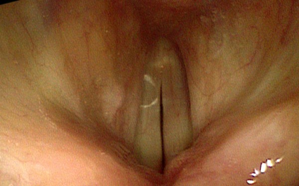
Cricopharyngeal Spasm
The cricopharyngeal spasm occurs in the cricopharyngeus muscle of the pharynx. This condition is often confused with a cancerous growth, as the person experiencing it will constantly describe the sensation of having a foreign body in the throat. However, it is important to understand that throat cancer is often detected at an advanced stage, when swallowing food becomes difficult. Even though it is quite a scary experience, the cricopharyngeal spasm is harmless. It will cause the patient to be quite uncomfortable but, given the fact that it is a self-limiting disorder, it will go away in a short period of time.
The symptoms of the cricopharyngeal spasm are not characteristic for the condition and this is why it can cause a lot of anxiety. If you are wondering why this condition occurs, you should know it appears when the esophagus valve does not function properly. In this article, you will find useful information presented, regarding the symptoms occurring with this condition, the causes that led to it in the first place and the available treatment. You will also be presented with information on how long does the condition last; in this way, you will know what to expect and be prepared in the future.
What are the Symptoms of Cricopharyngeal Spasm?
These are the most common symptoms of the cricopharyngeal spasm:
- ‘Lump in the throat’ sensation
- The sensation of foreign body or lump in the throat can cause a lot of discomfort for the patient; the lump sensation is not present at all times, as it comes and goes throughout the day
- The lump is present at the level of the cricoid cartilage
- Description of lump by the patient – phlegm, golf ball
- The patient constantly tries to eliminate the lump in the throat, without any success
- Choking or constriction sensation
- The patient describes his/her throat as being swollen
- Pain can accompany to feeling of ‘lump in the throat’ (constant or occasional)
- The symptoms of spasms are aggravated during the night
- Emotions, stress and anxiety are known to aggravate the symptoms of the cricopharyngeal spasm
- The patient experiences difficulties in swallowing saliva; the paradox is that the food is easy to swallow and it can actually contribute to reducing the intensity of the symptoms. The same goes for drinking water, as this is not prevented by the cricopharyngeal spasm
- The symptoms of the cricopharyngeal spasm can be presented for prolonged periods of time (months).
Cricopharyngeal Spasm Causes
These are the most common causes of the cricopharyngeal spasm:
- Excessive amounts of stress
- High levels of anxiety
- Positive and negative emotions
- Foods
- It is suspected that there are certain foods capable of triggering the cricopharyngeal spasm
- Among these foods, you will find nuts (peanuts in particular) and pumpkin seeds
- Affects susceptible individuals
It is important to mention that stress, anxiety and emotions can be triggers of the condition. However, when they are not direct causes, they can aggravate the cricopharyngeal spasm, making the condition worse. Patients need to learn how to handle their levels of stress and anxiety, in order to avoid such problems in the future.
Treatment
In the majority of cases, the cricopharyngeal spasm does not require any treatment. Being a self-limiting disorder, it will disappear on its own. However, these are the most common methods of symptomatic treatment for the cricopharyngeal spasm:
- Muscle relaxers
- Most commonly administered – benzodiazepines (Lorazepam, Diazepam)
- Reduce the feeling of throat constriction
- The frequency of the muscle spasms is reduced as well
- Should be administered with caution and only for short periods of time, as they can cause a person to become addicted to them
- Such drugs are often administered for diagnosis purposes – if they alleviate the symptoms, this means that the problem is related to stress and anxiety and not to something physical
- Relaxation and breathing therapy
- Recommended for the reduction of stress and anxiety
- The patients needs to control both stress and anxiety, as these can trigger the cricopharyngeal spasm and also aggravate the condition, once it occurs independently of them
- Patients are recommended to keep a diary of the moments when the cricopharyngeal spasm appeared, so as the triggering factors can be identified
- This therapy is provided by the speech language pathologist
- Physical therapy
- One can visit a physiotherapist who is specialized in such problems
- After a professional assessment, the physiotherapist can recommend a program that includes neck relaxation exercises
- Fluid drinking
- Drinking fluids can reduce the intensity of the symptoms
- It can also help the patient go better through the episode
- Keeps the throat tissues hydrated
- Warm fluids, such as tea, are highly recommended for this condition, as they comfort the throat
- Local anesthetic injections
- Improve the symptoms of the cricopharyngeal spasm in certain patients
- The results are positive but they only last for short periods of time
- Injections with the botulinum toxins
- Not all patients have received relief from the cricopharyngeal spasm with this treatment
- The relief was present in a number of patients and for a prolonged period of time, which is suggestive that this treatment could be quite efficient for all patient in the future
- Heat applications
- Placing a heated pad over the throat can help with the feeling of constriction and tightness
- The pad should be maintained on the throat only for a couple of minutes
- It can help the muscle relax, improving the symptoms of the cricopharyngeal spasm.
How long does the Cricopharyngeal spasm last?
While this condition is often presented as a self-limiting disorder, it is important that the symptoms of the cricopharyngeal spasm can extend over a period of several months. During this period, it is important to follow the recommended measures of treatment and learn how to properly control the levels of stress and anxiety. If you learn how to successfully manage stress, then you will reduce the duration of the cricopharyngeal spasm and be protected against future occurrences. The relaxation and breathing therapy is essential during such periods.