Diseases General Health Skin Conditions

Archive for February, 2016

Cutaneous Larva Migrans

Feb 10 2016 Published by under Skin Conditions

Cutaneous larva migrans is a type of dermatitis that occurs in people who have traveled to tropical and subtropical geographical areas. However, because more and more people travel in different areas on the globe, the infection is no longer confined to these areas. The skin presents an eruption that is accompanied by redness and intense pruritus. What happens is that the larvae of different parasites penetrate the skin and migrate from one areas of the body to the other. The eggs of the parasite are passed from the feces of animals into the soil and then the larvae start searching for a new host.

The infection with these larvae often happens when there is a breakdown in the skin, such as a cut or abrasion. However, the larvae can penetrate through the hair follicle and even through intact skin. Migration begins several days after the cutaneous penetration has occurred. This condition is more often encountered in animals and humans are only accidental hosts. This is a benign condition and it is strictly limited to the area of infection. The most disturbing symptom of cutaneous larva migrans is the intense itchiness. There is no gender prevalence and, when it comes to the age, it seems that children are more often affected than adults.

Symptoms of Cutaneous Larva Migrans

These are the most common symptoms of cutaneous larva migrans:

  • Tingling sensation
    • This appears half an hour after the skin penetration
    • It may also be described as a prickling sensation
    • There are also cases where the onset is late
  • Intense itchiness
  • The penetrated area is red
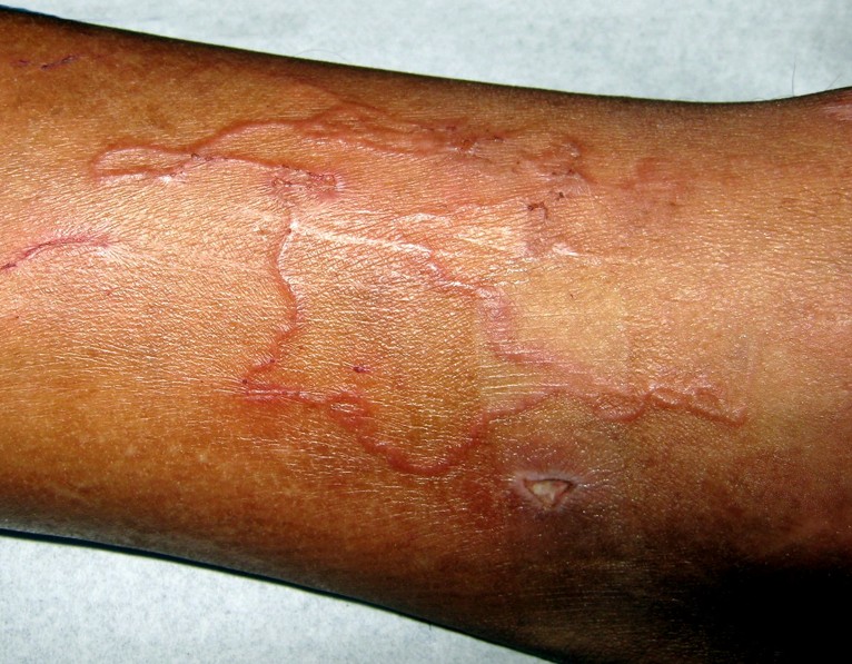
  • Lesions might form a straight line or even a serpiginous tunnel; when a tunnel is formed, it is a little bit elevated
  • Continuous advancement of the lesions (line or tunnel)
  • Lesions in the form of papules or vesicles
  • When vesicles form on the skin, these are filled with serous liquid
  • Systemic involvement:
    • Peripheral eosinophilia (Loeffler syndrome)
    • Migratory pulmonary infiltrates
    • Increased immunoglobulin E (IgE) levels
  • Areas that are most affected include:
    • Lower extremities
    • Dorsum of the feet
    • Interdigital space of the toes
    • Ano-genital region
    • Buttocks
    • Hands
    • Knees
  • Secondary infections can appear due to the excessive scratching

What are the Causes of Cutaneous Larva Migrans?

These are the most common parasites that lead to the appearance of cutaneous larva migrans:

Ancylostoma braziliense

  • Type of hookworm, commonly found in domestic animals
  • Present in the following geographical areas:
    • United States (central and southern)
    • Central America
    • South America
    • Caribbean

Ancylostoma caninum

  • Type of hookworm, commonly found in dogs
  • Present in Australia

Uncinaria stenocephala

  • Type of hookworm, commonly found in dogs
  • Present in Europe

Bunostomum phlebotomum

  • Type of hookworm, commonly found in cattle

Other parasites (rare cases):

  • Ancylostoma ceylonicum – type of hookworm, commonly found in cats and dogs
  • Ancylostoma tubaeforme – type of hookworm, commonly found in cats
  • Ancylostoma duodenale – type of hookworm, commonly found in humans, cats and dogs
  • Necator americanus – type of hookworm, commonly found in humans
  • Strongyloides papillosus – parasite commonly found in cattle, sheep and goats
  • Strongyloides westeri – parasite commonly found in horses
  • Pelodera strongyloides – parasite commonly found in dogs, cows, horses, sheep, guinea pigs and humans.

How is Cutaneous Larva Migrans Diagnosed?

These are the most common methods for the diagnosis of cutaneous larva migrans:

  • Clinical examination
    • Reveals characteristic appearance of the lesions
  • Travel history
    • Patients often declare having traveled in tropical or subtropical areas, performing different activities such as sunbathing or walking barefoot on the sand (exposure to the parasite eggs)
  • Complete blood count
    • Peripheral eosinophilia
    • Increased immunoglobulin E (IgE) levels
  • Skin biopsy
    • May identify the actual larva
    • Other modifications include:
      • Creation of basal layer tracts
      • Creation of under-the-skin burrows
      • Spongiosis – the epidermal layer of the skin presents vesicles
      • Keratin cells are necrotized
      • The epidermis and the upper layer of the dermis are inflamed and infiltrated with increased levels of eosinophils
  • Optical coherence tomography
    • The larva can be identified in the epidermis
    • Recommended for removal purposes

Differential diagnosis can be made with the following medical conditions:

  • Impetigo
  • Tinea pedis
  • Contact dermatitis
  • Dermatophytoses
  • Erythema chronicum migrans (commonly associated with Lyme disease)
  • Photoallergic dermatitis
  • Scabies

Treatment for Cutaneous Larva Migrans

The treatment is recommended in order to relieve the symptoms (itchiness) and also to treat the secondary infections. Prevention is recommended and people traveling to tropical and subtropical areas are indicated to avoid walking barefoot on different types of soil (because of the potential contamination with hookworm eggs).

The following courses of treatment are recommended:

Antiparasitic agents

  • Thiabendazole
    • Topical application – early and localized lesions
    • Oral administration – this is recommended in case of extensive lesions or if the topical treatment does not provide the desired results
  • Albendazole
    • Benzimidazole drug commonly recommended as treatment for infestations with parasitic worms
    • Better tolerated than Thiabendazole
  • Mebendazole
    • Generally recommended for parasitic infestations, including the one with hookworms
    • Oral administration
    • Low toxicity drugs, easily absorbed into the bloodstream
  • Ivermectin
    • Broad-spectrum anti-parasitic drug
    • Commonly recommended for parasitic infestations, including hookworms and other intestinal worms
    • Kills the larvae but not the adult worms
    • Potential side-effects include neurotoxicity (the central nervous system is depressed and ataxia can occur, among other serious symptoms)
    • Oral or injectable administration
    • Contraindicated in children and in women who are breastfeedings

Antibiotic therapy

  • Recommended in case of secondary infections caused by excessive scratching

Cryotherapy

  • Liquid nitrogen is used on the larval burrow
  • Other choices for freezing include: Ethylene chloride spray and Solid carbon dioxide
  • This procedure presents increased risk of blistering or ulceration

Anti-itchiness cream

  • Relieves the intense pruritus
  • Topical corticosteroids – hydrocortisone cream
  • Calamine lotion – this has a soothing effect for the skin, relieving the itchiness and also reducing the inflammation

Anti-histamines

  • Oral or topical administration
  • Recommended to relieve the intense itchiness and other upsetting symptoms

Cutaneous Larva Migrans Pictures

Collection of photos, images and pictures of Cutaneous Larva Migrans…

cutaneous larva migrans

cutaneous larva migrans pictures

cutaneous larva migrans pictures 2

cutaneous larva migrans pictures 3

cutaneous larva migrans pictures 4

cutaneous larva migrans pictures 5

cutaneous larva migrans pictures 6

No responses yet

Bump on Inside of Lip

Feb 08 2016 Published by under Oral Health

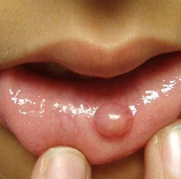
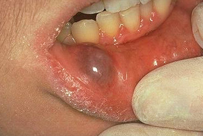
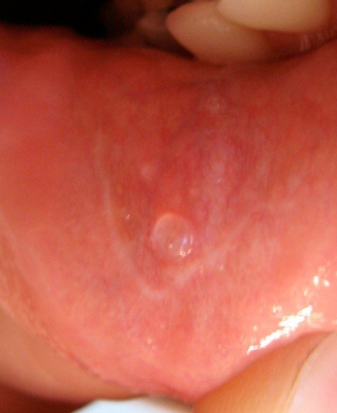
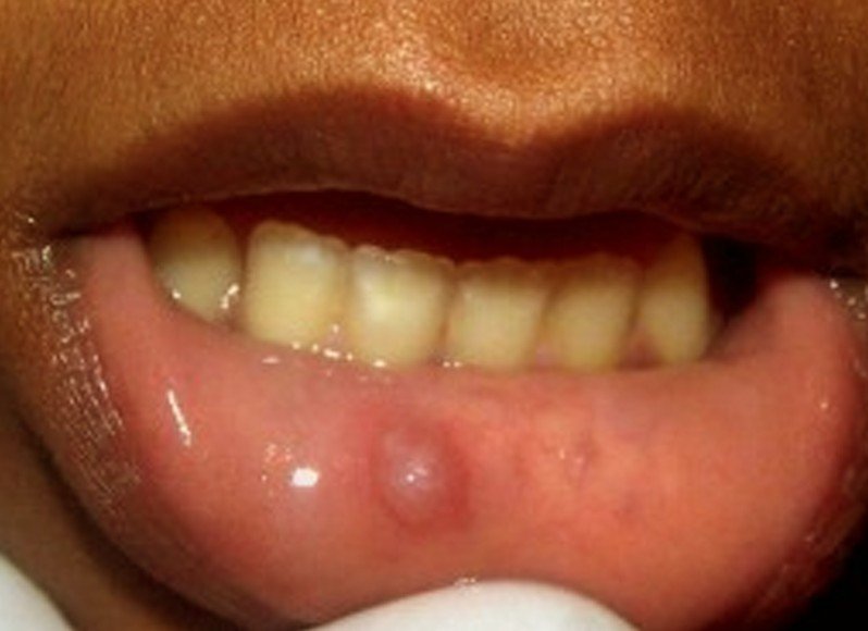
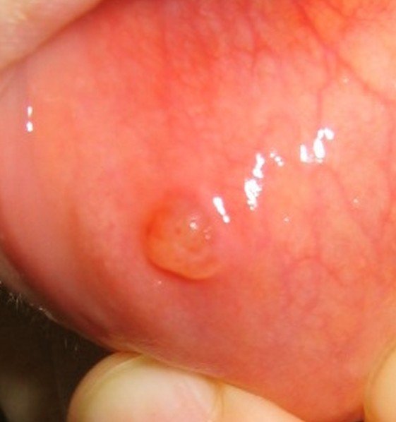
When a bump appears on the inside of the lip, it is often diagnosed as oral mucocele. This is a type of cyst that commonly appears on the inside of the lower lip. It is also known as the mucous retention cyst, mucous extravasation cyst or the mucous cyst of the oral mucosa. The oral mucocele is also presented in the medical literature as the mucous retention and the extravasation phenomena. There are two processes involved in the appearance of a bump on the inside of the lip. The first is represented by the mucous extravasation and the second is the development of the mucous cyst.

The bump on side of lip appears after the connective tissue has swollen and fluid collects in the cyst. The fluid contained in the mucocele is mucin. In case of mucous extravasation, local trauma or damage leads to the rupture of a salivary gland. The mucous cyst can occur when the salivary gland is either obstructed or ruptured. There is no sex prevalence but it seems that the condition is often encountered in children and young adults. The bump on side of the lip is often diagnosed as a disorder of the salivary glands. It is rarely found on the upper lip and it can also appear in other areas of the mouth, such as the cheek, tongue and mouth floor.

Causes of Bump on Inside of Lip

These are the most common causes that lead to the appearance of a bump on inside of the lip:

  • Ruptured or obstructed salivary gland due to:
    • Local trauma or damage, such as getting hit in the face
    • Repeated biting or chewing of the lower lip (constant aggression on the salivary gland)
    • Mechanical injury in the process of mastication
    • Chronic irritation of the oral cavity (excessive and prolonged smoking, eating spicy foods, drinking hot beverages)
    • Surgical intervention (trauma done to the salivary gland)
    • There is also a congenital variant (result of birth trauma)
    • Burns in the oral cavity
    • Oral infections
  • Aphthous ulcer
  • Lipoma
  • Salivary gland tumor (benign growth)
  • Submucosal abscess
  • Hemangiomas
  • Oral lichen planus
  • Medication – there are certain drugs that can have side-effects on the salivary glands
  • Lip piercings

Treatment

In a lot of patients, the bump on the inside of the lip disappears on its own, without requiring any specific treatment (the healing process lasts somewhere from three to six weeks). The healing period is dependent on the size of the bump and other pre-existing conditions. Clobetasol or gamma-linolenic acid topical applications are recommended for superficial bumps but these treatments do not prevent the risk of recurrence.

Chronic bumps on the inside of the lip require surgical removal. The procedure is simple: an incision is made on the inside of the lower lip and the bump (oral mucocele) is removed. In the first days after the operation, the patient is recommended to avoid moving the lips. The recovery period after the surgery is on average of two weeks. Also, in the initial period after the surgery, the patient is recommended to include only soft foods and liquids into the diet. In many patients, the removal does not guarantee the permanent treatment, as there is a high risk of re-occurrence. If the bump on the inside of the lip continues to re-appear, then the doctor might recommend the excision of the salivary glands.

There are different procedures (these are minimally-invasive, which translates into faster recovery time and less risk for side-effects) that are recommended for the surgical removal of the oral mucocele:

  • Laser removal – this is recommended for smaller cysts
  • Micro-marsupialization
  • Drainage and aspiration

There are also home remedies you can consider for the treatment of bumps on inside of the lip, including salt water rinses. This is recommended in cases of a bump that is small or one that has been recently identified. Patients are recommended to avoid constantly biting or chewing their lip, so as to reduce the risk of re-occurrence.

Applying alum to the bump has shown to reduce the inflammation and also to reduce the risk of secondary infections. The substance should be kept to the site of the mucocele for a quarter of an hour and then washed off with antiseptic mouthwash. Other home remedies for bump on side of the lip include: primrose oil, castor oil and honey. However, before trying out home remedies, it is recommended that you visit a doctor and see what treatment options you have available.

Other options of treatment include:

  • Cryotherapy – liquid nitrogen is used to eliminate the bump inside the lip; however, this procedure is not the first course of treatment, as there are serious side-effects to take into consideration (blistering, ulceration)
  • Intralesional injections with corticosteroids – this is one of the methods that allow for the inflammation to be reduced and the healing process to be accelerated.

Pictures

Here are some of the pictures of the Bump on Inside of Lip…

bump on inside of lip

bump on inside of lip pictures

bump on inside of lip pictures 2

bump on inside of lip pictures 3

bump on inside of lip pictures 4

bump on inside of lip pictures 5

No responses yet

Bacillary Angiomatosis

Feb 08 2016 Published by under Skin Conditions

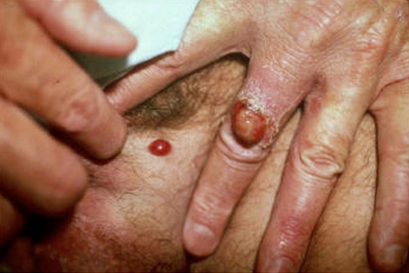
Bacillary angiomatosis is an infection of the skin and the subcutaneous tissue that also has a systemic involvement. This medical condition is caused by the infection with Bartonella, a gram negative bacteria and it often affects those who have a weakened immune system. Fortunately, there is a treatment for this condition and the majority of the patients respond well to the administered treatment. However, if this condition is left untreated, it has a high mortality rate. Bacillary angiomatosis is encountered in patients who are infected with HIV, being the second most common cause why angiomatous lesions appear on the skin.

This medical condition was first described in 1983, in a patient who was infected with the HIV virus. Today, the diagnosis of bacillary angiomatosis is also made in patients who have undergone organ transplantation and in those who have a weakened immune system because of other causes than HIV infection. In rare cases, it has been diagnosed in patients with a competent immune system. The systemic nature is one of the most important things to take into consideration, as the infection with Bartonella can lead to problems in various organs, including the brain, bone marrow, heart, lungs and kidneys.

What are the Symptoms of Bacillary Angiomatosis?

These are the most common symptoms of bacillary angiomatosis:

  • The lesions on the skin have the following characteristics:
    • A single lesion or multiple lesions can appear on the skin (papules)
    • Their color varies from one person to the other (purple, color of the flesh, no color)
    • Lesions resemble hemangiomas
    • Variable size (from 1mm to several cm)
    • Scaly nodules can also be present on the skin
    • Large masses are also encountered
    • Extensive plaques can affect the extremities
      • Excessive pigmentation
      • Excessive keratinization – this leads to induration of the plaque
  • Areas most affected include:
    • Oral mucosa
    • Tongue
    • Oropharynx
    • Nose
  • Pain
    • Areas most affected by pain include the forearms and legs (bone pain)
  • Systemic involvement:
    • Fever/chills
    • Sweating during the night
    • General consumption
    • Anorexia
    • Weight loss
    • Abdominal pain – This is often accompanied by nausea and vomiting
  • Mass in the abdomen – This mass is accompanied by bleeding inside the gastrointestinal tract
  • If the colon is affected, then the following symptoms will occur:
    • Abdominal cramps
    • Diarrhea with blood
  • If the central nervous system is affected, then the following symptoms are expected:
    • Depression
    • Psychosis
    • Changes in personality
    • Anxiety
    • Excessive irritability
    • Headache
    • Neuralgia (most common affected – trigeminal nerve)
    • Seizures
    • Back pain
  • The patient might have trouble breathing if the larynx is obstructed

Causes of Bacillary Angiomatosis

The most common causes that lead to the appearance of bacillary angiomatosis are:

Bacterial infection with the following organisms:

  • Bartonella henselae
    • Method of transmission – cat scratch/bite
    • Vector of transmission – ticks/fleas
  • Bartonella quintana
    • Method of transmission – from one human to the other
    • Vector of transmission – lice.

Treatment

These are the most common courses of treatment recommended for bacillary angiomatosis:

Antibiotic therapy

  • Erythromycin – patients respond the best to this treatment (oral administration)
  • Tetracycline – recommended in patients who do not tolerate the treatment with erythromycin
  • Cephalosporin/aminoglycoside – indicated in more severe cases
  • Doxycycline in combination with rifampin – recommended in patients  whose immune system is severely compromised
  • Doxycycline is not recommended in pregnant women
  • Other antibiotics that can be administered with promising results:
    • Clarithromycin
    • Azithromycin
    • Ciprofloxacin
    • Isoniazid
    • Gentamicin
  • The period for the antibiotic therapy lasts somewhere between eight and twelve weeks, depending on each patient
  • In case of systemic involvement, the treatment is administered for prolonged periods of time (three months)
  • In case of HIV infection, the treatment might be administered for the entire duration of life, concomitantly with antiretroviral therapy

Other therapy options, such as corticosteroids, cytotoxic agents and radiation are not effective in this medical condition.

Drainage

  • Lymph nodes that are filled with pus
  • Blisters

Supportive care

  • Administration of intravenous fluids for hydration
  • Anti-inflammatory medication – this can help with the pain and they can also reduce the fever

Adjuvant therapy

  • Cryotherapy
  • Electrodesiccation and curettage
  • Surgical excision – recommended for solitary cutaneous lesions

Preventive measures

  • These are recommended to avoid the infestation with the actual bacteria
  • Contact with unknown cats should be avoided
  • If you are the owner of the cat, it is recommended that you take care of the anti-flea treatment (flea and tick collar)
  • Clothing and bedding treatments are required as well to prevent the infestation
  • Delousing is also recommended in case of lice (there is a special powder that can administered directly to the skin)
  • Macrolide prophylaxis in HIV-infected patients.

Bacillary Angiomatosis Pictures

Collection of photos, images and pictures of Bacillary Angiomatosis…

bacillary angiomatosis

bacillary angiomatosis pictures

bacillary angiomatosis pictures 2

bacillary angiomatosis pictures 3

bacillary angiomatosis pictures 4

bacillary angiomatosis pictures 5

No responses yet

Gingival Hyperplasia

Feb 04 2016 Published by under Oral Health

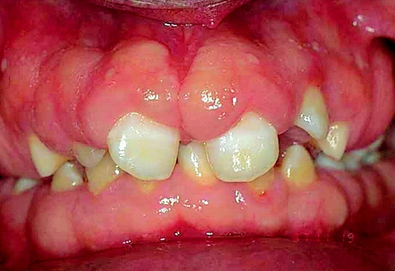
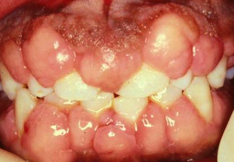
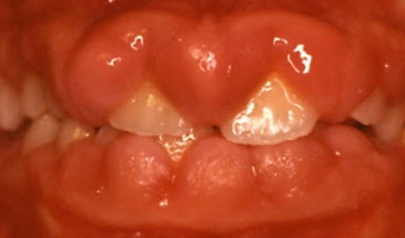
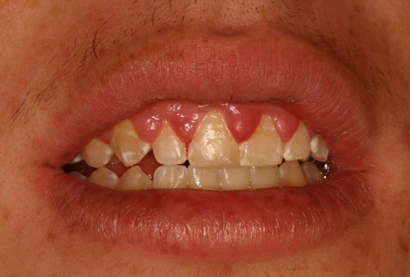
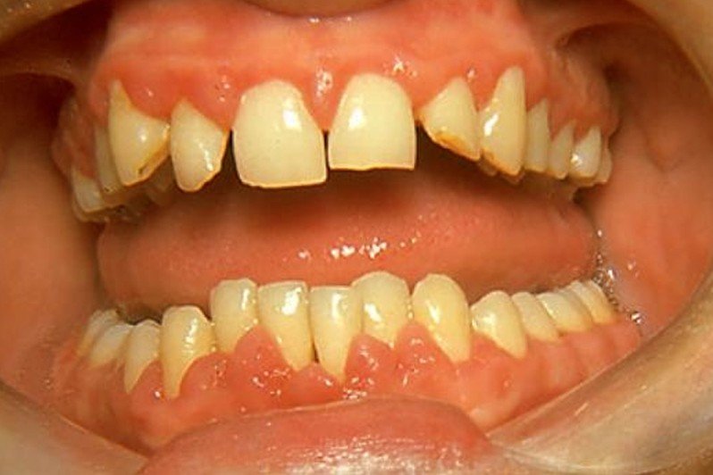
Gingival hyperplasia is a medical condition in which the gums increase in size. This condition is also known as gingival overgrowth, hypertrophic gingivitis or gingival hypertrophy. There are several types available, which will be discussed further down but two of the most important ones are the one induced by different medication and the one associated with inflammatory disorders. Gingival hyperplasia can also be congenital and it is considered to be a rare condition. This condition is often seen in children who are diagnosed with epilepsy but, otherwise, there is no race or sex prevalence.

Before proceeding with the different types, it is important to make the difference between hyperplasia and hypertrophy, as these terms are often used for this condition and they are not quite as similar as one might think. When we say hyperplasia, that means that the cells in the gums have increased in numbers. As for the hypertrophy, it means that the cells in the gums have maintained their number constant but their size has increased. This is why there are specialists who prefer not to use either of these two terms but rather choose gingival enlargement as the final diagnosis.

According to the medical literature, there are five different types of gingival hyperplasia:

  • Gingival enlargement associated with inflammation
  • Gingival enlargement induced by medication
  • Gingival enlargement associated with different medical disorders (systemic involvement)
  • Gingival enlargement associated with cancer
  • False gingival enlargement.

Determining the type of gingival enlargement is essential, because each type has its own set of causes. The moment the type has been identified, so will be the causes. Once the cause is identified, the proper treatment can be administered. Because, when it comes to gingival hyperplasia, the treatment depends on the cause.

Gingival Hyperplasia Symptoms

These are the most common symptoms of gingival hyperplasia:

  • In the majority of the cases, there is no other symptom than the actual gingival enlargement
  • When gingival hyperplasia is caused by inadequate oral hygiene, the following symptoms can accompany the gingival enlargement:
    • Dental plaque builds on the teeth, pushing into the gums
    • The gums are tender to the touch
    • Inflammation will occur in the affected area
  • Gums can bleed
  • Areas that are most affected include the labial gingival mucosa and interdental spaces
  • If there is also gum overgrowth, then the teeth might be covered by the over-grown tissues
  • If the gingival enlargement is caused by the administration of different medication, then the patient might experience the following symptoms:
    • Gums are firm yet swollen
    • They are not tender to the touch
    • They have a pink color
    • Bleeding does not occur
    • Gums can overgrow, covering the crown of the teeth and leading to periodontal disease
    • The overgrowth can also affect the eruption of the new teeth and even cause mal-alignment
  • In case of congenital gingival hyperplasia, the following symptoms might be encountered:
    • The inflammation can be only in a part of the gingiva or it can be generalized
    • The gums are pink in color and firm to the touch
    • Teeth might be covered by the overgrown gums, leading to displacement of new teeth.

Causes of Gingival Hyperplasia

These are the most common causes of gingival hyperplasia:

Medication

  • Gingival enlargement is encountered in people who are diagnosed with epilepsy and treated with phenytoin
  • The treatment with phenobarbital can also lead to this problem
  • Other drugs that can induce such problems are:
    • Cyclosporine (immunosuppressant)
    • Calcium-channel blocking agents (nifedipine, amlodipine)
  • There are also certain risk factors that can favor the appearance of drug-induced gingival enlargement:
    • Inflammation of the gums (gingivitis)
    • Poor oral hygiene
    • Excessive dental plaque
    • Nickel accumulation
    • Proliferation of epithelial cells
    • Periodontal disease
    • The depth of the periodontal pocket
    • Duration and dose of the administered treatment s

Congenital gingival enlargement can have the following causes:

  • Hereditary transmission
  • Metabolic disease – fetal valproate syndrome

Chronic inflammation of the gums

  • This is often present in the person who breathe through their mouths

Systemic causes

  • Pregnancy
  • Teenage (reaching puberty)
  • Vitamin deficiency (especially vitamin C)
  • Pyogenic granuloma
  • Leukemia
  • Granulomatosis
  • Sarcoidosis
  • Cancerous growths (benign or malign)

False enlargement of the gingiva occurs when there is a part of bone or teeth actually detached and causing the modifications

Treatment for Gingival hyperplasia

These are the most common courses of treatment and changes recommended for gingival hyperplasia:

  • Adequate oral hygiene
    • Dental plaque removal (thorough, professional dental cleaning)
    • Regular teeth brushing (improved technique)
    • Constant dental flossing
    • Using antiseptic mouthwash
  • Dental treatments for periodontal disease
    • Professional debridement with scaling and root planing
  • Gingivectomy
    • Surgical removal of the excessive gingival tissue
    • Often times, the surgical removal of the excessive tissue is performed because of the aesthetic concern rather than for functional purposes
    • The gingival enlargement recurs in patients who are of a younger age, those who suffer from chronic gingival inflammation or those who continue to maintain poor oral hygiene
    • Daily oral rinses with Chlorhexidine are recommended to prevent recurrences in patients who have undergone surgery for gingival enlargement.
  • Carbon dioxide laser therapy is also a solution for therapy, providing a very important advantage:
    • Reduced change of postoperative bleeding
  • In the situation of drug-induced gingival enlargement, it is recommended that the treatment with the said medication is stopped or an alternative to the medication is given
    • For example, instead of cyclosporin, one can administer tacrolimus. Even though the gingival overgrowth is more reduced in the case of tacrolimus treatments, the toxicity to the renal system is the same in both two cases
    • Phenytoin, the drug commonly administered for the treatment of epilepsy, can be easily substituted with another anticonvulsant
  • Providing adequate treatment for the systemic disease that led to the gingival hyperplasia in the first place
  • Ultrasonic treatments have also shown promising results when it comes to treating gingival hyperplasia
  • Antibiotic therapy is recommended in order to prevent or treat secondary infections

Gingival Hyperplasia Pictures

Here are some of the pictures collection of Gingival hyperplasia…

gingival hyperplasia

gingival hyperplasia pictures

gingival hyperplasia pictures 2

gingival hyperplasia pictures 3

gingival hyperplasia pictures 4

gingival hyperplasia pictures 5

No responses yet

Filiform Warts

Feb 01 2016 Published by under Skin Conditions

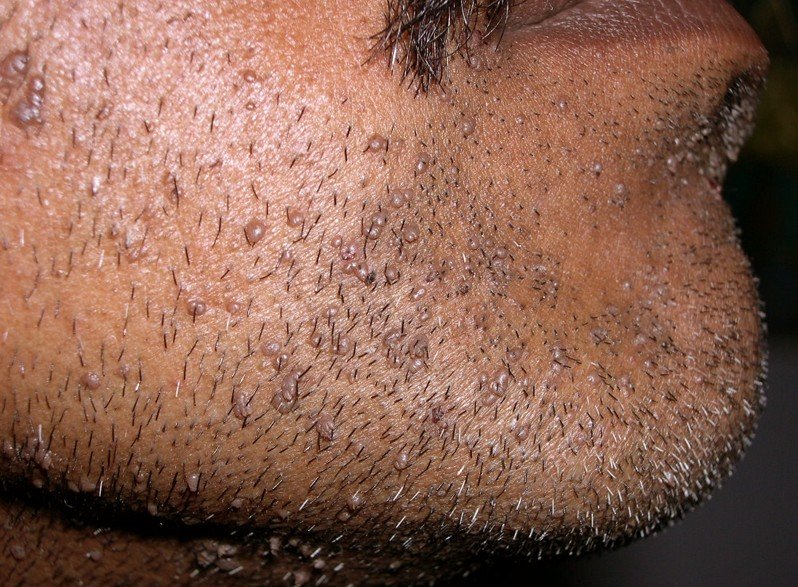
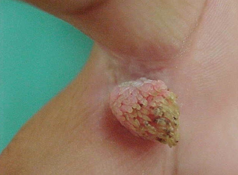
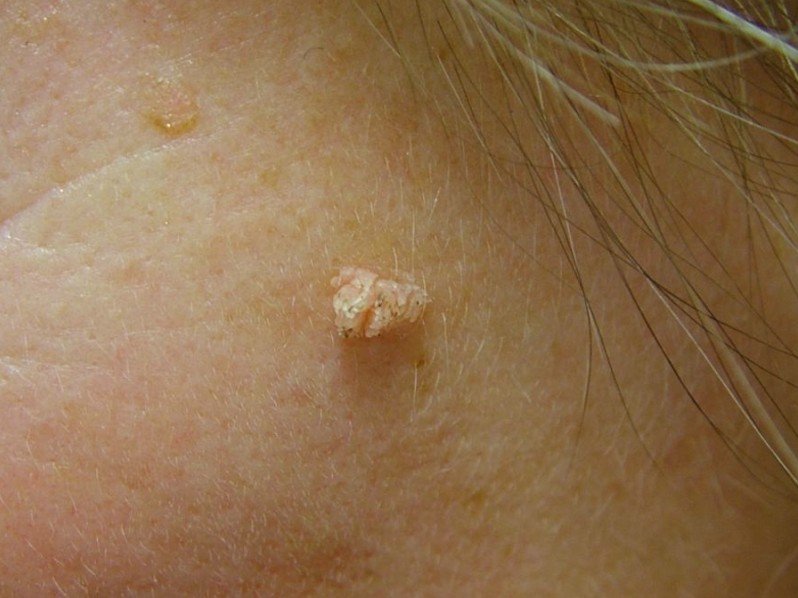
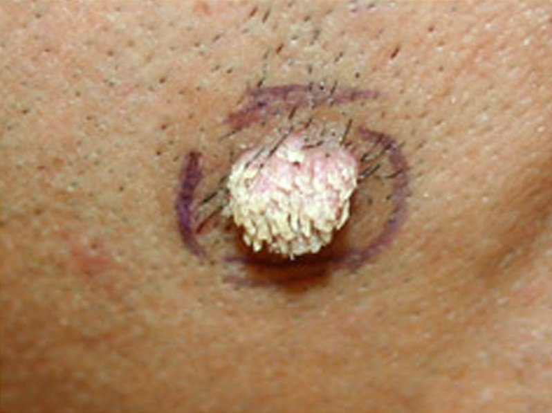
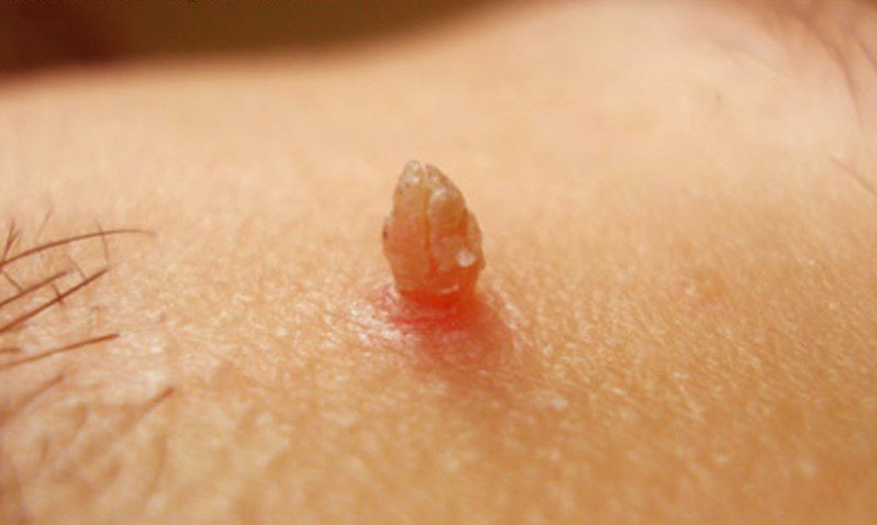
Filiform warts are benign fleshy growths on the skin, which appear in persons who have been infected with the human papilloma virus. In general, warts are named according to the way they look or according to the place where they appear on the body. When it comes to filiform warts, these are named like that because they are projections of filiform shape. They often appear on the face and especially on the eyelids and lips but they can also appear on the neck. However, it is important to remember that filiform warts are not strictly limited to the face. They can also be identified in the axillary region or on the legs.

The infection with the human papilloma virus stimulates the top layers of the skin to grow excessively and lead to the appearance of filiform warts. The need for treatment because obvious, as these warts are highly contagious and also because of the aesthetic concern. Other from that, they usually do not cause any discomfort or pain to the pain and they do respond well to the treatment. The important thing is not delay getting the treatment, especially if they cause a lot of embarrassment.

Symptoms of Filiform Warts

These are the most common symptoms of filiform warts:

  • Fleshy growths appear on the face, neck and other mentioned areas
  • These growth are benign
  • In some cases, they grow to be quite long
  • Usually asymptomatic
  • In some patients, the following symptoms may be however encountered:
    • Itchiness
    • Bleeding
  • Tendency to spread to other parts of the body
  • Painless
  • If they appear near the skin folds or in areas where the skin rubs, the filiform warts might become sore and irritated.

What are the Causes of Filiform warts?

Filiform warts are caused, as it was already mentioned, by the infection with the human papilloma virus. However, not all types of HPV can cause filiform warts. These are actually caused by the following strains: 1, 2, 4, 27 and 29. Below, you will find the most common ways through which this virus is actually transmitted:

Human contact

  • Touching the filiform warts is enough to become infected
  • The person can contribute to the spread of the filiform warts from the original area to another area (otherwise lesion free) by touch
  • If there is a breakdown in the skin, then the risk of becoming infected with the HPV is even higher.

Clothing

  • Sharing your clothing with a person who has filiform warts increases the risk of yourself becoming infected, especially if those clothes have touched the infected area

Objects of personal hygiene

  • Sharing your towels with a person who has filiform warts poses an increased risk for infection
  • Sharing skin care products also poses a risk for infection

Contact with potentially-infected surfaces

  • The HPV can be found on different surfaces, including shower floors or the surrounding surface of a pool

Shoes

  • It is recommended to avoid sharing shoes with a person who has been infected with HPV

Risk factors that can favor the infection with HPV

  • Breach in the skin – cut or abrasion
  • Weak immune system
  • Prolonged exposure to HPV
  • Carrier of the HPV
  • Warm and most skin.

Treatment for Filiform warts

These are the most common courses of treatment for filiform warts:

Topical irritants (active substance has a keratolytic effect)

  • Salicylic acid application
    • The active substances contained in this treatment will help the immune system to fight off the infection
    • Several applications are recommended before the first results are noticeable (the wart will disappear)
  • Trichloroacetic acid
  • 5-fluorouracil

Cryotherapy

  • Liquid nitrogen is used to freeze the fleshy growths
  • After the actual procedure, blisters may appear in the treated area
  • Patients are recommended to wear bandages in the treated area and remove the dead skin after a week
  • Several sessions of freezing are required for a wart to completely disappear
  • The sessions are repeated every two to four weeks; depending on the size, number and location of the filiform warts, there will be four to six therapy sessions
  • There are patients who develop a severe local reaction to cryotherapy, so the freezing process is performed in a gradual manner
  • Potential side-effects of cryotherapy include:
    • Pain
    • Tendon injury
    • Superficial nerve injury
    • Skin discoloration
    • Scars
    • Warts re-occur

Cantharidin

  • Similar to cryotherapy
  • Causes the skin to blister
  • The blister dries up and the dead tissue is then removed

Minor surgery

  • The procedure is performed using a scalpel or an electric needle
  • Local anesthetic is applied before the surgery
  • Immediate removal, no second procedure is necessary
  • The skin might be scarred in the affected area
  • Electrosurgery involves two procedures, meaning curettage and cautery
  • The base of the filiform wart is burned by using diathermy or cautery
  • After the surgery, it takes almost two weeks for the wound to heal
  • Wart recurrence is expected

Laser removal

  • Recommended for filiform warts that are difficult to remove through other methods
  • Higher costs
  • Risk of scarring

Other choices of treatment include:

  • Occlusion with plaster or duct tape
  • Applications of cytotoxic agents
  • Systemic retinoids – these are only recommended for extensive warts
  • HPV vaccine (not treatment, protects against infection with the HPV).

Are Filiform warts contagious?

Filiform warts are highly contagious and this is the reason why you should avoid touching them on another person. Also, it might be a good idea not to share clothes, personal items and objects of personal hygiene with a person who has this infection. You should always remember that the HPV can be taken from a public place as well, so, be sure to use personal slippers when using a public shower or when walking around the pool. And, if you have cuts or abrasions on the skin, be sure to protect those areas with a plaster. In this way, you reduce the risk of the HPV entering your body and causing filiform warts to appear.

Pictures

Collection of photos, images and pictures of filiform warts…

filiform warts

filiform warts pictures

filiform warts pictures 2

filiform warts pictures 3

filiform warts pictures 4

filiform warts pictures 5

filiform warts pictures 6

filiform warts pictures 7

No responses yet

« Prev

© 2011-2019 MDDK.com - Medical Tips and Advice. All Rights Reserved. Privacy Policy
The health information provided on this web site is for educational purposes only and is not to be used as a substitute for medical advice, diagnosis or treatment.