Cherry Angioma
What is Cherry Angioma?
Cherry angioma is the most common type of angioma and is a common skin growth. The term cherry in angioma describes the appearance of the papule which is cherry red in color. It is a benign skin growth that contains blood vessel proliferation.
Cherry angioma is also known as Campbell de Morgan spots and Senile angioma. The condition was first described by an English surgeon named Campbell de Morgan thus the term Campbell de Morgan spots.
Cherry angioma is known to affect people worldwide and without racial predilection although the disease is found to be more common among individuals with pale colored skin. It equally affects both men and women particularly those between the ages of 30 years and above. The frequency of cherry angioma increases with age.
Cherry angioma is not a harmless skin condition and is not potential for skin cancer. The main concern of the disease, however, is towards the cosmetic aspect wherein the disease has an unsightly effect. It can occur in any areas of the skin with common occurrences in the areas of the trunk and the extremities. The scalp, face and neck are also frequently affected with cherry angioma. It rarely develops in the mucus membranes, hands and feet. Cherry angioma on the other hand has a tendency to bleed especially when large enough to be pinched or punctured particularly when trying to remove the angioma. The development of the angioma may be single or in group although it often develops as a single angioma.
Symptoms of Cherry Angioma
Cherry angioma is composed of several capillaries or smallest blood vessels of the body. The reddish appearance or color of the angioma is due to the proliferation of the capillaries within the cherry angioma.
Cherry angioma occurs without warning and may happen to a healthy individual. During the initial development of cherry angioma, the lesion is usually small or about one tenth millimeter in diameter and is flat on the surface of the skin or like a small dot on the skin. The lesion tends to thicken as it gradually increases in size and becomes more prominent. It occurs in solace or may develop in groups or clusters of lesions.
What does Cherry Angioma look like?
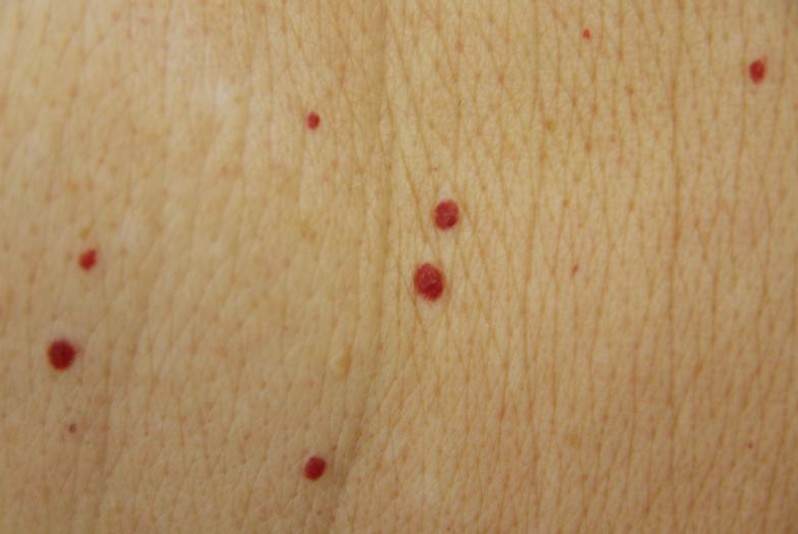
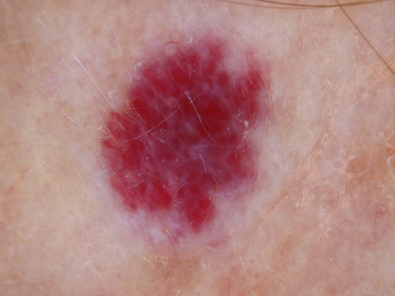
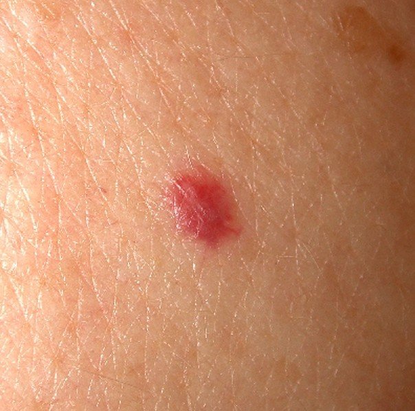
Cherry angioma is round to oval in shape and the size varies ranging from a pinpoint in size about 1mm to 3mm in diameter or more. When the angioma grows bigger, the lesion assumes a dome shape or much like a mushroom and is often spongy.
The lesion is of bright red color or may be more purplish or violet. The lesion seldom presents a dark brown to black in color. The Cherry angioma lesion is non-blanching and may be widespread, especially in elderly patients.
The onset of cherry angioma is often asymptomatic. It is generally harmless and causes no pain or discomfort that an affected individual can continue or go on with their daily routine without being bothered by the symptoms. Cherry angioma on the other hand can be unsightly especially if it grows in the area of the skin that is easily seen or in frequent exposure such as in the face and neck.
Cherry Angioma Causes
The exact cause of cherry angioma remains unclear probably due to the lack of significance as the disease generally does not involve malignancy. The onset is considered to have occurred in two different mechanisms known as angiogenesis and vasculogenesis. Angiogenesis is described as the formation of new blood cells. It plays an important role in growth and development and in shaping of granulation tissue. Angiogenesis also plays an important role in the process of wound healing. Vasculogenesis is described as the formation of new blood vessels in the absence of a pre-existing blood vessel.
Prichard’s structure
A recent study reveals the association of Prichard’s structure with the development of cherry angioma. Prichard’s structure is characterized by cellular growth within the tissue of the heart.
Chemicals
Exposure to certain chemicals and compounds are being considered to cause the cherry angioma. Mustard gas is a cytotoxic warfare agent that is yellowish-brown in color and a smell similar to the mustard seed. Exposure to mustard gas is high potential for blister formation in the skin and lungs.
Cyclosporine
Cyclosporine is a drug used for organ transplantation in preventing rejection of kidney, liver and heart transplant. It is an immunosuppressant drug that acts by impeding the activity and growth of T cells by diminishing the activity of the immune system. Cyclosporine is also used for the treatment of rheumatoid arthritis and psoriasis. The use of cyclosporine however, has various side effects and development of cherry angioma is believed to be among the side effects of this drug.
Bromide
Bromide is a chemical compound that naturally occurs in seawater. This compound is used as an ingredient in foods and is also used as a sedative. Consumption of food with high levels of bromide such as seafood is being considered to influence the development of cherry angioma.
Genetic factors
A genetic factor is also being considered to the onset of cherry angioma.
Treatment
Treatment of cherry angioma is not necessary as this skin condition is generally harmless and is not potential for malignancy. Medical intervention is not beneficial while cherry angioma is managed with surgical intervention.
Removal of Cherry Angioma
Surgical removal is the primary management for cherry angioma. The removal is based on the option of preference of the patient. The decision to have the cherry angioma remove is probably due to the unappealing appearance of the lesion which can bring significant embarrassment to the patient. Several methods of surgical removal can help in treating cherry angioma although the method of removal depends on the preference of the patient as explained by their doctors.
Laser surgery
Laser surgery is among the method of removing cherry angioma. This method involves the use of pulse dye laser that emits a sufficient amount of heat to destroy and remove the lesion.
Cryosurgery
Cryosurgery involves freezing of the cherry angioma with the use of liquid nitrogen. The liquid nitrogen will freeze the angioma which will later fall off.
Electrocauterization
Electrocauterization is a surgical method that involves the burning of the angioma. This method utilizes a tiny probe and an electric current to burn the angioma.
Cherry Angioma Pictures
Collection of Pics, Images and Pictures of the medical condition Cherry Angioma…


