Diseases General Health Skin Conditions

Archive for November, 2016

Central Pontine Myelinolysis

Nov 30 2016 Published by under Brain and Nervous System

Central pontine myelinolysis can be defined as a medical condition, in which the myelin sheath of the nerve cells in the brainstem become severely damaged. This is a neurological disease, with a specific location (area known as the pons) and iatrogenic etiology. Also known as osmotic demyelination syndrome or central pontine demyelination, this condition can lead to a wide range of symptoms. As you will have the opportunity to read below, one can suffer from acute paralysis, present difficulties swallowing and speaking. In some patients, the condition can appear outside the mentioned area.

This condition appears in the majority of cases as a complication to the treatment of patients who present life-threatening, low levels of sodium in the body. In order to prevent the appearance of this condition, one should correct these levels in a gradual manner (specific rate of sodium to be administered on a daily basis). There are other situations that may lead to central pontine myelinolysis, even though they are less common. Chronic alcoholism and the reduced functioning of the liver have been incriminated among the causes.

This condition was described for the first time in the 1950s, being observed in those who suffered from chronic alcoholism or malnutrition. As the studies extended, it was recognized that the primary cause was the too rapid correction of sodium levels in those who suffered from hyponatremia. In was only in the 1980s that the condition was established, with the guidelines we know today. Both adults and children are at risk of developing this condition, especially if there is a rapid rise in the serum sodium levels. It is important to understand that this condition never occurs on its own – as it was already mentioned, it appears as a complication of other medical problems or treatments.

Pathophysiology

The osmotic concentration is adjusted by the brain cells, with the levels of certain osmolytes being changed on a regular basis. When the sodium levels are dangerously low, the levels of the osmolytes are also affected and so is the capacity of absorbing free water. If the correction of sodium levels is too rapid, this means that the free water will also leave the brain cells. It is this change that leads to the appearance of central pontine myelinolysis, with paralysis and other neurological symptoms. Due to the lack of water, the brain appears as if it would shrink.

As the nerve fibers at the level of the brain suffer from demyelination, damage occurs. If the levels of sodium are chronically low, the brain can adapt to these changes and compensate by taking water from the interstitial area. On the other hand, if the correction of sodium is made with intravenous fluids, the brain might not have sufficient time to adapt to the changes. Basically, the more rapid the correction is made, the more water the brain will lose and suffer from additional damage. The damage is caused by the appearance of cerebral hemorrhage, which is a consequence of water moving into the cells.

Causes

These are the most common causes that lead to the appearance of central pontine myelinolysis:

  • Trying to correct the life-threatening low sodium levels too rapidly
  • Chronic alcoholism (especially in patients who are suffering from withdrawal symptoms)
  • In patients who have undergone hematopoietic stem cell transplantation
  • Reduced functioning of the liver due to severe disease
  • Transplantation of the liver
  • Patients who have suffered from severe burns
  • Eating disorders (malnutrition, anorexia, bulimia)
  • Severe disorders of the electrolytes
  • Excessive or prolonged usage of diuretics
  • AIDS
  • Hyperemesis gravidarum – rare complication of pregnancy
  • Peritoneal dialysis, leading to reduced sodium levels
  • Wernicke encephalopathy
  • Diabetes (very rare).
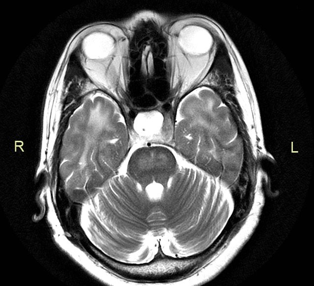
Diagnosis

Central pontine myelinolysis is difficult to diagnose using imaging studies. In the majority of the patients, the changes take several weeks after the onset of the symptoms before they appear visible on the MRI or CT. In general, the MRI is more recommended than the CT, due to higher fidelity of the investigation.

The physical examination can also reveal changes related to this condition, such as: abnormal reflexes, confusion, weakness or paralysis. Apart from the head MRI scan, the doctor can also order blood tests (sodium levels) and perform the brainstem auditory evoked response (not as faithful as the MRI). For the latter, electrodes are attached to the head and ears; then, a series of sounds are played through the headphones. The investigation determines the activity of the brain in response to those sounds.

Symptoms

These are the most common symptoms associated with central pontine myelinolysis:

Precursor symptoms

  • Seizures
  • Affectation of consciousness
  • Changes in the gait
  • Affectation of the respiratory function (decreased/ceased)

Acute symptoms

  • Paralysis (can affect two or all of the limbs)
  • Difficulties swallowing (dysphagia) and speaking (dysarthria)
  • Problems with the vision (diplopia)
  • Loss of consciousness
  • Confusion and delirium
  • Balance problems (unstable gait)
  • Hallucinations
  • Lethargy
  • Poor response
  • Tremors
  • Weakness in different parts of the body

Locked-in syndrome

  • Cognitive function is intact
  • All muscles are paralyzed (exception – eye blinking)
  • This syndrome appears in the situation that the corticobulbar and corticospinal tracts of the brainstem have suffered from rapid myelinolysis.

In some patients, this condition does not present any symptoms, which is suggestive of the fact that the lesion in the respective area of the brain is not that big. The symptoms mentioned above appear in patients who have suffered from bigger lesions.

Prevention and Treatment

The most certain way to prevent central pontine myelinolysis is to correct the reduced sodium levels in a gradual manner. If these are corrected at a slow pace, the risk of overcompensation is reduced and the brain has the necessary time to adjust to the changes. In case of patients who suffer from chronic alcoholism, vitamin supplementation can help prevent such problems.

Unfortunately, once central pontine myelinolysis has occurred, there is no cure. Patients can benefit from supportive care, with the main objective being the prevention of life-threatening complications, such as aspiration pneumonia or deep vein thrombosis. Vitamins can be administered to those who suffer from chronic alcoholism, so as to correct the other nutritional deficiencies. Corticosteroids have been used as a line of treatment (experimental phase) but their efficiency has proved out to be reduced.

Physical therapy and rehabilitation are essential during the recovery phase, as they can contribute to the overall improvement of the prognosis. The physical therapist can help with the reduction of spasticity, while working to maintain the strength and force of the muscles. As the patient recovers, the physical therapist will add exercises to increase mobility and function. The patient will be taught transfers to/from bed and to/from chair, learning how to sit and stand without getting dizzy. The last phase of the recovery process is going to concentrate on the improvement of the balance and walking (on his/her own or using assistive devices). Speech therapy is recommended to those who have trouble speaking and occupational therapy is essential for the daily living activities. The occupational therapist can teach the patient how to function on a daily basis, maintaining the highest level of independence that is possible.

In the situation that the patient has developed Parkinsonism, the treatment can include dopaminergic drugs (these are commonly recommended for those who have been diagnosed with Parkinson’s disease). One of the most recommended dopaminergic drugs is levodopa, providing successful results in patients who are suffering from tremors, associated with speech and swallowing difficulties.

Prognosis

In general, the prognosis of this condition is negative. There are patients who will not survive the changes brought on by this condition. Others can recover completely from the condition, while some will become disabled. Severe disability is a serious possibility – however, the disabilities can cover a wide range, starting with minor tremors or ataxia and going as far as spastic paralysis affecting all the limbs or even the locked-in syndrome. In general, the recovery process is influenced by how extensive the damage was at the level of the brainstem (how many axons have suffered from demyelination).

It is believed that the prognosis for this condition was improved due to the advancements of the imagining techniques and also due to the early diagnosis. The biggest challenges that come during the recovery period are related to a person’s degree of independence, as well as to the motor skills and speech. Often times, the recovery is prolonged, due to the patient suffering from emotional instability and also from forgetfulness.

Patients need to understand that the recovery from the central pontine myelinolysis is a long and difficult process. The changes that occur might be permanent, leading to chronic disabilities or they may take a very long time to subside. During this period, psychological counseling is highly important – it can help the patient maintain an optimist focus and fight to get better. Depending on the extent of the damage, some patients might require specialized assistance. Often times, the personal assistant will provide the necessary assistance with daily tasks, such as the ones related to personal hygiene, dressing and feeding.

Pictures

central pontine myelinolysis

No responses yet

Paraproteinemia

Nov 30 2016 Published by under Diseases and Conditions

Definition

Paraproteinemia can be defined as a medical condition, in which excessive quantities of paraprotein are present in the blood. This condition is also known as monoclonal gammopathy (excessive amounts of monoclonal gammaglobulin in the blood). As you will have the opportunity to read below, this condition is often encountered in those who have been diagnosed with immunoproliferative disorders. The paraproteins are actually immunoglobulins that are produced at the level of the bone marrow and they can cause damage to the kidneys, when excreted in excess through the urine. This condition is more common in the elderly population, rather than the young.

Types of Paraproteinemia

Paraproteinemia can be classified into different types, according to the paraprotein that is found in the blood. These are the three main types of paraproteinemia:

  • Light-chain paraproteinemia (the Bence Jones protein)
    • Often associated with immunoproliferative disorders, such as amyloidosis or multiple myeloma
  • Heavy-chain paraproteinemia
    • This condition is also known as the heavy chain disease
  • Whole-immunoglobulin paraproteinemia
    • M-proteins (monoclonal) are present in the blood
    • The condition can be influenced by the cold, being known as cryoglobulinemia (the immunoglobulins have a tendency to precipitate in the blood vessels, as a result of the cold temperatures)

It is important to understand that the above-mentioned types can appear on their own in an individual or combined. In general, the heavy-chain and the whole immunoglobulin proteins are restricted to the blood vessels. On the other hand, the light-chain immunoglobulins can escape from the blood vessels, being excreted by the kidneys together with the urine (these are known as the Bence Jones proteins).

In some patients, the whole immunoglobulins can bind with one another, leading to a condition known as macroglobulinemia. This condition can aggravate the already existent health problems. As it was already mentioned above, one can suffer from cryoglobulinemia, with the immunoglobulins precipitating in the blood vessels with cold temperatures. It is also possible that the whole immunoglobulins reduce the viscosity of the blood, leading to a condition known as Waldenström macroglobulinemia.

Causes

These are the potential causes that lead to the appearance of paraproteinemia:

  • Leukemia and lymphoma
    • B-cell non-Hodgkin lymphoma – type of cancer that derives from the lymphocytes (white blood cell)
    • Myeloma – cancer of the plasma cells, in which abnormal cells collect at the level of the bone marrow and interfere with the production of normal, healthy blood cells
    • Plasmacytoma – malignant plasma cell tumor, occurring either at the level of the soft tissues or within the axial skeleton
    • Lymphoplasmacytic lymphoma – cancer affecting the B white cells, with a tendency of growing and spreading slowly, in a gradual manner
  • Idiopathic (either no cause can be identified or the condition will develop into leukemia or lymphoma in several years)
    • Benign monoclonal gammopathy (also known as monoclonal gammopathy of undetermined significance) – the condition is identified through routine testing and, at first, it might resemble the multiple myeloma
    • Primary amyloidosis (light-chain paraproteinemia) – this is a rare medical condition, in which abnormal proteins accumulate throughout the body (these proteins are also known as amyloids).

Diagnosis

These are the most common methods used for the diagnosis of paraproteinemia:

  • Blood testing
    • Serum protein electrophoresis
      • Narrow band or spike – these are because of the excessive amounts of proteins (elevated serum total protein)
    • Serum immunofixation electrophoresis (further testing)
    • Serum free light chain – should be correlated with the urine analysis (24 hour collection)
    • Quantitative immunoglobulins
  • Determination of the hematocrit
  • Blood viscosity analysis – the immunoglobulins present in the blood can increase the level of viscosity
  • Urine analysis – for the best results, a 24-hour collection of urine is required
  • Immunoglobulin levels (IgA, IgG, IgM and IgD) – identification of normal and abnormal proteins
  • Identification of light and heavy chains
  • Other investigations
    • Skeletal investigation or survey
    • Bone marrow biopsy (suspicion of leukemia or lymphoma)
    • CT scan

Treatment

In order to improve the paraproteinemia signs, the treatment is usually concentrated on the associated disorder. These are the most common treatment approaches that can be taken in such cases:

  • Acute management
  • Supportive care (recommended for the more serious cases)
    • Bone directed therapy
      • Vitamin D and Calcium
      • Bisphosphonates
    • Medication to prevent neuropathy
    • Narcotics and other analgesics for the pain
  • Anti-cancer therapy
    • Radiation therapy
    • Chemotherapy
    • Biologic therapy
    • Autologous transplantation
  • Long-term therapy
    • Specific medication and management
  • Allogeneic transplantation
  • Medication administered in combination with the autologous transplant
  • In case of infections associated with myeloma – antibiotics or antivirals
  • Pain medication – helps the patient deal with the symptomatology
  • Plasma exchange in case of renal insufficiency
  • Corticosteroids – recommended for serious cases
  • Immuno-modulatory drugs – recommended choices: Thalidomide, Pomalidomide and Lenalidomide
  • Proteasome inhibitors – recommended choices: Carfilzomib, Bortezomib
  • Treatment measures for amyloidosis
    • Chemotherapy – targets plasma cells
    • The high-dose chemotherapy might be combined with the autologous stem cell transplantation
    • Anti-inflammatory medication
    • Organ transplantation in case of organ failure
    • Clinical trials – treatments in the experimental phase
  • Other treatments
    • Monoclonal antibodies
    • Immunotherapy
    • Hematopoietic stem cell transplantation

Disorders associated with paraproteinemia

These are the disorders commonly associated with paraproteinemia:

Non-malignant

  • Auto-immune disorders – systemic lupus erythematosus, rheumatoid arthritis, Hashimoto’s thyroiditis

Cutaneous disease

  • Pyoderma gangrenosum
  • Liver disease (hepatitis, cirrhosis)
  • Infectious disease (mycobacterium, bacterial endocarditis)

Pre-malignant

  • Monoclonal gammopathy of undetermined significance

Malignant

  • Non-Hodgkin lymphoma
  • Waldenström macroglobulinemia
  • Chronic lymphocytic leukemia
  • Amyloidosis
  • Plasma cell leukemia
  • POEMS syndrome
  • Myeloma
  • Solitary plasmacytoma.

In conclusion, paraproteinemia is a medical condition in which the paraprotein levels are elevated. This condition is often found in association with immunoproliferative diseases and especially cancers of the bone marrow. The diagnosis is made through serum electrophoresis and, often times, further methods of investigation are required for the confirmation of such health problems. The treatment is concentrated on the underlying condition and also on improving the survival rate; if the cancer is treated, the paraproteinemia is going to improve as well. However, in many situations, the supportive care might represent the only option.

No responses yet

Catatonic Schizophrenia

Nov 27 2016 Published by under Mental Disorders

Definition

Catatonic schizophrenia is a form of schizophrenia, in which the patient presents abnormal behavior (taken to the extreme). On one hand, the patient can enter into a state of catatonic stupor, in which he/she is unable to move, speak or respond. This is the stage in which all the movement is suspended. On the other hand, the patient can enter into a state of hyperexcitability, marked by agitation. When in the state of catatonic excitement, the patient can mimic the sounds or movements of other people, in an excessive manner.

Apart from the already mentioned changes, the patients diagnosed with catatonic schizophrenia might also present stereotypic behaviors. The assuming of bizarre positions is possible, as well as the abnormal movements of the limbs or the contortions that may occur at the level of the face. During the period of catatonic stupor, the patient might stay immobilized for a long period of time, resisting any attempt to be moved or re-positioned. This condition is considered a rare affliction nowadays, given the improvement in the available treatments.

Signs and Symptoms

These are the most common signs and symptoms of catatonic schizophrenia:

  • Lack of mobility
    • This appears in the catatonic stupor stage
    • The patient is unable to speak or move
    • The position might be assumed for prolonged periods of time, with the patient staring
    • The patient appears to be unaware of his/her surroundings
  • During the catatonic stupor stage, if any of the limbs of the patient are moved, they may remain in that respective position for a number of hours
  • In the catatonic excitement phase, the patient might exhibit excessive mobility
    • Energetic pacing
    • Walking in a circle
    • Speaking loud
  • Lack of cooperation
    • This appears in the catatonic stupor stage as well
    • The patient might resist any attempts of being moved or re-positioned
    • Lack of speech or unresponsive patient
    • May not pay attention or seem unaware of the given instructions
  • Abnormal movement
    • The patient might assume strange postures or perform abnormal movements
    • Among these, expect to see unusual grimaces or bizarre mannerisms
  • Abnormal behavior
    • Constantly repeating words
    • Obsession with lining things or other similar routines
  • The patient might mimic words or movements coming from other persons
    • Echolalia – word mimicking
    • Echopraxia – movement mimicking
  • Delusion
    • False belief of percussion
    • Guilt of grandeur
    • Belief to possess extraordinary powers
    • Someone plotting against them
    • Desire to hide in order to feel protected from potential persecutors
  • Hallucinations
    • Hearing voices
  • Thought disorder
    • Incoherent line of thinking and speech
  • Lack of motivation (this is also known as avolition)
    • Loss of drive
    • Abandonment of daily activities
  • Inappropriate expression of emotions – in situations that are either happy or sad
  • Social withdrawal
    • Reason – fear of being hurt
    • Poor social skills – reduced interest for human interaction
  • Unaware of illness
    • May refuse to take the medication
  • Cognitive difficulties
    • Lack of concentration
    • Memory is affected
    • Ability to organize compromised
    • Difficult communication
    • Incoherent speech
  • Inadequate hygiene
  • Angry outbursts
  • The patient’s coordination can be affected as well (often described as clumsy).

What Causes Catatonic Schizophrenia?

The exact cause that leads to the appearance of catatonic schizophrenia has yet to be identified. Recent research has found a connection between brain dysfunctions and catatonic schizophrenia. However, it is believed that genetic factors, as well as environmental triggers, contribute to this diagnosis. The dopamine imbalance, for example, is considered to play a part in the appearance of this condition. Other studies have shown that serotonin, another neurotransmitter at the level of the brain, might also be involved. A recent study, published in psychiatry journals, has revealed inadequate signals at the level of the brain, in patients who are diagnosed with catatonic schizophrenia.

How is Catatonic Schizophrenia Diagnosed?

These are the most common measures used for the diagnosis of catatonic schizophrenia:

  • Physical examination
    • Height, weight and heart rate
    • Blood pressure and temperature
    • Heart and lungs auscultation
    • Abdomen examination
  • CBC (complete blood count)
    • Identification of drugs or other medication in the blood
    • Thyroid hormones (check the functioning of the thyroid hormone)
  • Imaging studies – allow for the identification of brain abnormalities or different lesions that might cause the above-mentioned symptoms
    • MRI (magnetic resonance imaging)
    • CT (computed tomography)
  • EEG (electroencephalogram) – assessment of brain waves (brain function evaluation)
  • Psychological assessment
    • Behavior pattern
    • Thoughts and feelings
    • Symptoms, severity, interference with personal life and daily activities
    • Frequency and duration of the episodes
    • Suicide attempts or thoughts
    • Catatonic signs
    • Discussion with the other members of the family.

There are also specific diagnostic criteria for catatonic schizophrenia (DSM – Diagnostic and Statistical Manual of Mental Disorders, by the American Psychiatric Association):

  • Patient unable to move or to speak
  • Maintaining the same position for a prolonged period of time
  • Entering into a state of excessive agitation, with abnormal behaviors
  • Lack of cooperation, failure to respond to instructions or resisting attempts to be re-positioned
  • Abnormal postures or movements, grimaces or mannerisms
  • Mimicking different sounds (echolalia) and movements (echopraxia).

Risk factors

These are the risk factors commonly associated with catatonic schizophrenia:

  • Genetics
    • Family history of schizophrenia (schizophrenia gene)
  • Viral infection
    • Fetus exposed to different viruses – higher risk of developing schizophrenia later on in life
  • Fetal malnutrition
    • If the fetus does not get the necessary nutrients during the pregnancy phase, the risk of developing schizophrenia is higher
  • Severe stress (early stage of life)
  • Childhood abuse or trauma
  • Being born from parents who are older (in comparison to having younger parents)
  • Drugs (especially if those have been used during adolescence, affecting the development of the brain).

Treatment

These are the most common treatment measures taken for catatonic schizophrenia:

Medication

  • Benzodiazepines
    • Medication with sedative properties, commonly recommended for anxiety
    • May be administered intravenously (patients in catatonic state)
    • Long-time treatment leads to addiction
  • Barbiturates
    • Depressants of the central nervous system
    • They can have a wide range of effects, going from mild sedation to complete anesthesia
    • Not the routine choice for catatonic schizophrenia (can cause addiction as well)
  • Antidepressants
    • These are also known as mood stabilizers
    • May help with additional problems, such as depression or anger

Electroconvulsive therapy

  • Used in patients who do not respond to medication or in those with a high risk of suicide (immediate release of neurotransmitters into the brain)
  • Electric current is sent to the brain, in order to produce what is known as a controlled seizure
  • Short memory loss might occur as a side-effect

Other possible treatment approaches

  • Hospitalization – especially recommended during severe episodes
  • Psychotherapy – development of coping skills
  • Training of social and vocational skills (increasing the level of independence for the patient)
  • Support therapy – ensures that the patient follows the established treatment plan (lack of compliance – often a big problem).

No responses yet

Snapping Hip Syndrome

Nov 26 2016 Published by under Syndromes

What is Snapping hip syndrome?

This medical condition is one in which the person feels a snapping sensation or hear a snapping sound in their hip when they run, swing your leg around, walk, or get up from the chair. It is also referred to as a dancer’s hip. Snapping hip syndrome most often happens in people between the ages of fifteen and forty.

Symptoms

For most people with snapping hip syndrome the only symptom they experience is the snapping sensation or sound heard when you flex or extend your hip but with others, including athletes or dancers they may also experience:

  • Weakness when you try to lift your leg sideways or forward
  • Pain
  • Tightness in the back or front of your hip
  • Swelling in the side or front of your hip
  • Having difficulty performing daily activities like walking or getting out of a chair

With these additional two symptoms it could interfere with the dancers or athlete’s performance.

Causes

With most cases of snapping hip syndrome it is caused by a movement of the tendon or muscle over the bony structure in their hip. Some of these sites can include:

  • The most common area for this to occur is on the outside of the hip where the band of connective tissue called the Iliotibial band goes over part of your thighbone that sticks out called your greater trochanter. When a person bends their hip the band moves over and in front of your greater trochanter causing the snapping sensation or noise.
  • Your iliopsoas tendon, which can also cause the snapping sound or sensation when you move your hip and is a tendon that connects to the inner part of your upper thigh
  • The ball located at the top of your thighbone and fits into the socket in your pelvis and forms your hip joint, which happens when the rectus femoris tendon moves back and forth across this ball when you bend your hip and then straightened. This tendon runs from the inside of your thighbone up through your pelvis
  • Small pieces of broken bone or cartilage or cartilage tear in a joint space can cause a snapping sensation or sound but this is a less common cause
  • A piece of cartilage that is loose could cause your hip to lock up but this is also a less common cause
  • Physically and repetitive demanding movements in athletes and dancers
  • Military training
  • Any vigorous exercises
  • Repeated hip flexion
  • Excessive running or weightlifting due to the extreme thickness of the tendons in the hip area
  • Hip muscles that are used excessively and become tight, swollen and/or fatigued

Diagnosis

Snapping hip syndrome can be diagnosed using an ultrasound during hip motions to look any of the possible causes of this syndrome. The physician will also do a physical exam and ask questions such as:

  • Were there any swelling within the first two to three hours after the injury or the snapping sound or sensation
  • If there was a direct hit to your leg
  • Where you felt the pain or snapping sensation
  • If there is any pain when you lift your leg backward or forward, lifting your knee, walking, or changing directions when running or walking
  • If you participate in any forceful or repetitive sport activities or dancing

The physician may ask you to lift your leg quickly or ask you to push against their hand when they try to push your leg backward, outward, and forward to test your muscle strength.

Treatment

Most do not visit their physician for snapping hip syndrome or have any treatment unless it causes difficulty in their activities such as sports or is painful. For minor cases you can use home treatments for the first twenty-four to forty-eight hours such as:

  • Pain relievers and anti-inflammatory medications to reduce inflammation over-the-counter
  • Modifying or reducing activities
  • Applying ice every two hours for fifteen to twenty minutes at a time
  • Resting the injured hip

If home treatment does not help or there is moderate to severe pain you should see your physician. Sometimes physical treatment that includes stretching and alignment will help. Your physician may also give you a corticosteroid injection to help relieve the inflammation but these generally only last for weeks, maybe months. The downside of not having snapping hip syndrome treated is that the symptoms can last for months or years and in some cases it can be very painful.

Recommended exercises

Before you start any of these exercises for snapping hip syndrome you should check with your physician to make sure that they will not aggravate or prolong the situation.

  • Quadriceps stretch
  • Hamstring stretch
  • Piriformis stretch
  • Iliotibial band stretch

Ways to prevent Snapping hip syndrome

Before you start participating in a sport or dancing you should warm up by doing stretches for the muscles at the back, side, and front of your hip
Gradually increase the intensity of the sport or dance, making sure that you do not push too fast, too soon, or too hard
To maintain good physical conditioning follow a consistent flexibility and strength exercise program
Wear shoes that fit well and are in good condition.

No responses yet

Armpit Rash

Nov 23 2016 Published by under Skin Conditions

Armpit rash can be a little distressing due to the irritation and itching that go with it. If you are down with armpit rash, the tendency is that you keep scratching your armpit in public. This, of course, keeps people’s attention focused on you all of the times. But should you continue with such embarrassment? I don’t think so.

You may have believed there is no solution to your problems. You are dead wrong! Armpit rash has a cure. And that is exactly what we are going to show you in this post. Apart from showing you the cure, we will also let you know the causes, and why you used to have armpit rash. Just relax and enjoy the post, making sure you read it to the end.

Why do I have Armpit rash?

armpit rash

You may have armpit rash whether you are an adult (male or female) or kid/child (toddler or baby). But most times, you can be prone to armpit or underarm rash if you are an active athlete who is involved regularly in physical exercises, you have a sensitive skin type, or if you work in hot/humid weather conditions.

So, if you belong to any of the above-listed groups, you suffer more from armpit rash while others may not. It is relatively normal to see people who are involved in extremely demanding physical activities developing underarm rash even though the weather may not be too hot at the time. Some of the signs and symptoms of armpit rash include itching, rash, pain, bad odor, and skin discoloration.

Causes of Armpit rash

Armpit rash or underarm rash is caused by several factors. Some of the known causes include underarm shaving, infection, deodorants, extremely hot temperatures, acne, herpes, contact dermatitis, HIV infection, ringworm, and a host of others. So, let’s look at each of them and the role they play.

Underarm shaving

underarm shaving rash

Shaving of the armpit with a razor blade can lead to skin breakage, irritation, razor burn, ingrown hair, raised skin bumps, infection, sensitive armpit which can result in an armpit shaving rash. This is particularly made worse if the razor blade is an old one. Rashes can as well occur after epilating or plucking hairs from the armpit. There could also be armpit rash after waxing. It is always better to make use of laser hair removal, short strokes, or shaving creams to reduce damage to the skin.

Deodorants and Antiperspirants

Some deodorants and antiperspirants can make you become prone to armpit rashes. They cause this in three ways. First, they can alter the nature of the resident bacteria that inhabit the skin of the armpit thereby making them cause infections (they usually don’t cause any problem if left undisturbed).

The second way they cause rash in the armpit is that antiperspirants try to block the sweat pores so as to reduce perspiration. This invariably alters the body’s natural way of getting rid of byproducts and limiting bacterial growth. Alteration in this mechanism enables the bacteria to cause problems leading to rash in the armpit.

The third way is that you may be allergic to one or more of the ingredients in the deodorant or antiperspirant. The allergic reaction often leads to the development of itchy, reddish, bumpy rashes that can be accompanied with blisters, peeling, flaking or bad odor.

If your armpit rash is traceable to deodorants and antiperspirants, it is advisable to change them. You can make use of hypoallergenic ones like Stiefel B-Drier, Almay Hypo-Allergenic Fragrance Free Roll-On, Crystal Stick Body Deodorant (for sensitive skin), or Mitchum Roll-On Unscented (antiperspirant).

Ringworms

armpit ringworm infection

Underarm rash may as well be caused by Ringworm or tinea. Tinea axillaris produces a scaly, crusted rash with round, red, skin patches, especially on the armpit area. Other symptoms may include patchy hair loss, itching, and scaly, blister-like lesions.

Lymphoma cancer

The presence of Lymphoma occasionally leads to itchy rash in skin folds, including the armpits. This rash may come with swelling of the lymph nodes usually in the underarm.

Hidradenitis Suppurativa

This condition also referred to as acne inversa is an abscess of the skin which is accompanied with severe pain and scarring. It mostly affects the armpit, anus, groin, buttocks and breasts. It can also spread to the back, neck, face or legs. The condition is believed to be caused by the inflammation of hair follicles or blockade of the apocrine sweat gland, though its actual cause is unknown.

Hidradenitis Suppurativa can be cured with the use of retinoid (Vitamin A) based medications, corticosteroids, cyclosporin, biologics infliximab, adalimumab, surgical or laser treatments.

Fungal or yeast infection

Candida or yeast infection results in fungal armpit rash, though they infect any part of the body. Candida is mostly found in places where two skin areas rub against each other or touch. Such areas include the armpits, skin folds, groins, in between the toes and the fingers.

Candida albicans which causes Candida skin rash is known to flourish in warm, sweaty and moist environments. This is why it has a particular affinity for the armpits. Other conditions that help it to proliferate and spread include hot weather, wearing restrictive or tight-fitting clothing, and poor hygiene.

Certain conditions like obesity, diabetes, compromised immunity (such as in HIV infection), antibiotics treatment, pregnancy or some inflammatory disorders can make an individual to become predisposed to Candida infection. Most of the times, the disease condition presents with itchy rashes, scaling or flaking, soreness, pimples filled with pus, red purple patches, whitish or yellowish substances on the affected area, etc.

Heat Rash and Sweat Rash

Blocked sweat ducts can trap perspiration under the skin thereby leading to heat rash. With the increase in heat also comes an increase in moisture, friction and chafing in the armpit. These eventually result in rashes and discomfort. The rashes appear like pimples or may contain raised red bumps or even have some blisters and they are predominant during hot weathers like in the summer. Heat rash in armpits affects mainly children, though it can also affect adults.

Heat rash usually disappears on its own, when the hot temperature abates. So, you do not need any special cure for it. However, you can minimize the effect of heat rash on your skin by wearing loose and cool clothes during the hot weather. You can also apply hydrocortisone cream or calamine lotion which produces a relief or soothing effect on the skin.

Contact Dermatitis

Armpit rash periodically comes as a result of allergic reaction to different chemicals like shaving cream, cosmetics, detergents, certain synthetic clothing materials, soap, body lotions or fragrances. When any of these chemicals gets in contact with the armpit, it leads to allergic rashes.

Staphylococcal infection

Staphylococcus aureus mostly infects the skin through poor hygiene, contact with contaminated surfaces, intravenous catheters or surgical procedures.

Staph usually affects areas that are scratched, rubbed or broken. It also infects areas with plenty body hairs like the armpits, beard, buttocks and back of the neck. There is red and painful swelling of the infected area, filled with yellowish or whitish fluid.

Staph infection can be prevented by regular washing of the hands, personal hygiene, proper shower after athletic events, non-sharing of personal effects (like razors, towels, athletic equipment, bed sheets), and sanitization of linens.

Seborrheic Dermatitis

The excessive production of sebum or skin oil leads to seborrheic dermatitis, which is characterized by oily patches of flakes or scales with intense irritation. The rashes are mainly found in the armpits, scalp, face and ears. The excessive oil makes the flakes to look yellow or white. Sometimes, the rash can appear in infants and they are then referred to as cradle cap.

How to get rid of Armpit Rash

There are several ways to get rid of armpit rash. We are going to take a look at a few ones that you can easily employ to bring some comfort into your life.

Over-The-Counter Medications

Heat rashes can be relieved by the use of over-the-counter medications like Hydrocortisone cream and calamine lotion. They provide a soothing effect and help to reduce skin irritation.

Antifungal cream

The use of antifungal cream can help take care of Candida infection. Antifungal creams containing Nystatin, ketoconazole or Clotrimazole are particularly helpful in combating the fungi responsible for the infection. Fungal infection treatments usually last for several weeks in order to completely get rid of the spores.

Antibiotics treatment

Since some forms of underarm rashes are caused by Staphylococcus aureus, the best way to treat them is to make use of antibiotics.

See the doctor

If the rash becomes stubborn and refuses to respond to the above measures, it is time to visit the doctor who may carry out special tests or prescribe some drugs for you. The doctor (dermatologist) knows exactly what could be the cause of the rash and the best medication to use for its treatment.

Home remedies

No matter how funny your armpit rash could be, there is certainly something you can do to alleviate the pain, itching, irritation or swelling. Some of the home remedies to use include but not limited to the following:

  • Use Baking Soda: One good way to take care of the rash and make it heal faster is to make use of baking soda. Add some baking soda to your water and use it to bathe or simply apply the baking soda to your armpit without allowing it to get wet. It helps to maintain some dryness and reduce the spread of the underarm rash.
  • Ice pack: This can produce a soothing effect on heat rashes and minimize the level of itching, pain and irritation. Apply the ice pack to the armpit and leave it there for some time. Repeat this several times daily.
  • Apply Lemon: The citrus extract in lemon squeeze helps in destroying the bacteria in your armpit. Cut a fresh lemon and wipe the juice around your armpit. This acts as a deodorant and at the same time inhibits the growth of bacteria. But avoid applying the juice immediately after shaving, though you can apply it after sweating.
  • Tea Tree Oil: This is quite helpful in getting rid of the fungus responsible for the infection. Apart from getting rid of the fungus, Tea Tree oil also assists in reducing itching thereby bringing some skin relief. Dip a cotton ball in tea tree oil that is mixed in water. Apply it to the affected skin and leave it there for some time before washing it off with water.
  • Potassium Alum: The astringent and sterilization properties of Potassium Alum help in slowing down the growth and development of the bacteria responsible for underarm rash. Potassium Alum is found in stone-form and it’s quite affordable.
  • Alcohol Spray: Fill isopropyl alcohol into a bottle and spray it lightly under your armpits. If you wish to make a fragrance of it, add a few drops of essential oil such as mint or lavender. It will help to keep the armpit dry and also inhibit bacterial growth.
  • Other home remedies you can also use include Coconut oil, Vitamin C, Vitamin E, hot compresses, Aloe Vera, good personal hygiene, Neem oil, and a host of others.

Armpit Rash and HIV

Rash in the Armpit is usually present in the early stage of HIV infection, though the rash can also spread to other parts of the body like the shoulders, chest or back. This is common in the first two months of the infection, with the rash manifesting as barely raised or flat rashes with small reddish dots or spots. This looks like eczema in light-skinned individuals, or black/dark purple in dark-skinned people.
Usually, the armpit rash caused by HIV is hardly itchy and will go away within three weeks. But whereby there is itching, the use of Benadryl or Hydrocortisone cream can go a long way in relieving the itch.

Pictures of Armpit rash

Have a look at some of the pictures of armpit rash:
armpit rash pictures

armpit rash photos

Armpit rash after Shaving

Armpit rash after shaving is usually caused by the use of dull razor blades that causes skin irritation, razor burn or minor injury to the skin thereby allowing infective organisms to gain entry through the broken skin. Epilating or plucking of the hairs can also give rise to rash in the underarm.

Conclusion

Though armpit rash has different causes, a lot of treatments and home remedies are available to provide you some respite. If you are suffering from armpit rash make sure you apply the solutions we have provided here. Your rash would be gone in a matter of days or a few weeks.

No responses yet

Walking Corpse Syndrome

Nov 23 2016 Published by under Syndromes

What is Walking corpse syndrome?

It is a very rare neuropsychiatric disorder that affects a person’s normal thought patterns and causes them to believe that they are slowly decomposing, they are dead, or their organs and blood have been removed. It is also referred to as Cotard delusion and Walking dead syndrome. It is named after a French neurologist Jules Cotard in 1880. This syndrome is another type of delusional psychosis like schizophrenia or bipolar disorder. In some rare cases, people with walking corpse syndrome even think they are immortal and even claim they can even smell the rotting flesh.

Many people who are self-proclaimed zombies or vampires have walking corpse syndrome and have serious delusion about what or who they are. The notion that they are part of the undead underground is perpetrated through various sources like groups and cults. Both of these can be found around the world. According to studies done it appears that walking corpse syndrome is more prevalent in people who are older and have depression. It also appears that it affects women more than men. The prognosis of recovering from walking corpse syndrome depends on how severe the disorder is and what treatments are used to manage the syndrome.

Stages of Walking dead syndrome

  • First stage – this is called the germination stage, which includes symptoms of hypochondria and psychotic depression. Hypochondria is where a person worries about having a serious illness despite the absence of having an actual medical condition. Psychotic depression is where a person has a major depressive episode along with psychotic symptoms like hallucinations or delusions. They also suffer from extreme worry about being unwell.
  • Second stage – this is called the blooming stage and where you will see the delusions of negation and total development of walking corpse syndrome
  • Third stage – this is the chronic stage where the person experiences chronic psychiatric depression and severe delusions

Symptoms

This is a syndrome that has various symptoms associated with it. Some of these many symptoms can include:

  • The main one is that they have a feeling of being dead and will make different references to the illnesses and diseases that could have caused them to be this way. They actually believe they are dead
  • It causes the person to withdraw from other people
  • Neglecting their physical health and personal hygiene
  • The inability to make sense of external reality giving them a distorted view of the outside world and is normally found in a psychotic person who also has schizophrenia.
  • In the initial stages having a vague feeling of anxiety followed by the belief they are dead
  • Clinical features such as feeling of guilt, insensitivity to pain, depression, and negativity.
  • Feeling they are paralyzed
  • Having smell or auditory based hallucinations

Causes

Many of the cases of walking corpse syndrome can be attributed to various brain trauma incidents as this syndrome is based from the sections of their brain that control the emotions and face recognition that go along with walking corpse syndrome. These sections of the brain are:

Fusiform gyrus – this is the area of your brain that recognizes faces
Amygdala – these are a set of neurons that are almond-shaped and processes your emotions.

It makes them believe that they are really not in the world of the living because they have no feelings or recognition for any person around them. Many do not even recognize their own face. There is an indication that the occurrence of having walking corpse syndrome is associated with lesions in their parietal lobe, which is above your visual processing center (occipital lobe) and behind the front of your brain (frontal lobe) in the right hemisphere of your brain.

It can also be caused by an adverse physiological response to the medication acyclovir, which is an antiviral medication that is used for patients who have kidney failure. They are unable to excrete a metabolite of acyclovir called CMMG. It accumulates in their blood and causes the symptoms of walking corpse syndrome.

Although these might not actually cause walking corpse syndrome it does make them at risk for developing the syndrome if they have:

  • Brain atrophy
  • Seizure disorders
  • Brain tumors
  • Strokes
  • Migraine headaches
  • Parkinson’s disease

Diagnosis

Walking corpse syndrome is diagnosed based on the person’s symptoms. Physicians will generally run tests to rule out other medical conditions and diagnose associated diseases. The physician may order blood tests, MRI’s, EEG, or CT scan.

Treatment

There are a few treatments that any medical professional may attempt to cure a person of this syndrome but there is no true cure. Some physicians may prescribe antidepressant and antipsychotic medications along with mood stabilizer as they do seem to have some effect. Physicians may even try electroconvulsive shock treatment, which is when an electrical current is passed through their brain and intentionally triggers a brief seizure. It is done by placing electrodes on their head, administering small impulses. This treatment appears to cause a change in their brain chemistry to quickly reverse their symptoms of walking dead syndrome. It appears that using medications in combination with electroconvulsive shock therapy are more effective than using medications by themselves.

For some with walking dead syndrome attending therapy sessions with a trained therapist can help them to try to work the delusional thoughts out of their brain. The therapist will try to extract information they need to help them with getting rid of their delusional thoughts of being one of the undead.

Complications

Some who are suffering from walking corpse syndrome have died of starvation because they denied themselves food because they thought they were dead and did not need food to survive. People who have walking corpse syndrome also have the tendency to harm themselves or attempt suicide.

No responses yet

Periorbital Ecchymosis

Nov 22 2016 Published by under Skin Conditions

Periorbital ecchymosis can be defined as the condition in which dark circles surround the eyes. As you will have the opportunity to discover below, this condition can have different types of causes, the most common one being represented by the basal skull fracture. In the medical field, periorbital ecchymosis is also presented as panda or raccoon eyes, due to the obvious resemblance. If the condition was caused by a basal skull fracture, the patients are advised against coughing, sneezing or straining, as this can aggravate the condition (further tearing of the meninges). The periorbital ecchymosis forms as the blood from the skull fracture infiltrates into the soft tissues around the eyes.

Symptoms

These are the symptoms associated with periorbital ecchymosis:

  • Battle’s sign – this is also an ecchymosis, appearing behind the ear
  • Both ecchymosis appear several days after the injury
  • The ecchymosis can be unilateral or bilateral, the discoloration affecting both the upper and lower eyelids
  • Swelling can also be present as a symptom
  • Associated subconjunctival hemorrhage can be present in the situation that the periorbital ecchymosis is caused by blunt trauma
  • In case of neuroblastoma, the following symptoms might also be present:
    • Failure to thrive
    • Dysconjugate gaze (the sclera of the eyes remains normal)
    • The pupils are fixed and dilated
    • Abdominal mass present
    • Other changes include: subconjunctival hemorrhage, strabismus, bone pain
  • Superficial or subcutaneous hemangioma
  • In case of lymphangioma:
    • Vascular mass – present at the level of the orbit, eyelid or conjunctiva
    • Acute proptosis (usually triggered by an infection at the level of the upper respiratory tract)
  • In case of rhabdomyosarcoma:
    • Palpable mass
    • Periocular edema
    • Ptosis
    • Proptosis
    • Chemosis
  • Leukemia:
    • Retinal hemorrhage
  • Aplastic anemia:
    • Fatigue
    • Pallor of the skin
    • Recurrent infections
    • Mucosal hemorrhage
    • Menorrhagia
    • Petechiae
  • Thrombocytopenia:
    • Purpura
    • Petechial rash
    • Mucosal bleeding
    • Infection
    • Epistaxis
    • Gastrointestinal bleeding
  • Orbital myositis:
    • Diplopia
    • Painful proptosis
    • Ptosis
  • Amyloidosis:
    • Periocular pain
    • Palpable mass
    • Ptosis
    • Proptosis
  • Pertussis:
    • Coughing
    • Post-tussive vomiting
  • Blue rubber bleb nevus syndrome
    • Proptosis
    • Subconjunctival hemorrhage
    • Cutaneous lesions (blue rubber blebs)
    • Gastrointestinal bleeding.

Causes

These are the most common causes that can lead to the appearance of periorbital ecchymosis:

  • Basal skull fracture – leads to bilateral involvement (most common associated with fractures of the anterior cranial fossa)
  • Craniotomy (leading to the rupturing of the meninges)
  • Cancer (rare cases)
    • Disseminated neuroblastoma (commonly encountered in pediatric patients) – in neuroblastoma, the periorbital ecchymosis appears as the palpebral vessels are obstructed by tumor tissue, around the orbit area. The diagnosis of this condition is often delayed, due to it being mistaken for blunt trauma or physical abuse.
    • Amyloidosis (multiple myeloma)
    • Rhabdomyosarcoma
    • Leukemia
  • Hematologic disorders
    • Aplastic anemia
    • Thrombocytopenia
  • Benign vascular malformations (these can lead to spontaneous periorbital ecchymosis in children, requiring constant monitoring, so as to prevent potential visual complications)
    • Capillary hemangiomas
    • Orbital varices
    • Lymphangiomas
  • Blunt trauma to the eye due to falling, car accidents or physical force (for example, a fist), including perforating injuries
  • Intracranial hemorrhage
  • Cranial nerve injury
  • Orbital tumors
  • Giant cell arteritis
  • Orbital myositis
  • Pertussis
  • Blue rubber bleb nevus syndrome
  • Surgical intervention at the level of the ear (the periorbital ecchymosis appears as a complication of this intervention)
  • Fracture of the facial bones
  • Rhinoplasty
  • Endoscopic sinus surgery
  • Sinusitis
  • Superficial eyelid cellulitis
  • Preseptal infections
  • Allergic reactions.

Treatment

The periorbital ecchymosis can represent a medical emergency, especially if there is a suspicion of a basal skull fracture. Below, you will find the most common methods of treatment, according to the level of emergency and the seriousness of the condition that has caused the periorbital ecchymosis in the first place:

  • Surgical intervention
    • Recommended in patients who have suffered a basilar skull fracture or in those who have underwent craniotomy, suffering from a rupture of the meninges
  • Medical treatment
    • Indicated in patients diagnosed with cancer (neuroblastoma, amyloidosis)

This is the treatment according to each cause:

  • Capillary hemangioma:
    • Surgical intervention
    • Pulsed-dye laser
    • Steroids
    • Beta blockers
  • Lymphangioma:
    • Surgical debulking
    • Radiotherapy (local)
    • Intralesional sclerotherapy
  • Orbital varices:
    • Surgical debulking
    • Endovascular catheterization
  • Neuroblastoma:
    • Chemotherapy
    • Tumor resection
    • Stem cell therapy
  • Rhabdomyosarcoma:
    • Surgical debulking
    • Radiation therapy
    • Chemotherapy (systemic)
  • Leukemia:
    • Radiation therapy (local)
    • Chemotherapy (systemic)
  • Aplastic anemia:
    • Surgical decompression (orbital hemorrhage)
    • Bone marrow transplantation
  • Thrombocytopenia:
    • Intravenous immunoglobulins
    • Platelet transfusions
  • Orbital myositis:
    • Systemic steroids
    • Radiotherapy
    • Immuno-modulatory drugs
    • Surgical debulking
  • Amyloidosis:
    • Debulking
    • Steroids
    • Stem cell transplantation
  • Pertussis:
    • Systemic antibiotics
    • Supportive care (complications related to breathing)
  • Blue rubber bleb nevus syndrome:
    • Surgical debulking (keratopathy)
    • Endoscopic removal (GI bleeding)

In conclusion, periorbital ecchymosis often appears as the sign of an underlying condition. In the majority of the cases, it is caused by a fracture at the base of the skull and the patient has to be educated to avoid actions such as sneezing or coughing. The reason is that the force resulting from these actions could lead to the further tearing of the meninges, thus aggravating the condition.

There are also patients who suffer from different types of rare cancer who present raccoon eyes. These rare cancers are especially encountered in children and the diagnosis is often delayed, as it is mistaken with physical abuse or trauma. However, if the parents present to the emergency rooms, with the child having raccoon eyes and other related symptoms but no history of recent trauma, the diagnosis has to be guided towards cancer. The sooner the treatment options are analyzed, the better the prognosis will be.

Also, it is essential to understand that this condition does not appear on its own. It is a sign that somewhere a hemorrhage has occurred or that the tumor tissue is obstructing certain vessels, causing them to break. The surgical intervention might be the only alternative to solving the immediate hemorrhage.

Pictures

Here is how Periorbital Ecchymosis looks like…
periorbital ecchymosis

periorbital ecchymosis pictures

periorbital ecchymosis pictures 2

No responses yet

Kleine-Levin Syndrome

Nov 21 2016 Published by under Syndromes

The Kleine-Levin syndrome is a medical condition, in which the patient suffers from episodes of hypersomnia, accompanied by changes in the cognitive function or mood. This condition is also known as the Sleeping Beauty syndrome, being considered a rare sleep disorder. Apart from the changes already mentioned, many patients also present an enhanced appetite and frequent sexual urges. The episodes are recurrent and they may last for more than a decade. The duration of one episode varies from one person to the other (in general, it can last between one week and one month).

Due to the recurrent episodes, the patient’s quality of life is affected, including at personal and professional level. However, it should be mentioned that, in the majority of the cases, the condition goes away on its own and it does not cause permanent damage. As you will have the opportunity to read below, the prognosis of the condition depends on the severity of the symptoms. It has been estimated that one can suffer approximately twenty episodes in one decade. There are a lot of people who have an episode once every few months. Different factors can trigger the condition, including viral infections. The diagnosis is often made only after other potential causes have been excluded.

At the moment, the mechanism behind this condition is not thoroughly understood, even though it is believed that the thalamus might be involved. The condition is extremely rare, affecting approximately one in one million people. In general, it affects young man, although it has been diagnosed in older people and women as well. The condition cannot be cured and the treatment methods that are available might prove out to be limited. It was in the 20th Century that the condition was described for the first time, being added to the International Classification of Sleep Disorders in 1990. The syndrome was named after the two people who made the discovery, meaning Will Kleine and Max Levin.

Symptoms

These are the most common symptoms that appear in the patients diagnosed with this condition:

  • Hypersomnia (prolonged sleep)
  • Excessive tiredness (episodic)
  • Active episode – sleep duration varies between 15 and 21 hours per day
  • Excessive appetite – can be accompanied by cravings that unusual, affecting a large percent of the patients
  • Frequent sexual urges (hypersexuality – especially in males)
  • Mood swings
  • Changes in the cognitive function
  • The patient no longer perceives the world as real, suffering from extreme apathy
  • Hallucinations or delusions can also be present among the symptoms
  • Depression and anxiety (not in all patients)
  • No recollection of what happened during the episode
  • Repetitive behavior
  • Headaches
  • Improper behavior during the episodes (childlike)
  • During the episode, the communication abilities, as well as the coordination might be impaired
  • Malfunctioning of the autonomic nervous system
  • The first episode is accompanied by flu-like symptoms (viral infection)

Causes

The exact cause that leads to the appearance of this syndrome has yet to be identified. However, these are the causes that have been suggested so far:

  • Hypothalamic or circadian dysfunction
  • Dysfunction of the temporal lobe (especially in the patients who present cognitive impairment)
  • Frontal lobe dysfunction (in patients who are disinhibited or in those who present severe apathy)
  • Problems related to the brain neurotransmitters (dopamine, serotonin) – imbalance in the pathways through which these substances are transmitted
  • Viral infections – most commonly with: adenovirus, influenza A virus, herpes zoster, varicella zoster and Epstein-Barr
  • Genetics or environment (the condition has been diagnosed in members belonging to the same family)

These are considered to be triggers for the syndrome:

  • Alcohol consumption
  • Head injury
  • International travel
  • Not sleeping enough
  • Sleep

Diagnosis

These are the most common methods used for the diagnosis of the Kleine-Levin syndrome:

  • Diagnosis of exclusion – the patient is tested for other conditions and the diagnosis is confirmed only after excluding other potential causes
  • MRI – can be useful in determining whether the patient is suffering from stroke, multiple sclerosis or brain tumors
  • Lumbar puncture – exclusion of encephalitis diagnosis
  • Toxicology testing – exclusion of substance abuse diagnosis
  • Electroencephalography (EEG) – exclusion of temporal status epilepticus diagnosis
  • Other investigations
    • Sleep studies – multiple sleep latency test
    • CT scan
    • SPECT (Single-photon emission computer tomography)
    • Serum biology
    • C-Reactive protein
    • Lectins
    • Cerebrospinal fluid analysis

Treatment

These are the treatment measures that have been proposed for patients diagnosed with this syndrome:

  • Stimulants – modafinil (reduce the hypersomnia but can lead to behavioral problems)
  • Lithium – reduce the length, intensity and frequency of the episodes (may have side-effects, affecting the kidneys or the thyroid gland)
  • Antipsychotics and benzodiazepines – these are useful in alleviating the symptoms that may appear due to the psychosis or anxiety
  • Carbamazepine – not effective in all patients
  • Sleep hygiene

Facts

These are the most important facts you should know on the Kleine-Levin syndrome:

  • A person diagnosed with the Kleine-Levin syndrome can sleep more than 20 hours per day (when having an episode)
  • Teenage boys are commonly affected by this syndrome
  • The symptoms that were described above can re-appear all of a sudden, without any warning
  • In between episodes, the patients diagnosed with KLS present a normal state of health
  • The sleep pattern between the episodes is normal
  • Due to the increased sexual appetite, the patient might engage in excessive masturbation, being promiscuous or making inappropriate sexual advances
  • The condition is also known as the familial hibernation syndrome or the periodic somnolence and morbid hunger syndrome
  • During the episode, the individual might wake up only to eat and go to the toilet
  • The compulsive eating that appears during an episode can lead to weight gain on a long-term basis
  • There are approximately 500 cases of this syndrome recorded in the medical literature

Prognosis

If the condition appears in the adolescent period, it will generally disappear (with or without treatment) until the person reaches the 3rd decade of life. The patients who experience intense or frequent sexual urges do not have the best prognosis. If the patient hasn’t presented an episode in six years, he/she is considered to have been cured of this syndrome.

No responses yet

Hepatopulmonary Syndrome

Nov 20 2016 Published by under Syndromes

What is Hepatopulmonary syndrome?

This is an uncommon medical condition that involves both your lungs and liver. It normally affects the lungs of someone who has advanced liver disease. When someone has advanced liver disease it is thought by researchers that their liver starts to produce an increased amount of vasodilators, which are chemical compounds that are designed to widen your blood vessels. At the same time, in your lungs, these widened blood vessels inhibits gas exchanges, which is a process that allows your body to exchange waste gases for the gases it needs to function and is part of the respiratory process. This in turn limits the amount of oxygen in your blood and can lead to shortness of breath as they struggle for more breath.

Hepatopulmonary syndrome occurs in fifteen to twenty percent of people with cirrhosis of the liver. In adults with a liver disorder there is a five to twenty-nine percent possibility that they will develop this syndrome. In children who have a liver disorder this possibility is point five to twenty percent. If they have acquired childhood cirrhosis of the liver the possibility is two to four percent.

Types

There are two types of hepatopulmonary syndrome, which include:

  • Type one – with this type it involves the right to left shunting of your lung and is the most common form of this syndrome. It involves dilation of the capillaries, which are the smallest blood vessels in your body, during the exchange of oxygen. When this happens there is less oxygen contained in your alveoli, which are tiny air sacs in your lungs, where the gas exchange is the primary role. Here the ventilation process is altered.
  • Type two – this type accounts for approximately ten percent of all cases of hepatopulmonary syndrome. In this type the oxygen exchange is still not normally attained because the arteriovenous shunts, which is a connection between a vein and artery, in your circulatory system has not attained their purpose and causes ineffective oxygen exchanged.

Symptoms and Characterizations

Hepatopulmonary syndrome is characterized by difficult breathing and decreased oxygen levels in your blood. Other symptoms can include:

  • Having periods of anxiety
  • Having cyanosis, which is a bluish discoloration of their skin.
  • Fingertips enlarged referred to as digital clubbing
  • Having a spider-like appearance of the blood vessels that are visible on your skin, referred to as spider naevi
  • Increased levels of Nitric oxide levels
  • Increased heart rate

If a person does not already have an underlying liver disease there are no symptoms.

Causes

It is caused by a combination of decreased arterial oxygen concentration, advanced liver disease, and dilation of the blood vessels of your lungs.

Diagnosis

If you have liver disease and see your physician with symptoms of shortness of breath and low blood oxygen levels they may suspect that you have hepatopulmonary syndrome so to find out if you do they will recommend that you have an echocardiogram of your heart. This is a test that will look for signs of dilated blood vessels and check on the function of your heart. Another diagnostic test that can check the blood flow in your lungs is called a nuclear medicine scan. You will be injected with a radioactive contrast agent to highlight the blood flow. Both of these diagnostic tests can help to confirm if you have hepatopulmonary syndrome.

The physician will also check your level of oxygen referred to as levels of partial oxygen (PaO2). If this level is less than seventy millimeters Hg it indicates that you have hypoxemia, which is having an abnormally low level of oxygen in your blood. When a person has hepatopulmonary syndrome it is essential that his PaO2 level be checked with they are in an upright position when their PaO2 level decreases more than three millimeters Hg. This decrease is an indication of orthodeoxia, which is a decrease in your arterial blood oxygen.

Treatment

This is a medical condition that can be managed but the only effective treatment is to have a liver transplant. The treatment team for someone who has hepatopulmonary syndrome includes a physician who specializes in liver care referred to as a hepatologist, along with other surgeons and pulmonary specialists. A pulmonary specialist is one who deals with the care of your lungs.

When it has been confirmed that you have hepatopulmonary syndrome the immediate treatment needed is supplemental oxygen, which will help you get more oxygen into your blood. Sometimes standing or sitting upright can make it the situation worse so your physician may suggest that you change certain body positions to address the symptoms. Many times when a person has liver disease they are already on a transplant list but developing hepatopulmonary syndrome may push them up higher on the transplant list.

If there are other complications present you may need to be hospitalized so you can receive a higher level of care. You may also be able to stay at home while waiting for a liver transplant but using supplemental oxygen. While you are preparing for your lung transplant you will learn about the anti-rejection medications that you will need to take to keep your new liver alive and healthy.

Prognosis

The prognosis varies with a lot depending on the person’s level of health. In general, when a person has hepatopulmonary syndrome the prognosis is poor. When diagnosed with this syndrome it is very important that you have check-ups every two to three years.

Life expectancy

When a person has hepatopulmonary syndrome and undergoes a liver transplant they have a seventy-four percent chance of reaching a survival of five years. On average there is a two to five year survival rate if you have a liver transplant but is much lower if you do not have the transplant.

No responses yet

Benign Fasciculation Syndrome

Nov 20 2016 Published by under Syndromes

The Benign fasciculation syndrome is a medical condition, in which fasciculation or twitching occurs at the level of voluntary muscles, due to a neurological dysfunction. Even though the fasciculation can appear in any muscle of the body, the most common areas in which it happens include: feet, legs, arms and eyelids. Some people might experience twitching at the level of the tongue as well. The fasciculation can appear from time to time, the frequency of appearance varying from one patient to the other. Interesting enough, the movement of the respective muscle causes the fasciculation to stop; however, the fasciculation might return, as soon as the movement has stopped.

Symptoms

These are the most common causes and symptoms that can appear with the benign fasciculation syndrome:

  • Fasciculation at the level of the voluntary muscles
  • The twitching is common at the level of the above-mentioned areas (varying from one patient to the other)
  • The patient might experience overall fatigue and pain in the respective muscles
  • One can become anxious, considering that the twitching is a symptom of more serious neurological disorders, such as ALS
  • Intolerance to physical exercise can appear as one of the symptoms
  • Sensation of having something in the throat (due to the anxiety as well)
  • Numbness or tingling at the level of the affected muscles
  • Cramps or rigidity in the respective muscles
  • Exaggerated tendon reflexes
  • Tremors or itchiness
  • Sudden muscle contractions (also known as myoclonic jerks)

It is important to keep in mind that the symptoms of the benign fasciculation syndrome (especially the fasciculation) become obvious when one is resting, being stopped with active movement. The fasciculation can move from one muscle group to the other, being felt in the leg, then in the arm or at the level of the tongue. These symptoms are often mistaken for the ones of amyotrophic lateral sclerosis, causing the patient to become anxious.

Causes

The exact cause of the benign fasciculation syndrome has yet to be identified. However, these are the causes that have been suggested:

  • Overexertion
  • Chronic organophosphate poisoning (further research is necessary to confirm that cause)
  • Administration of certain drugs, such as: anticholinergics (diphenhydramine), opiates (morphine)
  • Magnesium deficiency – patients who suffer from such problems present both the symptoms of the benign fasciculation syndrome and the ones of anxiety
  • Neuropathies – gluten neuropathy.

Diagnosis

These are the most common methods used for the diagnosis of the benign fasciculation syndrome:

  • Differential diagnosis – neuropathy, Lyme disease, motor neuron disease (ALS)
  • Electromyography – important diagnostic tool  (if the patient suffers from the benign fasciculation syndrome, the EMG should be normal)
  • Clinical examination of the patient
    • Identification of muscular weakness
    • Strength tests – clinical weakness (the patient might be asked to walk on his/her heels or toes)
    • Resistance strength tests
    • Examination of tendon reflexes

Treatment

These are the most common methods used for the treatment of the benign fasciculation syndrome:

  • Medication commonly administered for the essential tremor, such as anti-seizure medication and beta-blockers
  • Psychological counselling and medication for the symptoms of anxiety
  • Magnesium supplements or special diet in order to reduce the symptoms presented by the patients with magnesium deficiency
  • Anti-inflammatory medication – this is recommended in the patients who also suffer from pain or inflammation; the indicated choices include ibuprofen and acetaminophen

These are the methods that can be used in order to reduce the daily stress, commonly associated with the benign fasciculation syndrome:

  • Physical exercise
  • Sleeping at least eight hours per night
  • Reducing the working hours
  • Practicing meditation or yoga on a regular basis
  • Eliminating caffeine from the diet (caffeine-based drinks, sodas or chocolate)

Prognosis

Given the fact that this is a benign condition, the prognosis is quite good, if not excellent. It is important to keep in mind that the benign fasciculation syndrome does not cause physical changes, despite the constant twitching of the muscles. The highest level of discomfort comes from the associated anxiety, the patient constantly worrying about having a more serious neurological disorder.

Psychological counselling is of essential importance, as it can help the patient discover coping strategies and reduce the level of associated stress. The patient has to be taught that the anxiety can be reduced and he/she suffers from a benign condition, one that does not present any associated health risks. Medication might be prescribed in order to keep the anxiety under control.

Some patients have experienced spontaneous remission, the most successful results being related to the elimination of the anxiety symptoms. Once again, the psychological counselling is the best option available for an excellent prognosis. Few patients report a worsening of their symptoms after the psychological counseling has started.

Is there any cure for the benign fasciculation syndrome?

Despite the extensive research in the field, there is no cure for the benign fasciculation syndrome. As it was already mentioned, there are some patients who have experience a spontaneous remission. The treatment solutions can be used in order to keep the symptoms under control. A diet that is rich in magnesium and iodine salt can improve this condition. Psychological counselling is also useful for anxious patients, allowing them to make the difference between a benign and a malign condition.

Why is the management of anxiety so important? When a person is anxious because of the benign fasciculation syndrome symptoms, the anxiety and the negative emotions can lead to re-occurrences. The sooner you learn to control your anxiety, the more reduced the change of the fasciculation reappearing are going to be.

In conclusion, the benign fasciculation syndrome does not present any serious concerns but it can cause the patient a lot of discomfort. However, it is important to make the difference between this benign condition and other neurological disorders, such as ALS. Lifestyle changes, including those related to the diet, are essential in keeping this condition under control. Psychological counselling is, as it was already mentioned, also useful in reducing the anxiety related to this condition. Patients are advised with go to the doctor for regular check-ups, so as to verify the symptoms and especially the functioning of the muscles.

No responses yet

Next »

© 2011-2019 MDDK.com - Medical Tips and Advice. All Rights Reserved. Privacy Policy
The health information provided on this web site is for educational purposes only and is not to be used as a substitute for medical advice, diagnosis or treatment.