Diseases General Health Skin Conditions

Archive for July, 2016

Pleuritic Chest Pain

Jul 20 2016 Published by under Pain Management

Definition

The pleuritic chest pain can be defined as one the symptoms of pleurisy. Pleurisy is a medical condition, in which the membrane that surrounds the lungs and the chest cavity becomes inflamed (pleura). The inflammation occurs, as you will see, due to an infectious process or as a direct result of different damaging factors. The pleuritic chest pain is often the first symptom of pleurisy.

What does pleuritic chest pain feel like?

The pleuritic chest pain is intense and sharp, appearing when the patient is trying to breathe. Some patients describe the pleuritic chest pain as stabbing or they compare it to a burning sensation. Others present the pain as rather dull, being located in either the right or the left side of the chest (depending on the part of the body that was affected). The pleuritic chest pain can be aggravated by breathing, coughing or even sneezing. The pain can irradiate from the chest to the shoulder or back area (referred pain).

Signs and Symptoms

These are the signs and symptoms of pleurisy that can accompany the characteristic pleuritic chest pain:

  • Coughing – most common, the patients suffer from a dry cough (no expectoration)
  • Fever and chills – these occur as the body is trying to fight the infection
  • The breathing can become impaired – shallow and rapid
  • One might experience shortness of breath
  • Increased cardiac rhythm is also present (also known as tachycardia)
  • If the infection is caused by a bacterial agent, the patient might experience a sore throat, with arthralgia and inflammation
  • Overall fatigue or weakness
  • Cyanosis in case of severe breathing difficulties

Causes

These are the most common causes that lead to the appearance of pleurisy, and thus to the pleuritic chest pain:

  • Infection with different viruses
    • Most common viral infections include the following microorganisms: influenza and parainfluenza, EBV, adenovirus, cytomegalovirus, RSV and coxsackievirus
  • Other infections
    • Fungal
    • Parasitic
  • Aortic dissection – tear in the interior wall of the aorta (emergency intervention required)
  • Autoimmune disorders
    • Systemic lupus erythematosus
    • Drug-induced lupus erythematosus
    • Autoimmune hepatitis
    • Rheumatoid arthritis
  • Pneumonia
  • Tuberculosis
  • Injury to the chest – caused by blunt force (penetrating into the chest)
    • Fracture of the ribs
    • Rupturing of the esophagus
  • Familial Mediterranean fever
    • Inherited condition
    • Can lead to the appearance of the pleuritic chest pain, along with the high-running fever and abdominal swelling
  • Surgical intervention on the heart
    • Bypass grafting – surgical intervention performed at the level of the coronary arteries
  • Heart disease
    • Ischemia
    • Inflammation of the pericardium (pericarditis)
    • Congestive heart failure
  • Spastic colon (inflammatory bowel disease)
  • Neoplastic growths
    • Cancer of the lungs
    • Lymphoma (cancer at the level of the lymphatic system)
  • Lung disease
    • Cystic fibrosis
    • Sarcoidosis
    • Asbestosis
    • Lymphangioleiomyomatosis
    • Mesothelioma
    • Pneumothorax (collapsed lung – emergency medical intervention is required)
    • Pulmonary embolism – Caused by a blood clot in one of the lungs
  • Idiopathic pain
    • No identifiable cause identified, despite the patient presenting the above-mentioned symptoms
  • Pancreatitis
  • Cirrhosis of the liver
  • Kidney failure
  • Adverse reaction/side-effect of medication
    • Medication for tuberculosis – isoniazid
    • Cancer medication – methotrexate, procarbazine
    • Medication for autoimmune disorders – hydralazine, procainamide, phenytoin, quinidine.

Treatment

The treatment for the pleuritic chest pain and the other symptoms presented by patients diagnosed with pleurisy has the following objectives: relief from the symptoms mentioned above, removal of the fluid/air/blood that has occupied the pleural space (leading to the inflammation) and the treatment of the underlying conditions that has led to such problems in the first place. It is important to remember that the removal of the fluid/air/blood is essential, so as to prevent the collapse of the respective lung.

These are the most common procedures used for the removal of the fluid/air/blood from the pleural space:

  • Thoracentesis
    • Insertion of a small tube through the wall chest
    • The doctor will then attach a syringe to the respective tube, drawing the fluid out of the chest cavity
    • Allows for the removal of approximately 1.5 L of fluid at once
  • Chest tube insertion
    • The chest tube in inserted in the wall chest as well
    • Recommended for the removal of larger quantities of fluid from the chest cavity
    • The chest tube is connected to a special device that removes the fluid through regular suction
    • A chest X-ray might be performed in order to determine whether the chest tube has been correctly placed or not
    • The chest tube can be used for the removal of blood
  • Special chest tube insertion
    • Recommended for the removal of blood or air from the chest cavity
    • Hospital procedure (not outpatient or emergency)
  • Administration of medication through the chest tube
    • Recommended in case of fluid in the pleural cavity that contains pus or blood clots (hard to remove)
    • The medication administered allows for the efficient removal of the pus/blood clots (fibrinolytics)
    • Surgical intervention is required if the medication does not provide the desired results.

The medication that can be administered to those who suffer from pleuritic chest pain and pleurisy includes:

  • Anti-inflammatory medication
    • Purpose – pain-relief and reduction of inflammation
    • Recommended choices – acetaminophen (paracetamol), ibuprofen, naproxen, indomethacin
  • In case of coughing – anti-cough syrup (preferably with natural ingredients); the products with codeine should be reserved only for the more severe cases
  • Corticosteroids
    • In case of pleuritic chest pain in patients diagnosed with tuberculosis
    • Cannot be administered for prolonged periods of time (negative consequences over the health)
  • Other medications
    • Tacrolimus
    • Methotrexate.

The doctor might also be able to make a series of recommendations that are going to help you deal with the pain. For example, you can ease the pain by actually lying on the side on which the pain has appeared. Try to breathe as deep as you can and do not avoid to cough; you need to cough, in order to remove the excess mucus. Also, get plenty of rest and eat healthy (with plenty of hot liquids), so that you can recuperate from the infection.

The treatment of the underlying conditions includes:

  • Antibiotics in case of bacterial infection
    • The antibiotics should be taken for as long as they are prescribed, otherwise the bacteria will develop resistance to the treatment
    • During the period of the treatment, you should consider taking probiotics (maintain a healthy intestinal flora)
  • Antifungal medication (topical or oral administration) for fungal infections
  • Pleurodesis – the first step of this procedure involves the drainage of the fluid present in the pleural space; for the second step, the doctor will use a special substance in order to seal up the pleural space (no more risk for fluid buildup)
  • In case of cancer
    • Removal of the cancerous growths
    • Chemotherapy
    • Radiotherapy
  • Medication for heart disease
    • Reduce the risk of heart failure
    • May include diuretics
  • Anti-tuberculosis medication
  • Alternative treatments
    • Acupuncture
    • Natural expectorants
    • Traditional Chinese herbs (Echinacea)
    • Contrast hydrotherapy
    • Nutritional therapy (vitamin supplements)
    • Homeopathy.

How long does the pleuritic chest Pain last?

If there are no complications associated, you can expect the pleuritic chest pain to last for approximately two weeks. However, in case of chronic conditions, such as tuberculosis or cancer, the pleuritic chest pain can become chronic as well.

No responses yet

Psoas Sign

Jul 16 2016 Published by under Bone, Joints and Muscle

Definition

The Psoas sign is an indication that the iliopsoas muscle is irritated. The iliopsoas muscle is responsible, along with other muscles, for the flexion of the hips, being located at the level of the abdomen. In the medical literature, it is also known as the Obraztsova’s sign. The psoas test can be performed in order to identify the inflammation of the appendix or in case of pelvic pain. The doctor might also decide to perform the psoas test in patients who complain of low back pain.

How to perform Psoas sign?

In order for the psoas test to be performed, the patient has to lie on his/her side, with the knees extended. From this position, the doctor will try to passively extend the thigh. The doctor can also make the patient to perform the active flexion of the thigh on the hip.

psoas sign

Positive Psoas sign

The psoas sign is considered positive if abdominal pain results from the above mentioned movements. The pain occurs as the iliopsoas muscle is located in the retroperitoneal cavity; given that fact, the movements suggested earlier – the hyperextension of the hip performed passively or the active flexion of the thigh on the hip – will contribute to the appearance of pain, as the tissues in the area are inflamed.

As you will see below, the positive psoas sign is an indication of appendicitis. However, this sign is encountered in other medical problems as well, such as the abscess at the level of the psoas. If there are other factors leading to the irritation of the retroperitoneum, such as the hemorrhage of a major blood vessel (iliac vessel), the psoas sign might be positive as well. Among the other factors that contribute to the appearance of a positive psoas sign, you can also find: extrapelvic abscess, adnexal changes and different adhesions at the levels of the bowels.

If the patient suffers from peritonitis (caused by the rupture of the inflamed appendix), the doctor will also search for other signs. The abdomen might be rigid upon palpation and the patient might also suffer from high-running fever. In case of an iliac abscess, apart from the positive psoas sign, the doctor can also palpate a mass in the abdomen or in the pelvis.

In children, the psoas test can be performed by asking the child to raise their head, while applying pressure to their forehead. It is important to remember that the psoas sign is difficult to be elicited in patients who are older; in some of these patients, it does not appear. The doctor performing the psoas sign test should be experienced enough, so as to be able to make the difference between the positive psoas sign and the joint or musculoskeletal pain due to aging.

Psoas sign in Appendicitis

The psoas sign can be used to determine the inflammation of the appendix. The right iliopsoas muscle is located right under the appendix, with the patient maintaining the supine position. The identification of a positive psoas sign might mean that the patient is suffering from acute appendicitis, requiring surgical intervention.

Video

No responses yet

Esophageal Stricture

Jul 15 2016 Published by under Ear, Nose and Throat

The Esophageal stricture is a medical condition in which the esophagus narrows, a modification which leads to the appearance of certain symptoms, such as swallowing difficulties.

Symptoms of Esophageal Stricture

These are the most common symptoms that are encountered in patients diagnosed with esophageal stricture:

  • Difficult swallowing (dysphagia)
    • This symptom is progressive, being more obvious when the patient is trying to swallow something solid
    • In more severe cases, the patient might even have difficulties trying to swallow liquids
  • Pain upon trying to swallow (odynophagia)
  • Weight loss – this occurs as the patients avoids eating because of the pain and difficult swallowing
  • Food regurgitation
  • Heartburns
  • Modified taste in the mouth – bitter or acidic
  • Choking
  • Constant and chronic coughing
  • Shortness of breath
  • Belching
  • Hiccupping
  • Vomiting (digested food or blood)
  • Pain in the chest
  • Asthma – this is developed as a complication to the constant aspiration of food or acid.

What Causes Esophageal Stricture?

The causes that lead to the appearance of esophageal strictures can be classified into three main categories:

  • Medical conditions in which the esophagus is narrowed by inflammatory processes, excess growth of fibrous tissue or cancerous growths
  • Medical conditions in which the esophagus is narrowed by the enlargement of the lymph nodes in the area
  • Medical conditions in which the smooth muscles and the innervation of the esophagus are affected, with a negative effect on the peristalsis and overall functioning.

These are the most common types of health problems that can cause the appearance of the esophageal stricture:

  • Ingestion of toxic substances
  • Cancerous growths (neoplastic syndrome)
    • Squamous cell carcinoma
    • Adenocarcinoma
  • Therapy for neoplasm (radiation therapy)
  • Inflammation of the esophagus
    • Often caused by infectious microorganisms, such as: HIV (human immunodeficiency virus), CMV (cytomegalovirus), HSV (herpes simplex virus) and candida
    • Idiopathic – increased levels of eosinophils
  • AIDS (acquired immunodeficiency syndrome)
  • Reduced functioning of the immune system (such as the immunosuppression, which is characteristic for the patients who have received a transplant)
  • Medication
    • NSAIDs
    • Alendronate
    • Ferrous sulfate
    • Phenytoin
    • Potassium chloride
    • Quinidine
    • Antibiotics (tetracycline)
    • Ascorbic acid
  • Dermatological conditions
    • Pemphigus vulgaris
    • Epidermolysis bullosa dystrophica
  • Graft versus host disease
  • Compression of the esophagus (extrinsic)
  • Dissection of cancerous growth (squamous cell carcinoma)
  • External force in trauma or injury
  • Foreign body stuck in the esophagus
  • Post-surgical complication
  • Congenital causes
  • Peptic stricture
    • GERD (gastroesophageal reflux disease)
    • Zollinger-Ellison syndrome
  • Collagen vascular disease
    • Scleroderma
    • Systemic lupus erythematosus
    • Rheumatoid arthritis
  • Reflux following the partial resection of the stomach
  • Sclerotherapy
  • Prolonged period of intubation (nasogastric)
  • Crohn disease
  • Endoscope injury
  • Treatment for esophageal varices.

Treatment

These are the most common methods of treatment recommended for the esophageal stricture:

  • Aggressive acid suppression (PPIs)
    • Omeprazole
    • Ranitidine
    • Considered more effective and cost-efficient than H2 blocker therapy
  • H2 blocker therapy
  • Mechanical dilation (endoscopic)
    • Maloney/Hurst dilators (mercury-filled bougies)
      • Recommended if there are no complications associated with the strictures
      • Indicated in strictures that have a diameter which goes over 1-1.2 cm
      • Cheap method of treatment
      • Performed with the help of the fluoroscope (guidance)
      • Light sedative or no sedative can be administered
      • Can be performed at home by the patient himself/herself
    • Savary-Gilliard dilators (wire-guided bougies)
      • Main characteristics of these dilators – stiff, reusable
      • Recommended for patients who were diagnosed with esophageal strictures that are long, tight and irregular
      • The usage of fluoroscopic guidance is optional
      • Available range varies between 5 and 20 mm
      • Disadvantages include: damage of the wall of the larynx, discomfort
      • Alternative – American dilators – advantages: shorter size, not so much tapered and they are coated with barium (allows for better guidance with the fluoroscope)
    • Balloon dilators
      • Used through the endoscope
      • Direct visualization of the esophageal stricture
      • Disadvantages – high price, one-time usage
      • Fluoroscopic guidance is generally used for the cases that are more difficult
  • Corticosteroid injections
    • Beneficial for peptic strictures
    • Cannot be administered for prolonged periods of time, due to the negative effects on the overall health
  • Endoscopic stricturoplasty
    • The incisions made at the level of the esophagus are followed by the usage of the Savary-Gilliard dilators
  • Pharyngoesophageal puncture
    • Endoscopic dilation technique
    • Used in the esophageal strictures induced by radiation therapy (complete or severe esophageal stricture)
    • It is based on the usage of:
      • Savary-Gillard dilators
      • Endoscopic balloon dilators
      • Puncture
      • ERCP techniques
  • Stent
    • Recommended in patients who have failed to obtain positive results from the dilation therapy
    • Temporary placement
    • Risk – recurring of the strictures after the removal of the stent
  • Anti-reflux surgery
  • Esophageal lengthening
  • Esophageal resection and reconstruction (parts from the colon are used for that purpose)
  • Minimally invasive procedures
    • Laparoscopic transhiatal esophagectomy
    • Laparoscopic Collis gastroplasty.

Diet

These are the most important recommendations for the diet that the patients with esophageal strictures should follow:

  • Avoiding the foods that cause the reflux of the acid into the mouth (spicy, greasy, acidic)
  • No alcohol is allowed
  • No smoking
  • Chocolate and peppermint are forbidden, as they can have negative effects on the already-damaged esophagus
  • Smaller meals are recommended to be eaten
  • The patient is educated to eat the food slowly, chewing it really well (so as not to add further stress on the esophagus)
  • In case of excessive weight, a diet with the purpose of weight loss is going to be established
  • If the patient has dental problems, these should be solved (so as to allow for the correct and complete chewing of the esophagus)
  • Going to sleep with at least two or three hours after a meal
  • Avoiding lying down after a meal (increased risk for acidic reflux or food regurgitation)
  • No carbonated beverages are allowed on the diet
  • Other forbidden products include: caffeine-based beverages, tomatoes (and products that are made from tomatoes) and citrus fruits.

No responses yet

Herpes Gladiatorum

Jul 12 2016 Published by under Skin Conditions

Herpes gladiatorum is an infectious condition, caused by the herpes viruses and easily transmitted from one person to the other (skin-to-skin contact). The condition was first described more than fifty years ago, being presented in medicine journals. The main culprit behind this infectious condition is the human herpes simplex virus type 1 (commonly causes oral herpes or cold sores, as they are also known). The infection can also be caused by the human herpes simplex virus type 2, which is commonly responsible for the appearance of genital herpes. However, it is important to remember that both types of viruses can contribute to the appearance of herpes gladiatorum.

Given the increased contagiousness through skin contact, herpes gladiatorum is a common complaint in sport communities. It is frequently encountered in those who play contact sports, in rugby players and wrestlers. The association with those who play sports has caused this condition to receive other names, such as herpes rugbiorum or scrumpox. Herpes gladiatorum is also known as the herpes of the wrestlers or mat pox.

On average, those who are diagnosed with herpes gladiatorum experience between one and ten outbreaks each year. However, it is believed that there are certain factors that can trigger a herpetic outbreak, such as intense stress, anxiety or a compromised immune system due to different illnesses (temporary or permanent). Some people become infected with the strains of the herpetic virus, without exhibiting any symptoms. These are the carriers of the virus and they can easily transmit it to other people, through a process which is known as asymptomatic shedding. This means that the virus is active on the skin but there are no obvious symptoms present, such as the characteristic skin rash or the liquid-filled blisters.

Is Herpes gladiatorum an STD?

It is important not to mistake herpes gladiatorum for a sexually transmitted disease. There are a lot of people who believe that this condition is transmitted only through sexual contact but, in reality, it is the skin-to-skin contact that favors the transmission of the virus from one person to the other.

Symptoms

These are the most common symptoms that are encountered in patients diagnosed with herpes gladiatorum:

  • Rash on the skin
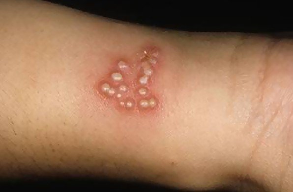
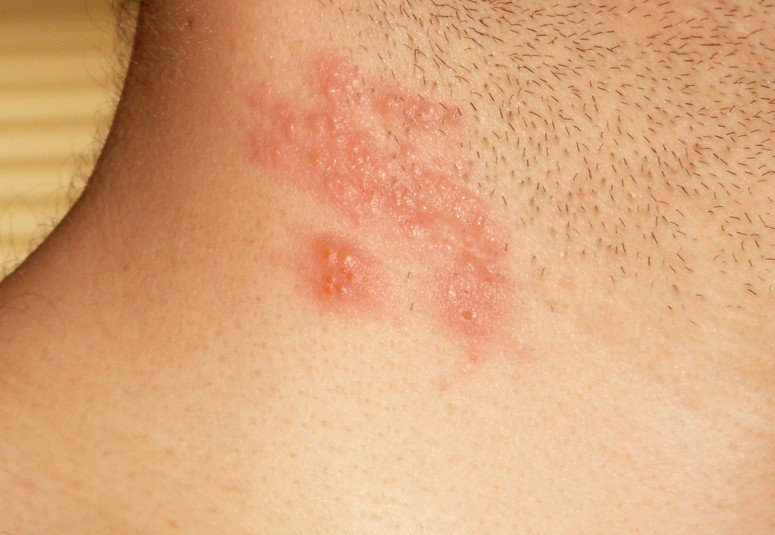
  • The lesions appear formed in clusters
  • Among the lesions, blisters filled with liquid can appear on the skin, causing a lot of pain (these blisters contain small infectious particles – in contact sports, these are easily broken and the infection is thus transmitted from person to the other)
  • Most common regions where the lesions appear include: neck area, thorax, face, abdomen and inferior limbs
  • Systemic symptoms are also present:
    • Swelling of the lymph nodes (lymphadenopathy appears as the body tries to fight the infection)
    • Fever (moderate to high-running)
    • Sore throat
    • Nausea or vomiting
    • Headaches
  • Symptoms last for a couple of weeks
  • The symptoms are more intense at the first outbreak of viral infection
  • In case of recurrent herpetic infections, the lesions heal faster and scabs are formed at approximately three days (however, it should be mentioned that even though the lesions heal, the patient will remain infectious for a couple of days after starting the treatment with antiviral medication)
  • The lesions heal on the skin without scarring
  • In some patients, there are no symptoms present (asymptomatic viral infection).

Causes

As it was already mentioned, herpes gladiatorum is caused by the infection with the herpes simplex virus (more commonly type 1 and rarely type 2).

Treatment

The infection caused by the herpes simplex virus is treated with antiviral medication. The most commonly recommended medication chosen for herpes gladiatorum includes: famciclovir, valacyclovir and acyclovir. For the best outcome, the medication is administered orally, as tablets. In many patients, antiviral medication is administered orally, with the purpose of prophylaxis. Studies have demonstrated that the administration of this medication can reduce the risk of recurring herpetic infections. Plus, this medication reduces the chances of transmitting the virus to another person – there are a lot of people who are carriers of herpetic viruses, presenting no symptoms whatsoever (except for the outbreak periods).

Is herpes gladiatorum contagious?

Herpes gladiatorum is a highly contagious condition, being transmitted through skin-to-skin contact. As it was already mentioned, the blisters on the skin contain small viral particles. Upon forceful contact, these blisters break and the virus is transmitted to the skin of the other person. The level of contagiousness is just as increased in those who are carriers of the virus – not presenting any symptoms, these people easily transmit the infection to other people. It is also important to remember that a person remains contagious even after the lesions on the skin have healed. During the outbreak, the level of contagiousness is even more increased, so you need to protect yourself and not come into contact with those who have been infected.

Herpes gladiatorum vs Shingles

The difference between herpes gladiatorum and shingles is given by the type of virus that is behind the actual infection. Herpes gladiatorum is caused by the infection with the human herpes simplex virus, type 1 and type 2. Shingles, on the other hand, are caused by a reactivation of the varicella zoster virus. This is also a herpetic virus, which is known to cause varicella during the first impact. Once infected, the virus will remain for the rest of the life in the body. During periods of low immunity, the virus can become re-activated and causing the condition known as shingles.

Patients who are diagnosed with shingles present really painful lesions on the skin, commonly following a nerve pathway. As opposed to herpes gladiatorum, this condition can become quite debilitating – because of the intense pain, patients can enter into depression and the overall quality of life can become really affected. Also, it should be mentioned that shingles does not present such a high risk of contagiousness, especially in comparison to the herpes gladiatorum condition. However, outbreaks in both cases can be triggered by intense stress, low immunity or other similar factors. Taking vitamin C and zinc supplements can improve the strength of the immunity system, reducing the risk for outbreaks in both cases.

Herpes Gladiatorum Pictures

Here are some of the pictures of Herpes Gladiatorum…
herpes gladiatorum

herpes gladiatorum pictures

herpes gladiatorum pictures 2

No responses yet

Viscosupplementation

Jul 10 2016 Published by under Treatments

Definition

Viscosupplementation is a treatment procedure recommended in patients who are suffering from degenerative conditions such as osteoarthritis. For this procedure, the doctor will inject hyaluronic acid directly into the affected joint. It is known that the patients who suffer from osteoarthritis have reduced levels of hyaluronic acid in the affected joints (this substance commonly exists in the joints of the body, contributing to the lubrication process). Apart from lubricating the joints, the hyaluronic acid also acts as an absorber of shocks, protecting the respective joints.

This procedure is recommended as a conservative treatment in patients who have been diagnosed with osteoarthritis of the tibio-femoral joint. It is the preferred method of symptomatic treatment when aspiration and corticosteroid injections have failed to provide the desired results. In deciding to recommend viscosupplementation, the doctor will take into consideration a series of factors, such as: there are no other joint problems causing the pain, the patient is not scheduled to undergo total knee replacement procedure (for the following six months) or there are no contraindications for this procedure (infection in the respective joint, hemorrhage or infection of the skin in the respective area).

How does Viscosupplementation work?

As it was already mentioned, viscosupplementation works by lubricating the affected joints with hyaluronic acid. The substance is directly injected into the joints, which allows for its rapid absorption and immediate effects. The hyaluronic acid lubricates the joints, improving the symptoms experienced by the patients diagnosed with osteoarthritis and other degenerative conditions.

Before actually injecting the hyaluronic acid, the doctor will check to see if there is excess fluid in the respective joint. If there is excess fluid in the joint, the doctor will first remove it and then inject the hyaluronic acid. Several visits to the doctor will be required, so as to obtain an improvement of the symptoms you experience. On average, between three to five injections with hyaluronic acid are required. Keep in mind that the injections are going to be made one week apart, so as to provide the best outcome for your condition.

Is it effective?

It is important to understand that viscosupplementation cannot cure osteoarthritis. But, when it comes to improving the symptoms experienced by the patients diagnosed with osteoarthritis, viscosupplementation is highly effective. Studies performed on patients who have received injections with hyaluronic acid have clearly demonstrated its efficiency. Viscosupplementation seems to be the most effective in patients who suffer from mild or moderate cases of osteoarthritis.

The effectiveness also varies from one patient to the other. Some patients might notice an improvement of their symptoms in the first weeks, while for other this treatment might not work at all. In general, the procedure can be repeated every six months.

One of the most important benefits of viscosupplementation is the pain relief brought to the patients suffering from osteoarthritis. In the majority of the patients who undergo such procedures, the pain relief is obtained in a couple of weeks (so one should not expect an immediate relief from the pain). Relief from the pain can be obtained between the third and the fifth injection course; however, most of the patients declare that relief from the pain has been obtained between eight to twelve weeks after the treatment has started.

On a long-term basis, the injections with hyaluronic acid will reduce the pain experienced, improving the overall quality of life for the patient with osteoarthritis. Apart from the analgesic properties, it seems that the hyaluronic acid is also capable of reducing the associated inflammation in the affected joint. The effects of the viscosupplementation procedure last a couple of months, this being the reason why it is recommended to repeat the procedure every six months. It is recommended that, for the next couple of days after the injections, you avoid intense physical effort or bearing weight on the respective joint.

Even though this is an effective procedure, you must always keep in mind that it cannot reverse the cartilage tear or damage in the affected joint, nor can it stimulate the cartilage to grow back. This is a procedure used for the improvements of the symptoms caused by osteoarthritis; it is not meant and cannot cure the condition.

Cost

Depending on the brand of the hyaluronic acid used for the viscosupplementation procedure, you can expect to pay around $300 – $700. Apart from that, you will have to add the fees for the doctor to the overall cost. The overall cost is also influenced by the number visits you will make to the doctor’s office. You can check your insurance policy and see if you are covered for such procedures (partial or complete coverage).

Viscosupplementation Side effects

These are the most common side-effects that can appear after the viscosupplementation procedure:

  • Pain to the site of injection
  • The area is red and warm to the touch
  • Inflammation can be noticed in the affected joint (temporary)
  • Skin rash and mild to moderate itchiness
  • Bruising around the joint
  • Allergic reaction – increased local temperature, redness, intense pain and fluid accumulation in the respective joint (emergency treatment is necessary)
  • Rarely – infection or hemorrhage.

With the exception of the rare complications, all of the above mentioned side-effects go away on a couple of days. However, if you are experiencing an allergic reaction, you should not wait for it to disappear on its own but rather seek out emergency treatment. The treatment administered in the emergency room is going to provide immediate relief from the upsetting symptoms you have experienced due to the allergic reaction.

In conclusion, viscosupplementation can improve the symptomatology of osteoarthritis, bringing the necessary pain relief and increasing the range of motion in the affected joint. At the moment, it is applied in patients who were diagnosed with osteoarthritis of the tibio-femoral joint but further research is made to extend this conservation treatment to other joints in the body (shoulder, hip etc.). Studies are also undertaken in order to determine whether viscosupplementation would be a suitable treatment alternative for other orthopedic conditions and degenerative diseases.

No responses yet

Cachexia

Jul 08 2016 Published by under Diseases and Conditions

Definition

Cachexia is a medical condition in which severe weight loss occurs without the person intending to achieve such an objective. The syndrome is accompanied by a wide range of other symptoms, as you will have the opportunity to find out in this article. It should be noted that the weight loss that results from cachexia cannot be recuperated through nutrition. Even if the patient is administered an increased number of calories, the loss of lean body mass is going to continuously occur, which is significant of an underlying pathology.

Cachexia Symptoms

These are the most common symptoms that are encountered in patients with cachexia:

  • Significant weight loss (body fat)
  • Atrophy of the muscles
  • State of general fatigue and weakness
  • Severe appetite loss
  • Asthenia
  • Early satiety sensation
  • Anorexia
  • Nausea
  • Changes in the taste
  • Anemia symptoms
  • Hormonal imbalances
  • Patient – characteristic appearance for chronic illness (emaciated)
  • In terms of fever, the patient can be normothermic, febrile or hypothermic
  • Changes of the skin – modified texture, loose folds
  • Bony protuberances become more obvious, as the body fat is lost
  • Gum atrophy
  • Sagging and shriveled breasts (due to the loss of fat)
  • High heart rates
  • Eschars and ulcerations appear on the skin, due to the loss of fat and circulatory problems
  • Slow growth of the nails and hair.

Causes

These are the most common causes that lead to cachexia:

  • Different types of cancer
    • Common seen in patients who are terminal (end-stage cancer)
  • HIV/AIDS
    • Involuntary weight loss (wasting)
    • Severe protein energy malnutrition
    • Cachexia can contribute to the progression of the condition
  • Chronic obstructive lung disease
    • Patients experience shortness of breath, cough and excess mucus
    • Systemic inflammation is present, contributing to the appearance of cachexia
    • The metabolism is affected by the inflammation at the level of the lungs
  • Multiple sclerosis
    • Cachexia is present in those with advanced stages of multiple sclerosis
  • Congestive heart failure
    • Cardiac cachexia syndrome
    • Cachexia appears as a complication of the chronic congestive heart failure
    • The nutrition and metabolism are affected, with severe weight loss and other symptoms of cachexia becoming obvious
  • Tuberculosis
    • Cachexia – pathognomonic sign of pulmonary tuberculosis
    • Poor prognosis
  • Familial amyloid polyneuropathy
    • Rare autosomal dominant disease
    • Affects the autonomic nervous system
    • Cachexia is one of the symptoms
  • Gadolinium poisoning
    • Can cause severe toxicity, interfering with the metabolism and leading to the appearance of cachexia symptoms
  • Mercury poisoning (acrodynia)
    • Mercury cachexia leads to systemic exhaustion and changes in the mental status
  • Hormonal deficiency
    • Often encountered in those who also suffer from congestive heart failure
  • Metabolic acidosis
    • Decrease protein synthesis and increased protein consumption
    • Encountered in patients diagnosed with rare types of cancer
  • Chronic pancreatitis
    • Loss of pancreatic function can impair the overall metabolism and lead to the appearance of cachexia symptoms
  • Autoimmune conditions
    • Rheumatoid arthritis
    • Autoimmune arthritis
  • Amphetamine addiction
    • Cachexia appears due to the physical wasting caused by the excess amphetamine consumptions
  • Chronic kidney disease
    • Systemic inflammation – cachexia symptoms appear
  • Cystic fibrosis
    • Cachexia symptoms appear in the more severe stages of the disease
  • Motor neuron disease
    • Cachexia may be caused by the appearance of symptoms such as dysphagia
  • Neurodegenerative diseases
    • Parkinson’s disease
    • Dementia.

Treatment

First of all, it is important to understand that the treatment of cachexia depends on the cause that led to its appearance. In many patients, given the fact that they are terminally ill, it is more about the management rather than the treatment. If there are certain causes that can be reversed, these will be treated, improving the symptoms of cachexia and the overall quality of life for the patient. Apart from the administration of the medication, the treatment of cachexia and its symptoms includes nutritional counselling, physiotherapy and psychological counselling.

It is believed that a single course of therapy is not efficient in treating the symptoms of cachexia. This is why the recommended therapeutic approach for cachexia includes the administration of medication, nutritional counseling and other non-drug therapies. Corticosteroids can be administered as a short-term therapy (increasing the appetite) but their administering cannot be prolonged, as it leads to serious side-effects. For example, methylprednisolone might be used for a period of 2 weeks, in a patient who suffers from refractory cachexia. If a person suffers from cachexia, with anorexia as the primary concern, the symptoms can be ameliorated through the administration of synthetic progestogens (progestins). Studies have shown that megestrol, a synthetic progestogen, may contribute to an increase in both the appetite and the overall weight.

The medication that can be administered in patients suffering from cachexia includes but is not limited to the following choices:

  • Thalidomide (cytokine antagonist)
  • Cannabinoids (dronabinol)
    • Increase the appetite but not the overall weight
  • Omega-3 fatty acids – eicosapentaenoic acid
    • Reduce the release of cytokines
  • NSAIDs
    • Recommended in those who present high levels of CRP (C-Reactive protein)
    • More useful in patients who suffer from cachexia caused by other medical problems than cancer
    • Not beneficial in patients who suffer from refractory cachexia
  • Prokinetics
  • Ghrelin + ghrelin receptor agonists – stimulate the appetite
  • MT-102 (anabolic catabolic transforming agents)
  • Selective androgen receptor modulators
  • Cyproheptadine
  • Hydrazine

In some parts of the United States, it is acceptable for marijuana to be used as a treatment for the symptoms of cachexia, as it improves the appetite like the other cannabinoids. Oral fish oil has also been proposed as a potential natural remedy but studies demonstrating its efficacy remain inconclusive.

Parenteral nutritional support is recommended to reduce the risk of morbidity in patients who are suffering from cachexia. The decision to provide such kind of support depends on the swallowing abilities. Normal eating is encouraged in terminal patients, so as to reduce the level of stress. Antioxidants might be used in order to reduce the levels of the oxidative stress. Vitamin and mineral supplements may be administered in less severe cases of cachexia. Physical therapy might reduce the wasting of the muscle mass, reducing the risk of eschars and ulcers due to the prolonged decubitus.

No responses yet

Garcinia Cambogia

Jul 06 2016 Published by under Fitness and Diet

Garcinia Cambogia is a tropical fruit that is commonly found in regions of Indonesia and other countries of South Asia, being often consumed for its weight loss properties. It is said that the fruit contributes to the weight loss process by blocking the production of fat and reducing the appetite. Other health benefits are also presented for both the real fruit and the supplements or pills available on the market, such as the maintaining of the blood sugar at normal levels and the reduction of bad cholesterol.

The fruit is also known by other names, such as: Malabar tamarind, Pot tamarind, kudampuli or brindleberry. It is small and shaped like a pumpkin, being sweet to the taste. Its color ranges from green to pale yellow. It was around the 1960s that studies showed that the fruit contains an active acid, similar to the one found in other citrus fruits, such as lemons or oranges. The identified acid was hydroxycitric acid (abbreviated HCA) and soon was used for the different weight loss supplements.

garcinia cambogia

In the paragraphs below, you will discover how Garcinia Cambogia works and how it promotes the weight loss. At the same time, you will be given useful information regarding the other benefits and recommended dosage. Make sure that you read the safety and side-effects paragraphs as well. Read about the pills and the different places from where to buy the supplements. And, most important, try to decide whether this is a suitable weight loss solution for you or not. Enjoy reading!

What does Garcinia Cambogia do to your body?

As it was already mentioned, the active ingredient of Garcinia Cambogia is represented by the hydroxycitric acid. This works by stimulating the burning of the fat and reducing the appetite. Once the amount of fat is reduced and the sensation of hunger is reduced, the weight loss process is on the right path. The acid will block the citrate lyase enzyme, which the body commonly uses for the production of fat. As for the reduction of the appetite, this occurs as Garcinia Cambogia contributes to an increase of the serotonin levels. This brain chemical is known as the happiness hormone, reducing the appetite among other positive effects.

Garcinia Cambogia seems to be a suitable choice for those who are suffering from diabetes, as it helps one to keep the blood sugar at stable levels. Other positive effects on the health for Garcinia Cambogia include: reduction of bad LDL cholesterol, increase of good HDL cholesterol and reduction of triglycerides.

Garcinia Cambogia for Weight loss

Garcinia Cambogia supplements and pills have been recommended to those who are interested in losing weight. As it was already mentioned, Garcinia Cambogia is known to block citrate lyase, this enzyme being responsible for the transformation of the sugar and starch into fat. Once the enzyme is blocked, the carbs are rather used to provide the body with energy rather than be accumulated as fat or increase the sugar levels.

The suppression of the appetite is another reasons why Garcinia Cambogia has been recommended for weight loss purposes. The increase in serotonin levels contributes to the increased satiety – this means that you will feel a reduced need to eat. The elevated levels of serotonin contribute to the reduction of hunger, promoting weight loss among other beneficial effects.

Even though Garcinia Cambogia has been often presented a solution for excess weight, the research is still under way. It is not certain whether the side-effects of Garcinia Cambogia are more harmful than the benefits offered. It is for the best to take these supplements or pills only under the guidance of a specialized doctor or dietitian, so as to make sure they work to your benefit.

Does Garcinia Cambogia work?

The studies that have been performed on rats have shown that Garcinia Cambogia indeed promotes the weight loss process. These studies have demonstrate both the reduction of appetite and the blockage of the citrate lyase in rats.

For the studies that were performed on humans, it should be mentioned that the people who took part in the study took Garcinia Cambogia extracts for three times a day, with half an hour before the eating. All of the participants to the study were overweight and interested in losing weight. Apart from taking the Garcinia Cambogia extract, they also had to change their diet, reducing their caloric intake and increasing the amount of fiber. The study was undertaken for a period of 12 weeks and it was later published in The Journal of The American Medical Association.

In the respective study, the participants were split into two groups – one group received the Garcinia Cambogia extract, while the other one was administered placebo. The results of the study demonstrated that both groups lost weight; however, the group that received the Garcinia Cambogia extract managed to lose more weight than the one receiving placebo.

Other studies have demonstrated that, by taking Garcinia Cambogia extracts, one can achieve short-term weight loss. However, more studies are required to study the long-term effects of Garcinia Cambogia and the possible side-effects that might occur with taking such supplements for prolonged periods of time.

Dosage

The recommended dosage of Garcinia Cambogia varies between 1500 mg/day and 2800 mg/day, depended on the product and the manufacturer. In general, it is recommended that you check out the bottle and see the recommended dosage.

Garcinia Cambogia extract can be taken as pills, which are recommended to be taken with half an hour or an hour before eating. You need to take these pills with plenty of water. It is recommended that you take the pills three times a day, each pill representing approximately 500 mg of active substance.

You can also consider taking liquid Garcinia Cambogia. The recommended dosage for the Garcinia Cambogia in the liquid form is one or two drops. The drops should be placed under the tongue, for the best absorption and they should be maintained as such for about a minute. Just as the pills, the drops should be taken with 30 or 60 minutes before eating.

Benefits

These are the benefits of taking Garcinia Cambogia:

  • Prevention of fat storage (blockage of citrate lyase enzyme)
  • Reduction of the appetite through the increase of serotonin levels
  • Increased exercise performance (especially in women)
  • Treatment for infestations with different parasites or worms
  • Constipation
  • Diarrhea
  • Stable blood sugar levels (especially in patients who are suffering from diabetes)
  • Improvement of cholesterol (reduction of bad LDL cholesterol levels and increase of good HDL cholesterol levels)
  • Reduced risk of atherosclerosis or heart disease (reduction of triglycerides)

Is Garcinia Cambogia safe?

Taking Garcinia Cambogia supplements is safe, as the reported side-effects are mild and they do not represent a subject of concern. However, if you want to be absolutely certain that this is the right product for you, talk about the possibility of taking it with a doctor or a dietitian. In deciding whether Garcinia Cambogia is a safe choice for you or not, the doctor or dietitian will take into account other pre-existing medical conditions you are suffering from, your age and overall physical health. However, in general, for the majority of the people, Garcinia Cambogia is a safe choice.

Side effects

The studies that have been conducted for Garcinia Cambogia have only been short term, so it is quite difficult to assess the side-effects on a long term basis. However, when taking Garcinia Cambogia for under 4 months, the possible side-effects are possible to appear: nausea, discomfort or pain in the abdomen and headaches. It is also recommended to avoid taking Garcinia Cambogia during pregnancy or while you are breastfeeding, as it is not certain how the active ingredients of these supplements might affect the growing baby. A study performed on mice in 2005 has demonstrated that the long-term administration of high doses of Garcinia Cambogia might have a negative impact on the health of the testicles. However, further research did not manage to prove the effectiveness of the previously-mentioned study.

Other side-effects of Garcinia Cambogia might include: vertigo, dry mouth and toxicity of the liver. The FDA (Food and Drug Administration) prevented the usage of a product that was advertised as containing Garcinia Cambogia, linking it to many case of hepatic toxicity. However, this product also contained other active ingredients, so it is not certain whether it was Garcinia Cambogia actually causing the problems.

When taking Garcinia Cambogia, you must also take into account that the extracts might interact with the different medication you might be taking. Among the medication that can interact with the Garcinia Cambogia extracts, there are: drugs for respiratory conditions (such as asthma) and allergies; medication for diabetes (insulin included); iron supplements for different types of anemia; anti-inflammatory medication; drugs for different psychiatric conditions; medication for lowering cholesterol levels (such as statins) and blood thinning medication (such as warfarin).

Pills

As it was already mentioned, Garcinia Cambogia is available, among other forms, as pills. If you have decided to take Garcinia Cambogia pills, remember to take only the recommended dosage (see the dosage paragraph). The pills have to be taken three times a day, with a tall glass of water. It is also recommended that the pills are taken with thirty or sixty minutes before eating.

You need to avoid the products that have a high content of calcium salts, as these are not good for you. In general, you should search for pills that contain at least 50% of Garcinia Cambogia extract and preferably other healthy minerals, such as potassium or magnesium. Choose the pills that have a low lactone content, as this is not good for you either.

The pills are recommended to be taken on an empty stomach, so as to maximize the appetite suppression and also be to be 100% absorbed. If you were to take them with food, their effect would not be as powerful as it should. When food is present in the stomach, the Garcinia Cambogia extract might bind to the food molecules and lose some of its properties, becoming in activated.

Where to buy Garcinia Cambogia?

If you are interested in buying Garcinia Cambogia extracts for weight loss and other health related benefits, there is no better way to do that than the Internet. As you will have the opportunity to discover, there are many online stores that sell such products at attractive prices. However, you must do a thorough research and find a store that is both reputable and reliable. You can determine whether that store is a good choice or not by reading the reviews of other customers – if there are a lot of positive reviews, this means that you have found the right one. On the other hand, if there are a lot of people complaining about the service or the quality of the Garcinia Cambogia extracts, close the page and move on.

A reliable online store should provide detailed information on the Garcinia Cambogia extract that you want to purchase. It should clearly state the percentage of active substance contained in the product, as well as the rest of the ingredients. You ought to be provided with information on how to take the products, what is the recommended dosage and the potential side-effects. Do not be quick to choose an online store just because it has reduced prices; it is better to pay a little bit extra and be certain that you are purchasing Garcinia Cambogia extract and not some bogus product presented under this name.

In conclusion, Garcinia Cambogia extract can be used with the purpose of weight loss but, remember, it will never work like magic, taking your extra weight in the blink of an eye. You need to start working out and change your diet, including only healthy choices. Leave the couch and go outside, start jogging and being physically active. Say goodbye to fast-food, sugary drinks and refined desserts. Change your life and never forget that product such as Garcinia Cambogia are supplements to a healthy diet. Good luck and stay healthy!

No responses yet

Mastoidectomy

Jul 05 2016 Published by under Ear, Nose and Throat

What is Mastoidectomy?

Mastoidectomy is a surgical intervention in which the mastoid air cells (portion of the mastoid bone) are removed due to their being infected or damaged.

Types

There are five different types of mastoidectomy:

  • Simple mastoidectomy
    • The mastoid cortex is opened and the aditus ad antrum is identified
  • Complete mastoidectomy (canal up)
    • Removal of all the mastoid cells from the following areas: tegmen, presigmoid dural plate, sigmoid sinus and external auditory canal (posterior wall)
    • Even though the mastoid cells from the posterior wall of the external auditory canal are removed, the wall itself is maintained
  • Complete mastoidectomy (canal down)
    • The mastoid cells are removed from all the above mentioned areas
    • Apart from that, parts of the external auditory canal are going to be removed as well (posterior wall and superior osseous part)
    • The intervention requires the reconstruction of the tympanic membrane, so as to guarantee a separation between the middle ear and the mastoid cavity, with the ear canal
  • Modified radical mastoidectomy
    • Procedure similar to the one of canal down mastoidectomy
    • Difference – no manipulation of the middle ear space or of the tympanic membrane
    • Recommended – no extension of cholesteatoma (squamous epithelium) in the middle ear space or in patients who have this problem in the ear with the better hearing
  • Radical mastoidectomy
    • Procedure similar to the one of canal down mastoidectomy
    • Tympanic membrane and ossicles are not reconstructed
    • The middle ear and the mastoid remain exteriorized
    • Soft tissue is used for the obliteration of the Eustachian tube, so as to reduce the risk for chronic conditions, such as otorrhea
    • Skin grafts are often placed at the level of the middle ear, thus reducing the risk for otorrhea or other chronic problems, such as mucosalization.

Why is Mastoidectomy performed?

These are the most common reasons why mastoidectomy is performed:

  • Infection of the air cells that are part of the mastoid bone
    • Infection spread from the middle ear
    • Mastoiditis
    • Chronic otitis media (with or without cholesteatoma)
  • Cholesteatoma (inflammatory disease)
    • Excess growth of epithelium in the middle ear
  • Repairing of the paralyzed facial nerves
  • Placement of a cochlear implant
    • Purpose – rehabilitate the acquired or congenital hearing loss
    • Mastoidectomy – step of the overall procedure
  • Removal of lateral skull base neoplasms (epidermoids, paragangliomas of the temporal bone, meningiomas and vestibular schwannomas)
    • Mastoidectomy – step of the overall procedure
  • Complications of otitis media
    • Intratemporal/intracranial suppuration
    • Lateral venous sinus thrombosis.

Mastoidectomy Procedure

The procedure is performed under general anesthesia. The patient is placed in a supine position, with the head rotated at an angle of 30-45 degrees. After the anesthesia has kicked in, the head of the patient is going to be rotated at an even higher angle (180 degrees).

For the procedure, the surgeon will access the infected or damaged part of the mastoid bone through the ear or by making a cut behind the ear. Small surgical instruments are going to be used in order to access the actual mastoid bone. Suction irrigation is going to be used to remove any small bone particles, after the surgeon has drilled into the mastoid bone. The infected mastoid air cells are going to be removed and the surgeon is going to close the surgical field. The operation site is going to be covered with a gauze, allowing for the excess fluid to be drained. During the procedure, the surgeon might use a special monitor, so as to prevent the damaging of the facial nerve.

How long does a mastoidectomy take?

On average, if there are no complications, the procedure takes approximately 3 hours.

Cost

In calculating the costs of the procedure, you will have to take several factors into account, such as: anesthesia fees, doctor fees and the clinic in which the intervention is performed. Also, if there are any complications during the surgery or you may need to spend some time in the ICU, this might also translate into higher overall costs. You will also have to check your insurance policy, so see if this procedure is covered or not. In general, you can expect to pay around $7000 for a procedure, with a discount given by the insurance. Keep in mind that this cost refers strictly to the mastoidectomy – if you are having a cochlear implant, for example, you will have to pay extra.

Complications

These are the complications that can occur with the mastoidectomy procedure:

  • Paresis or paralysis of the facial nerve (nerve injury)
  • Hearing loss (occurs in 6% of the patients)
  • Vertigo (temporary complications, goes away on its own a few days after he surgery)
  • The taste can be affected (patients describe having a metallic or a sour taste; others complain of their taste being reduced in intensity – temporary complication as well, goes away in a couple of months)
  • Ringing sound in the ears (tinnitus) or other abnormal noises (popping or hissing)
  • Mastoid cutaneous fistula – rare complication
  • Dural injury – occurs during the drilling of the mastoid bone
  • Vascular injury – often at the level of the sigmoid sinus (large bleeding).

Recovery

The recovery process is slow and there are some patients who might require a second intervention in the next six to twelve months. The recovery process can be shortened by following the advice of the doctor and protecting yourself against the risk of infection.

What to expect after mastoidectomy surgery?

After the surgery, the doctor will recommend that you use antibiotic ear drops, so as to reduce the risk of postoperative infection. Pain medication might also help with the pain and inflammation during the recovery period (avoid taking aspirin as this can increase the risk for bleeding). During bathing, you will need to cover the surgical site. For the first weeks after the surgery, you will have to avoid the excess physical exercise. You must not place pressure on the ear and it is important to avoid flying until you get the okay from the doctor. Swimming should also be postponed until the doctor says it is alright.

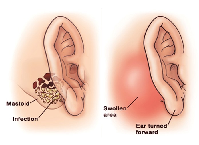
Mastoidectomy Anatomy Pictures

mastoidectomy pictures

No responses yet

© 2011-2019 MDDK.com - Medical Tips and Advice. All Rights Reserved. Privacy Policy
The health information provided on this web site is for educational purposes only and is not to be used as a substitute for medical advice, diagnosis or treatment.